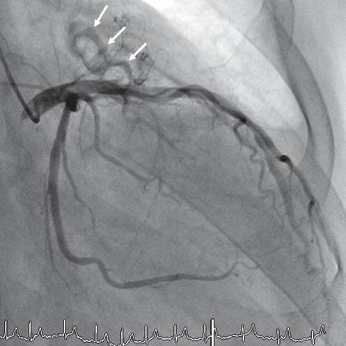
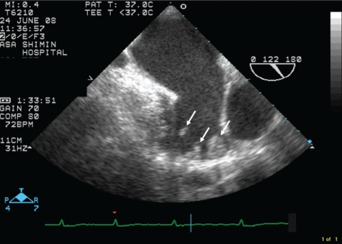
A 77-year-old male patient was admitted to the hospital because of chest pain. Electrocardiography revealed atrial fibrillation with high voltage. Coronary angiography (CAG) revealed left atrial appendage (LAA) thrombi with abnormal neovascularization arising from the left atrial branch of the left circumflex artery in the right anterior oblique projection (arrows in Figure 1). There were no angiographical findings indicating severe stenosis or embolic occlusion. Transesophageal echocardiography also showed severe spontaneous echocardiography contrast and hypoisoechoic thrombi in the LAA without mitral stenosis (arrows in Figure 2). Although previous studies (1–3) have reported that CAG is useful for the diagnosis of LAA thrombi in patients with mitral stenosis, it is rare that both CAG and transesophageal echocardiography clearly reveal the thrombi without mitral stenosis.
Figure 1.
Figure 2.
REFERENCES
- 1.Fu M, Hung JS, Lee CB, et al. Coronary neovascularization as a specific sign for left atrial appendage thrombus in mitral stenosis. Am J Cardiol. 1991;67:1158–60. doi: 10.1016/0002-9149(91)90888-r. [DOI] [PubMed] [Google Scholar]
- 2.Sakamoto I, Hayashi K, Matsunaga N, et al. Coronary angiographic finding of thrombus in the left atrial appendage. Acta Radiol. 1996;37:749–53. doi: 10.1177/02841851960373P264. [DOI] [PubMed] [Google Scholar]
- 3.Bochna AJ, Falicov RE. Diagnosis of intracardiac thrombi in mitral stenosis and left ventricular dysfunction. Use of selective coronary arteriography. Arch Intern Med. 1980;140:759–62. [PubMed] [Google Scholar]


