Figure 1.
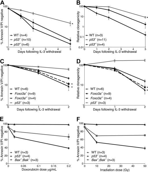
Loss of p53 but not loss of FoxO3a confers short-term as well as clonogenic survival advantage to IL-3–dependent HoxB8-immortalized myeloid progenitor cells after cytokine deprivation. (A) Multiple independent IL-3–dependent HoxB8-immortalized myeloid progenitor lines (number of individual clones tested indicated as n) of the indicated genotypes were deprived of IL-3 over a 5-day time course. Cell viability was determined by flow cytometry analysis after staining with FITC-conjugated Annexin V plus PI. The results show means ± SEMs of 2 independent experiments. Asterisk denotes a P value between WT and p53−/− cells of .0012 at day 1, .0018 at day 2, and .044 at day 5 with t test (2-tailed, 2-sample equal variance). (B) Cells from panel A were plated in soft agar in the presence of IL-3 after the indicated times of culture in the absence of IL-3 (n refers to the number of independent clones analyzed). Colonies were counted after 14 days, and their clonogenicity was determined. The results show means ± SEMs of 2 independent experiments. Asterisk denotes P value between WT and p53−/− cells of .019 at day 1, .0051 at day 2, and .023 at day 5 with t test (2-tailed, 2-sample equal variance). (C) Multiple independent FDM lines (numbers indicated as n) of the indicated genotypes were deprived of IL-3 over a 2-day time course. Cell viability was determined as described in panel A. The results show means ± SEMs of 4 independent experiments. Asterisk denotes a P value between WT and FoxO3a−/− cells of .0025 and between WT and FoxO3a+/− cells of .0023 at day 2 with t test (2-tailed, 2-sample equal variance). (D) Clonogenicity of cells from panel C was determined as described in panel B. Asterisk denotes a P value between WT and FoxO3a−/− cells of .052 and between WT and FoxO3a+/− cells of .015 at day 2 with t test (2-tailed, 2-sample equal variance). The results show means ± SEMs of 4 independent experiments. (E) Multiple clones (n) of the indicated genotypes were cultured in the presence or absence of doxorubicin for 24 hours before viability was analyzed as described in panel A. The results shown are means ± SEMs of 2 independent experiments. (F) Multiple clones (n) of the indicated genotypes were subjected to irradiation were then cultured for 24 hours. Viability was analyzed as described in panel A. The results shown are means ± SEMs of 4 independent experiments.

