Abstract
Context:
Knowledge of the kinetic changes that occur during sloped running is important in understanding the adaptive gait-control mechanisms at work and can provide additional information about the poorly understood relationship between injury and changes in kinetic forces in the lower extremity. A study of these potential kinetic changes merits consideration, because training and return-to-activity programs are potentially modifiable factors for tissue stress and injury risk.
Objective:
To contribute further to the understanding of hill running by quantifying the 3-dimensional alterations in joint kinetics during moderately sloped decline, level, and incline running in a group of healthy runners.
Design:
Crossover study.
Setting:
Three-dimensional motion analysis laboratory.
Patients or Other Participants:
Nineteen healthy young runners/joggers (age = 25.3 ± 2.5 years).
Intervention(s):
Participants ran at 3.13 m/s on a treadmill under the following 3 different running-surface slope conditions: 4° decline, level, and 4° incline.
Main Outcome Measure(s):
Lower extremity joint moments and powers and the 3 components of the ground reaction force.
Results:
Moderate changes in running-surface slope had a minimal effect on ankle, knee, and hip joint kinetics when velocity was held constant. Only changes in knee power absorption (increased with decline-slope running) and hip power (increased generation on incline-slope running and increased absorption on decline-slope running in early stance) were noted. We observed an increase only in the impact peak of the vertical ground reaction force component during decline-slope running, whereas the nonvertical components displayed no differences.
Conclusions:
Running style modifications associated with running on moderate slopes did not manifest as changes in 3-dimensional joint moments or in the active peaks of the ground reaction force. Our data indicate that running on level and moderately inclined slopes appears to be a safe component of training regimens and return-to-run protocols after injury.
Keywords: biomechanics, decline running, incline running, joint moments, joint power
Key Points.
Running style modifications on moderate slopes did not manifest as changes in 3-dimensional joint moments or active peaks of the ground reaction force.
However, changes in knee power absorption (increased on the decline slope) and hip power (increased generation on the incline slope and increased absorption on the decline slope in early stance) were seen. The impact peak of the vertical ground reaction force increased only during running on the decline slope.
Running on level and moderately inclined slopes appears to be a safe component of training regimens and return-to-run protocols after injury.
Incline and decline hill training is regularly used by distance runners to improve cardiovascular conditioning and to increase strength.1 However, this training can bring about kinetic changes (in ground reaction forces [GRFs], joint moments, and joint powers) that may be related to the onset or exacerbation of injuries in runners. Knowledge of the kinetic changes that occur during sloped running is important in understanding the adaptive gait-control mechanisms at work and can provide additional information about the poorly understood relationship between injury and changes in kinetic forces in the lower extremity. By first establishing a profile of normal kinetic changes incurred during decline, level, and incline running in the uninjured runner, we hope to provide a reference point for practitioners seeking to design appropriate training regimens for healthy runners as well as suitable return-to-activity programs for injured athletes.
Changes in lower extremity joint mechanics and physiology during sloped running have been previously reported in the literature. Increased oxygen consumption, heart rate, blood lactate concentration, and lower limb muscle activity have been associated with incline running.2–7 As Yokozawa et al7 suggested, these findings imply that the mechanical load and energy cost to the lower extremity are greater during incline running than during level running. Using a musculoskeletal model to compare level and incline kinetics, Yokozawa et al7 found that muscle activation of the hip extensors and hip flexors was augmented during incline running at high speeds. Furthermore, knee extension torques were greater during incline running than during level running at all speeds.7 Researchers8,9 have demonstrated important biomechanical changes during decline running as well, finding that the vertical impact force peak increased by as much as 14%. Additionally, Buczek and Cavanagh10 measured increased power absorption at the ankle and knee joints during decline running. Gottschall and Kram,9 studying the effects of sloped running on GRFs, noted that neither decline nor incline running affected normal active force peaks but that the impact peak of the vertical GRF increased during decline running. No authors have found a direct correlation between sloped running and injury risk, but it is worthwhile to recognize that changes in force demands on level surfaces have been suggested11–20 to play a role in the development or progression (or both) of joint injury.
Although select kinetic factors have been previously analyzed during sloped running, no researchers have focused on investigating the complete 3-dimensional kinetic changes at the ankle, knee, and hip during decline, level, and incline running. A global analysis of the biomechanical compensations occurring throughout the gait cycle during decline, level, and incline running may reveal alterations in joint kinetics similar to those previously theorized to increase injury risk. Specifically, these alterations may correlate with changes in the need for joint stabilization under different running conditions. In addition, changes in joint kinetics may provide insight into redesigning training and return-to-activity programs to reduce the risk of running-related injury. We believe it prudent to examine these kinetic alterations with changes in surface slopes typically encountered by the majority of runners. Our aim in this study was to contribute further to the understanding of hill running by quantifying the 3-dimensional alterations in joint kinetics during moderately sloped decline, level, and incline running in a group of healthy runners.
METHODS
Participants
Twenty-one healthy young runners or joggers between 18 and 36 years of age were recruited from the local population; 19 (9 females, 10 males) of these runners were ultimately studied. Two of the original 21 volunteers were excluded as a result of data problems: a data set that we were unable to debug in one case and lack of complete data in another. The participants were regular runners who ran or jogged at least 20 miles (32.19 km) weekly. Additionally, they were free of chronic musculoskeletal conditions and had experienced no running-related injuries within 6 months of testing. The protocol was approved by the Institutional Review Board for Health Science Research, and informed consent was obtained from each individual before testing. Mean age, height, and mass were 23.9 ± 2.5 years, 166.4 ± 6.2 cm, and 56.2 ± 4.8 kg, respectively, in the females and 26.6 ± 5.9 years, 180.5 ± 5.2 cm, and 74.2 ± 8.1 kg, respectively, in the males.
Protocol
The same laboratory technician placed all retroreflective markers used for motion capture, and marker placement remained the same during the treadmill slope conditions. Specifically, markers placed on the left and right anterior and posterior superior iliac processes defined the motion of the pelvis. The motion of each lower limb segment was tracked by markers on the lateral femoral condyles, lateral mid-thighs, lateral mid-shanks, lateral malleoli, posterior calcanei, and second metatarsal heads. This set of 16 retroreflective markers defined the 3-dimensional kinematics of the pelvis and the left and right thighs, shanks, and feet.
After 3 to 5 minutes of practice running on the instrumented treadmill, participants ran at 3.13 m/s (7 mph) on the treadmill, approximating an 8.5-minute mile, on the following 3 surface slopes: 4° (6.98% grade) decline, level, and 4° (6.98% grade) incline. The running speed was selected to be consistent with that in the established running-related literature7,9,21 and was commensurate with the abilities of our volunteers, as determined through a face-to-face screening performed by the study coordinator. Decline and incline slopes were chosen to emulate pitches commonly encountered by runners in real-world conditions. Participants ran in their personal running shoes, and the sequence of running slopes was randomly selected for each person.
Measurements
Kinematic data were recorded using a 10-camera motion capture system (model 624; Vicon Peak, Lake Forest, CA) operating at 120 Hz. Ground reaction force data were acquired at 1000 Hz in synchrony with the motion capture data and were collected using a compound instrumented treadmill (AMTI, Watertown, MA). Paolini et al22 previously reported on the characteristics of the treadmill force plates. Briefly, the instrumented treadmill is an assembly of 3 treadmill force plate units. Two smaller units (0.33 m × 1.40 m) sit side by side behind a larger unit (0.66 m × 1.4 m), providing a total running surface that is 0.66 m wide by 2.80 m long. Running data were captured from the largest of the 3 treadmill force plate units to ensure participant comfort. The existence of a flight phase in running allowed for the collection of right-side and left-side GRF data from a single force plate unit.
The treadmill GRF data were preprocessed using in-house algorithms, implemented in LabVIEW (National Instruments Corp, Austin, TX). The preprocessing software detected heel strikes and toe-offs using a 60-N (approximately 5%–10% body weight) threshold for the vertical component of the force vector of GRF. The 60-N threshold was necessary to distinguish characteristic gait cycle events (eg, heel strike, toe-off) as a result of the increased noise levels inherent in the treadmill signals.22,23 Treadmill GRF data were filtered using a low-pass Butterworth filter with a cutoff frequency of 30 Hz. A Woltring filtering technique was applied to marker data, with a predicted mean squared error value of 20. The preprocessed, filtered treadmill data were combined with the filtered motion capture data, and 3-dimensional kinetics were calculated through a full inverse dynamic model implemented using the Vicon Plug-in Gait.
Analysis
Individual and group means were obtained using in-house algorithms developed with LabVIEW. Participant average maximums and minimums for each kinetic variable during the stance phase of gait only were extracted from the average curve of 10 consecutive gait cycles, each of which was first normalized to 100% of the gait cycle. The average overall cycles included in the analysis were reported for each condition. Ground reaction forces and joint powers were normalized to body mass. Joint moments were normalized to standing height and body mass and were reported as external moments. Variables under analysis included the peak 3-dimensional joint moments; peak sagittal-plane joint powers at the ankle, knee, and hip; and the 3 components of the GRF. From among these factors, we evaluated a total of 20 kinetic maximums and minimums during the stance phase of gait. A secondary examination of temporal and spatial factors was performed, including measures of cadence and stride length. The sloped (decline and incline) running conditions were compared with the condition of the level surface. The significance of group mean differences in the maximums and minimums was evaluated using a 1-way analysis of variance for repeated measures, followed by the Fisher least significant difference test for pairwise comparisons. After we applied a Bonferroni adjustment for the 21 primary kinetic comparisons, significance was defined as P < .002 (0.05/21). Significance for all secondary comparisons of stride length and cadence was left at P < .05.
RESULTS
The average speed of all participants was identical during the sloped conditions, in accordance with the protocol design. One volunteer had a running speed of 7.5 mph (12.07 km/h) as a result of a miscalibration of the treadmill running speed. While running at this constant velocity, the individual's cadence increased (170.5 ± 7.9 versus 168.5 ± 8.1 steps/min, P = .01) and stride length decreased (1.26 ± 0.1 versus 1.28 ± 0.1 m, P = .05) for incline running compared with level running. No statistically significant differences in these variables were observed between level running and decline running (decline cadence: 167.6 ± 7.7 steps/min, P = .20; decline stride length: 1.28 ± 0.6 m, P = .26).
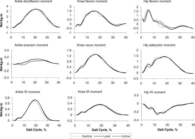
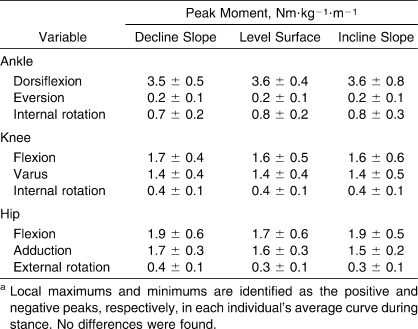
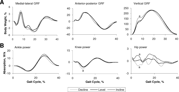
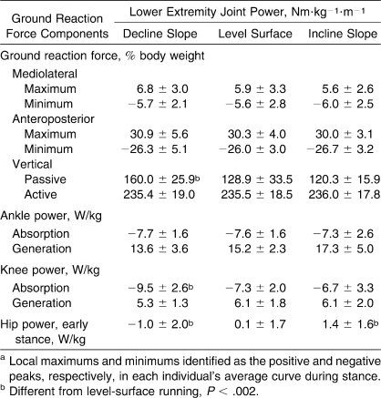
No differences were noted in the 3-dimensional joint moments at the ankle, knee, and hip during moderate-slope running when holding velocity constant (Figure 1; Table 1). Changes were seen, however, in knee and hip power (Figure 2; Table 2). The general behavior of the hip power curve during stance was altered with changes in running slope. Further, only the characteristic peaks of the hip power curve in early stance were consistently identifiable among the population for each condition. In early stance, hip power generation increased during incline running, whereas hip power absorption increased during decline running. Also, power absorption increased at the knee during decline running. Ankle power did not demonstrate statistically significant differences among running conditions. We observed an increase only in the impact peak of the vertical GRF during decline running, whereas the nonvertical GRF components exhibited no differences (Figure 2; Table 2).
Figure 1.
Three-dimensional joint moments during stance for decline (gray), level (black), and incline (dotted) running normalized to body mass. No differences were found. Abbreviation: IR, internal rotation.
Table 1.
Peak Joint Moments Normalized by Standing Height and Body Mass (Mean ± SD)a
Figure 2.
Three-dimensional (A) ground reaction force (GRF) and (B) joint powers during stance for decline (gray), level (black), and incline (dotted) running normalized to body mass. a Indicates running peaks different from level running at P < .002. For (B), a value greater than 0 reflects power generation; a value less than 0 reflects power absorption.
Table 2.
Peak Ground Reaction Force Components and Joint Powers Normalized to Body Mass (Mean ± SD)a
DISCUSSION
Our aim in this study was to contribute further to the understanding of hill running by investigating 3-dimensional alterations in joint kinetics, which may reflect differences in joint stabilization and the risk of injury during decline, level, and incline running. We hypothesized that alterations in running slope would affect joint kinetics at the lower limb. Surprisingly, we found no differences in joint moments at the ankle, knee, and hip in the sagittal, coronal, or transverse planes during moderate-grade decline, level, and incline running. Although no previous authors have reported on 3-dimensional changes in lower extremity joint moments during decline, level, and incline running, a comparison of our results with the literature supports the general kinetic patterns and peak magnitudes previously shown for the ankle, knee, and hip during level running.21 The absence of comprehensive alterations in lower extremity joint moments during incline running may indicate changes in cadence and stride length as a consequence of runners' efforts to maintain a constant velocity with changes in surface slope according to the study design, although we observed no changes in these variables for decline running.
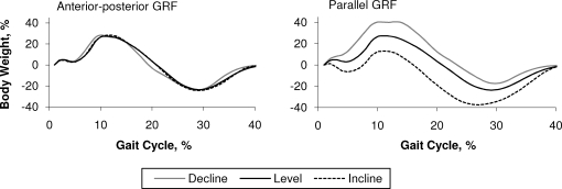
The unexpected uniformity in joint moments across decline, level, and incline running is consistent with the absence of changes observed in the peak magnitudes of the 3 components of the GRF. Earlier researchers9,24 identifying differences in GRF components with decline and incline running have done so by examining the parallel component of the GRF. Determining this component from the net GRF in our data set reveals a pattern similar to the patterns previously presented for running on decline and incline slopes (Figure 3). Although this component may provide insight into the interaction between the runner and the surface during sloped running, it is not explicitly used in the inverse dynamic calculation of the lower extremity joint moments that form the basis of the present study. We did, however, observe an increase in the impact peak of the vertical GRF during decline running. This finding corroborates the ideas of Gottschall and Kram,9 who suggested that these impact force changes may reflect changes in foot strike during decline running, such that participants contact the ground with the rearfoot rather than the midfoot, as in incline running.10 Some authors9,25 have speculated that decline running, associated with an increase in the impact peak of the vertical GRF, may increase the risk of impact-related injury. However, well-designed research25 has not demonstrated isolated impact peak events to be the single causative factor in running injury. As Nigg and Wakeling26 suggested, the effects of impact loading are participant specific and depend on the muscle tuning characteristics unique to the individual runner.
Figure 3.
Comparison of the anterior-posterior ground reaction force (GRF) with the parallel GRF during stance for decline (gray), level (black), and incline (dotted) running normalized to body mass. The parallel GRF is calculated as the component of the net GRF vector running parallel with the inclination of the instrumented treadmill force plate.
The increase in hip power absorption observed during early stance in decline running indicates an increase in the rate of eccentric loading at the hip. Similarly, the increase in hip power generation during early stance in incline running indicates an increase in the rate of concentric loading. These results corroborate the findings of Yokozawa et al27 with regard to power absorption during decline running as well as those of Swanson and Caldwell28 and Roberts and Belliveau29 with regard to increased hip power generation with incline running, albeit on a lower-grade slope. It should be noted that we held speed constant to standardize findings. As discussed, while maintaining a constant running velocity, changes in cadence and stride length in response to the changes in slope may have influenced the observed consistency in peak joint moments. The changes in power generation and absorption at the knee and hip could have been similarly affected. Specifically, an increase in cadence could lead to an increase in joint angular velocity and, subsequently, in the observed joint power absorption or generation (or both). Winter30 has written about the influence of cadence on knee power absorption, finding that greater absorption at the knee was associated with increasing running velocity. In the community, individuals may increase or decrease their speed to maintain effort in response to inclines and declines. Nonetheless, by controlling for speed, we were able to isolate specific kinetic changes associated with a runner's response to alterations in the decline and incline conditions.
CONCLUSIONS
Our data indicate that running style modifications associated with moderate-slope running did not manifest as changes in 3-dimensional joint moments or in the active peaks of the GRF. We did, however, note changes in impact forces during decline running and in joint powers at the knee and hip with changes in slope. Alterations in joint powers have not been implicated in running-related injury. Although some speculate that higher impact forces contribute to musculoskeletal injury, the evidence supporting this theory is disputed. Considering the controversy surrounding the relationship between impact-related events and musculoskeletal injury, our results allow us to support the conclusion that both level and moderate-incline running are safe components of training regimens and return-to-run protocols after injury. As greater slopes may further exacerbate the observed increases in impact forces, future authors may consider studying greater degrees of decline and incline grades to assess differences in joint kinetics.
REFERENCES
- 1.Tulloh B. The role of cross-country in the development of a runner. New Stud Athl. 1998;13(4):9–11. [Google Scholar]
- 2.Gregor R. J., Costill D. L. A comparison of the energy expenditure during positive and negative grade running. J Sports Med Phys Fitness. 1973;13(4):248–252. [PubMed] [Google Scholar]
- 3.Pivarnik J. M., Sherman N. W. Responses of aerobically fit men and women to uphill/downhill walking and slow jogging. Med Sci Sports Exerc. 1990;22(1):127–130. [PubMed] [Google Scholar]
- 4.Staab J. S., Agnew J. W., Siconolfi S. F. Metabolic and performance responses to uphill and downhill running in distance runners. Med Sci Sports Exerc. 1992;24(1):124–127. [PubMed] [Google Scholar]
- 5.Sloniger M. A., Cureton K. J., Prior B. M., Evans E. M. Lower extremity muscle activation during horizontal and uphill running. J Appl Physiol. 1997;83(6):2073–2079. doi: 10.1152/jappl.1997.83.6.2073. [DOI] [PubMed] [Google Scholar]
- 6.Swanson S. C., Caldwell G. E. An integrated biomechanical analysis of high speed incline and level treadmill running. Med Sci Sports Exerc. 2000;32(6):1146–1155. doi: 10.1097/00005768-200006000-00018. [DOI] [PubMed] [Google Scholar]
- 7.Yokozawa T., Fujii N., Ae M. Muscle activities of the lower limb during level and uphill running. J Biomech. 2007;40(15):3467–3475. doi: 10.1016/j.jbiomech.2007.05.028. [DOI] [PubMed] [Google Scholar]
- 8.Dick R. W., Cavanagh P. R. A comparison of ground reaction forces (GRF) during level and downhill running at similar speeds. Med Sci Sports Exerc. 1987;19:S12. [PubMed] [Google Scholar]
- 9.Gottschall J. S., Kram R. Ground reaction forces during downhill and uphill running. J Biomech. 2005;38(3):445–452. doi: 10.1016/j.jbiomech.2004.04.023. [DOI] [PubMed] [Google Scholar]
- 10.Buczek F. L., Cavanagh P. R. Stance phase knee and ankle kinematics and kinetics during level and downhill running. Med Sci Sports Exerc. 1990;22(5):669–677. doi: 10.1249/00005768-199010000-00019. [DOI] [PubMed] [Google Scholar]
- 11.Stefanyshyn D. J., Stergiou P., Lun V. M., Meeuwisse W. H., Worobets J. T. Knee angular impulse as a predictor of patellofemoral pain in runners. Am J Sports Med. 2006;34(11):1844–1851. doi: 10.1177/0363546506288753. [DOI] [PubMed] [Google Scholar]
- 12.Kerrigan D. C., Johansson J. L., Bryant M. G., Boxer J. A., Della Croce U., Riley P. O. Moderate-heeled shoes and knee joint torques relevant to the development and progression of knee osteoarthritis. Arch Phys Med Rehabil. 2005;86(5):871–875. doi: 10.1016/j.apmr.2004.09.018. [DOI] [PubMed] [Google Scholar]
- 13.Schipplein O. D., Andriacchi T. P. Interaction between active and passive knee stabilizers during level walking. J Orthop Res. 1991;9(1):113–119. doi: 10.1002/jor.1100090114. [DOI] [PubMed] [Google Scholar]
- 14.Winter D. A. The Biomechanics and Motor Control of Human Gait: Normal, Elderly And Pathological. 2nd ed. Waterloo, ON, Canada: University of Waterloo Press; 1991. [Google Scholar]
- 15.Perry J. Gait Analysis: Normal and Pathological Function. Thorofare, NJ: Slack; 1992. [Google Scholar]
- 16.Reilly D. T., Martens M. Experimental analysis of the quadriceps muscle force and patello-femoral joint reaction force for various activities. Acta Orthop Scand. 1972;43(2):126–137. doi: 10.3109/17453677208991251. [DOI] [PubMed] [Google Scholar]
- 17.Fredericson M., Weir A. Practical management of iliotibial band friction syndrome in runners. Clin J Sport Med. 2006;16(3):261–268. doi: 10.1097/00042752-200605000-00013. [DOI] [PubMed] [Google Scholar]
- 18.Kortebein P. M., Kaufman K. R., Basford J. R., Stuart M. J. Medial tibial stress syndrome. Med Sci Sports Exerc. 2000;32(suppl 3):S27–S33. doi: 10.1097/00005768-200003001-00005. [DOI] [PubMed] [Google Scholar]
- 19.Campbell K. M., Biggs N. C., Blanton P. L., Lehr R. P. Electromyographic investigation of the relative activity among four components of the triceps surae. Am J Phys Med. 1973;52(1):30–41. [PubMed] [Google Scholar]
- 20.Rupani H. D., Holder L. E., Espinola D. A., Engin S. I. Three-phase radionuclide bone imaging in sports medicine. Radiology. 1985;156(1):187–196. doi: 10.1148/radiology.156.1.2860698. [DOI] [PubMed] [Google Scholar]
- 21.Novacheck T. F. The biomechanics of running. Gait Posture. 1998;7(1):77–95. doi: 10.1016/s0966-6362(97)00038-6. [DOI] [PubMed] [Google Scholar]
- 22.Paolini G., Della Croce U., Riley P. O., Newton F. K., Kerrigan D. C. Testing of a tri-instrumented-treadmill unit for kinetic analysis of locomotion tasks in static and dynamic loading conditions. Med Eng Phys. 2007;29(3):404–411. doi: 10.1016/j.medengphy.2006.04.002. [DOI] [PubMed] [Google Scholar]
- 23.Riley P. O., Dicharry J., Franz J. R., Croce U. D., Wilder R. P., Kerrigan D. C. A kinematics and kinetic comparison of overground and treadmill running. Med Sci Sports Exerc. 2008;40(6):1093–1100. doi: 10.1249/MSS.0b013e3181677530. [DOI] [PubMed] [Google Scholar]
- 24.Devita P., Janshen L., Rider P., Solnik S., Hortobagyi T. Muscle work is biased toward energy generation over dissipation in non-level running. J Biomech. 2008;41(16):3354–3359. doi: 10.1016/j.jbiomech.2008.09.024. [DOI] [PMC free article] [PubMed] [Google Scholar]
- 25.Hreljac A., Marshall R. N., Hume P. A. Evaluation of lower extremity overuse injury potential in runners. Med Sci Sports Exerc. 2000;32(9):1635–1641. doi: 10.1097/00005768-200009000-00018. [DOI] [PubMed] [Google Scholar]
- 26.Nigg B. M., Wakeling J. M. Impact forces and muscle tuning: a new paradigm. Exerc Sport Sci Rev. 2001;29(1):37–41. doi: 10.1097/00003677-200101000-00008. [DOI] [PubMed] [Google Scholar]
- 27.Yokozawa T., Fujii N., Ae M. Kinetic characteristics of distance running on downhill slope. Int J Sport Health Sci. 2005;3:34–45. [Google Scholar]
- 28.Swanson S. C., Caldwell G. E. An integrated biomechanical analysis of high speed incline and level treadmill running. Med Sci Sports Exerc. 2000;32(6):1146–1155. doi: 10.1097/00005768-200006000-00018. [DOI] [PubMed] [Google Scholar]
- 29.Roberts T. J., Belliveau R. A. Sources of mechanical power for uphill running in humans. J Exp Biol. 2005;208(pt 10):1963–1970. doi: 10.1242/jeb.01555. [DOI] [PubMed] [Google Scholar]
- 30.Winter D. The Biomechanics and Motor Control of Human Gait. Waterloo, ON, Canada: University of Waterloo Press; 1987. pp. 38–43. [Google Scholar]


