Abstract
Currently, researchers can use the Actigraph 7164 or one of three different versions of the Actigraph GT1M to objectively measure physical activity.
Purpose
To determine if differences exist between activity counts from the Actigraph 7164 and the three versions of the GT1M at given walking and running speeds.
Methods
Ten male participants (23.6 ± 2.7 yrs) completed treadmill walking and running at ten different speeds (3-minute stages) while wearing either the Actigraph 7164 and the latest GT1M (GT1M-V3) or GT1M version one (GT1M-V1) and GT1M version two (GT1M-V2). Participants walked at 3, 5, and at 7 km˙hr−1 followed by running at 8, 10, 12, 14, 16, 18, and 20 km˙hr−1. The accelerometers were worn on an elastic belt around the waist over the left and right hips. Testing was performed on different days using a counterbalanced within-subjects design to account for potential differences attributable to accelerometer placement. At each speed, a one-way repeated measures ANOVA was used to examine differences between activity counts in counts˙min−1(cpm). Post-hoc pairwise comparisons with Bonferroni adjustments were used where appropriate.
Results
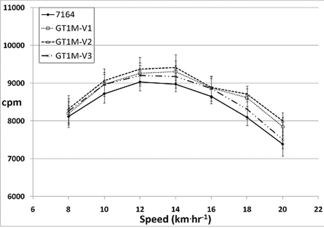
There were no significant differences between activity counts at any given walking or running speed (p<0.05). At all running speeds, activity counts from the Actigraph 7164 and GT1M-V2 displayed the lowest and highest values, respectively. Output from all accelerometers peaked at 14 km˙hr−1 (mean range: 8974 ± 677 to 9412 ± 982 cpm) and then gradually declined at higher speeds. The mean difference score at peak output between the Actigraph 7164 and GT1M-V2 was 439 ± 565 cpm.
Conclusions
There were no statistically significant differences between outputs from all the accelerometers indicating that researchers can select any of the four Actigraph accelerometers to do research.
Keywords: Accelerometry, Activity Counts, Objective Monitoring, Physical Activity
Introduction
Physical activity monitors that sense body accelerations, commonly known as ‘accelerometers,’ are often used to assess physical activity and energy expenditure. In 1961, Cavagna, Saibene, and Margaria built one of the earliest accelerometers to study human movement consisting of strain gauges that sensed accelerations in the forward, lateral, and vertical directions (4). Several years later, piezoelectric ceramic transducers replaced strain gauges and the current generation of physical activity accelerometers use this technology (5, 23). Of the commercially available accelerometers, those manufactured by Actigraph (Pensacola, Florida) are commonly used in physical activity research. Actigraph introduced the 7164 activity monitor in the 1990s. Since its introduction, the Actigraph 7164 has been used extensively by researchers in laboratory and field-based studies to derive energy expenditure prediction equations and establish cut-points to distinguish between light, moderate, and vigorous activity (3, 7, 9, 11, 14, 16, 20, 24). These cut-points in turn have been used in several intervention studies and also in a national surveillance study in the U.S. (7, 9, 21). Actigraph discontinued the 7164 and replaced it with the GT1M in the early 2000s. With technological advancements, the GT1M was further remodeled to be smaller in size. As a result of repeated modifications, currently there are 2 different models (Actigraph 7164 and GT1M) and 3 different versions of the GT1M model being used in research today.
Both the Actigraph 7164 and GT1M models have a built in single axis (vertical) accelerometer. The main technological difference between the two devices is in the type of accelerometer they contain. The Actigraph 7164 uses a cantilever beam sensor while the GT1M uses a Micro-Electro-Mechanical-System (MEMS) capacitive accelerometer. Additionally, the Actigraph 7164 has a lower sampling frequency (10 vs. 30 Hz) and memory capacity (64 kb vs. 1 Mb) as compared to the GT1M (5, 22). Physical differences between the accelerometers are described in the methods section of this paper. All four monitors provide output in the form of activity counts. Activity counts are raw accelerations filtered, digitized, and integrated over a given sampling period.
Actigraph claims that only a negligible difference in activity counts exists when comparing inter-model or inter-version activity counts (personal communication with Actigraph Vice President for research & development, John Schneider, February 2008). However, studies that compared activity counts from the Actigraph 7164 and the GT1M have shown varying results (6, 10, 18). A study by Fudge et al. (2007) compared activity counts from several objective monitors including the Actigraph 7164 and the GT1M on endurance trained male participants who performed an exercise protocol comprising of walking and running at various speeds. Although the researchers did not test for statistical differences between mean activity counts from the Actigraph 7164 and the GT1M, they found that the Actigraph 7164 had a substantially lower peak activity counts as compared to the GT1M (approximately 10,000 cpm vs. 16,000 cpm) (10). In 2007, Corder et al. examined differences between the Actigraph 7164 and the GT1M in free-living Indian adolescents (6) Although the investigators reported that the GT1M recorded 9% lower activity counts as compared to the Actigraph 7164, there were no statistical differences between the two accelerometers (6). However, contrary to the free-living condition, Corder et al. observed a significant difference between the accelerometers during mechanical spin testing at a frequency of 3 Hz in the same study (6). A more recent study by Rothney et al. used an orbital mechanical shaker to compare activity counts from three Actigraph accelerometer models, i.e., the Actigraph 7164, the GT1M, and a rarely used intermediate Actigraph 71256 model (18). While examining the activity counts from the Actigraph 7164 and the GT1M at different frequencies, these researchers found no significant differences between activity counts from the two devices (18).
Although methodologically different, a few inconsistencies between findings from the aforementioned studies can be observed. A peak output of 10,000 cpm for the Actigraph 7164 was consistently seen by both Fudge et al. and Rothney and colleagues. However, the peak GT1M activity counts observed by Fudge et al. were much higher (around 16,000 cpm ) as compared to findings by Rothney et al. (approximately 9200 cpm).
Although the study by Rothney et al. showed no statistical differences between activity counts obtained from the Actigraph 7164 and the GT1M during mechanical shaker testing, it is important to document whether this holds true during human walking and running trials. Additionally, it has not been established that activity counts from the three versions of the GT1M are comparable to each other or to those from the Actigraph 7164. This information is needed to allow researchers to use multiple models and versions of Actigraph accelerometers in a single study and allow direct comparisons of findings from studies that have used one or all of the aforementioned Actigraph accelerometers. Thus, the purpose of our study was to determine if counts obtained from the Actigraph 7164 and the three versions of the GT1M were different from each other at various walking and running speeds.
Methods
Instrumentation
The Actigraph 7164 (37.8 g, 5.1 × 4.1 × 1.5 cm) is lighter and smaller than the GT1M-V1 (40.2 g, 5.1 × 4.1 × 1.8 cm) but is larger and heavier than the GT1M-V2 (26.7 g, 4.0 × 3.9 × 1.8 cm). The GT1M-V3 is physically identical to the GT1M-V2, except for the absence of a marker button available on the GT1M-V2. Previous studies have shown excellent intra model reliability (average ICC for Actigraph 7164: r=0.98, 95% CI=0.97 to 0.98, p<0.001; GT1M: r=0.94, p<0.05) among Actigraph accelerometers at various running and walking speeds (3, 16, 19). Thus, data on all participants were collected using a single accelerometer of each model/version.
Prior to data collection, the Actigraph 7164 used in this study was calibrated by the manufacturer at a reference gain factor of 0.60 g˙sec−1 at a mid-scale voltage of 127 (associated with zero acceleration) on the A/D conversion scale. This was done to minimize the systematic error that could be attributable to the device (15). Following the completion of data collection, the unit was returned to the manufacturer to verify that there were no changes in calibration, thus ensuring consistent calibration of the device during the data collection. Unlike the Actigraph 7164, the GT1M does not require calibration after it leaves the factory. This is primarily because the latter is a MEMS accelerometer that uses solid state circuitry; i.e., it has a low number of individual components. Another feature that reduces error is the firmware digital filter in the GT1M as compared to a more error prone hardware digital filter in the Actigraph 7164. The piezoelectric MEMS devices also have low temperature-related drifts in sensitivity (25).
Participants
Ten endurance trained male participants (23.6 ± 2.8 yrs), all able to run one mile in less than 5 min, were recruited from the University of Tennessee and the surrounding community. The rationale for the above selection criterion is explained later. A health history questionnaire was used to screen for the presence of any contraindications to exercise, and participants were not included in the study if any contraindications were present. All participants provided written informed consent and had prior experience with treadmill running. The experimental protocol was approved by the University of Tennessee Institutional Review Board. Participant characteristics are shown in Table 1.
Table 1.
Mean ± SD of participant demographics.
| Characteristic | |
|---|---|
| Age (yrs) | 23.6 ± 2.7 |
| Height (m) | 1.83 ± 0.04 |
| Weight (kg) | 72.1 ± 6.4 |
| BMI (kg˙m−2) | 21.57 ± 1.76 |
Testing Protocol
Prior to testing, the participants’ height and weight were measured using a stadiometer and a physician’s scale, respectively. All participants did two trials of an incremental treadmill (Quinton Q65 Series 90, Vaerloese, Denmark) exercise protocol each separated by at least two days. The exercise protocol is explained below. During the first exercise test, participants wore an elastic belt secured with the Actigraph 7164 and the GT1M-V3 around the waist such that the accelerometers were located on the anterior axillary line above the left and right hip, respectively. During the second test, the exercise protocol was performed wearing a similar belt secured with the GT1M-V1 and GT1M-V2 on the anterior axillary line above the left and right hip, respectively. A within-subjects counterbalanced design with respect to accelerometer placement was used to avoid confounding due to left/right side placement effects. All accelerometers were initialized and synchronized to a digital timer prior to being worn by the participants.
The exercise protocol consisted of a brief warm up, followed by 3-minute stages consisting of level walking and running. Participants first completed the walking stages continuously at 3, 5 and 7 km˙hr−1. Participants then ran at 8, 10, 12, 14, 16, 18, and 20 km˙hr−1. A two-minute break was given between walking and running stages. Additionally, participants were allowed to take a three to five minute recovery break between running stages if desired. Speed during walking and running was monitored at the beginning of each stage using a tachometer (Shimpo DT 107, Nidec-Shimpo America Corporation, Itasca, IL). Data were downloaded using the Actilife 3.5.0, software (Actigraph, Fort Walton Beach, Florida). Data from the last two minutes of each stage were averaged and used to represent 1-minute activity counts.
The protocol used in this study was previously used by Fudge et al. to predict VO2 using accelerometry and heart rate, during fast running up to world-record marathon pace (10). Those investigators recruited endurance trained participants with an average peak VO2 of 60.3 ± 4.2 ml˙kg−1˙min−1. Although we did not intend to measure VO2, we desired participants with a similar fitness level as those in the study by Fudge and colleagues. Therefore, we predicted the equivalent one mile run time using a VO2 value of 60.3 ml˙kg−1˙min−1 and used it as a selection criterion to ensure that participants would be able to complete the exercise protocol in this study (8, 10). The predicted time of 4 min and 56 s was rounded off to 5 min and participants were included in the study only if they could run one mile under 5 min.
Data Analysis
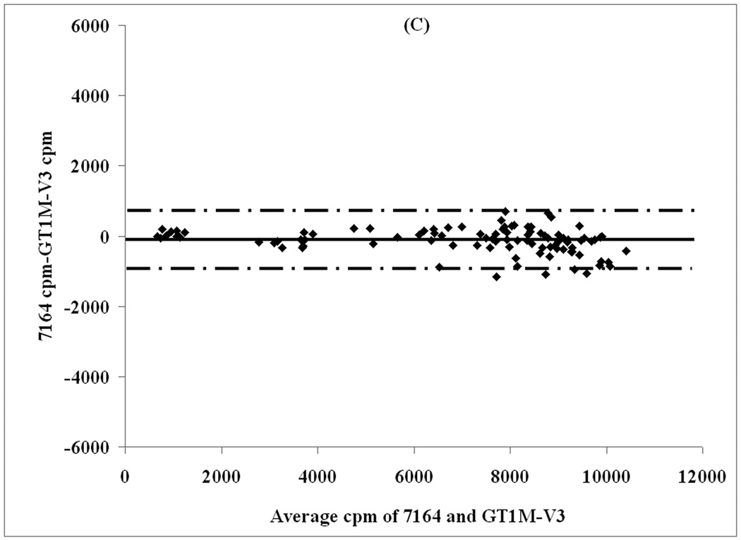
Statistical analyses were conducted using SPSS version 17 for windows (SPSS ,Chicago, IL). One-way repeated measures ANOVAs at each speed were performed to determine if activity counts obtained from the four accelerometers at various speeds were statistically different from each other. If the ANOVAs revealed significant differences, post-hoc pairwise comparisons with Bonferroni adjustments were used to identify the differences. Additionally, Bland-Altman plots were used to examine the agreement between activity counts from the Actigraph 7164 and each of the three GT1Ms. Limits of agreement were set at ± 2 standard deviations of the difference scores. For these comparisons, activity counts from the Actigraph 7164 were chosen as the true value because this accelerometer has been used to establish energy expenditure equations and cut-points for physical activity (light, moderate, and vigorous) (3, 7, 9, 11, 14, 16, 20, 24). In addition, the Actigraph 7164 was also selected for use in the 2003–2004 National Health and Examination Survey (NHANES) (21).
Results
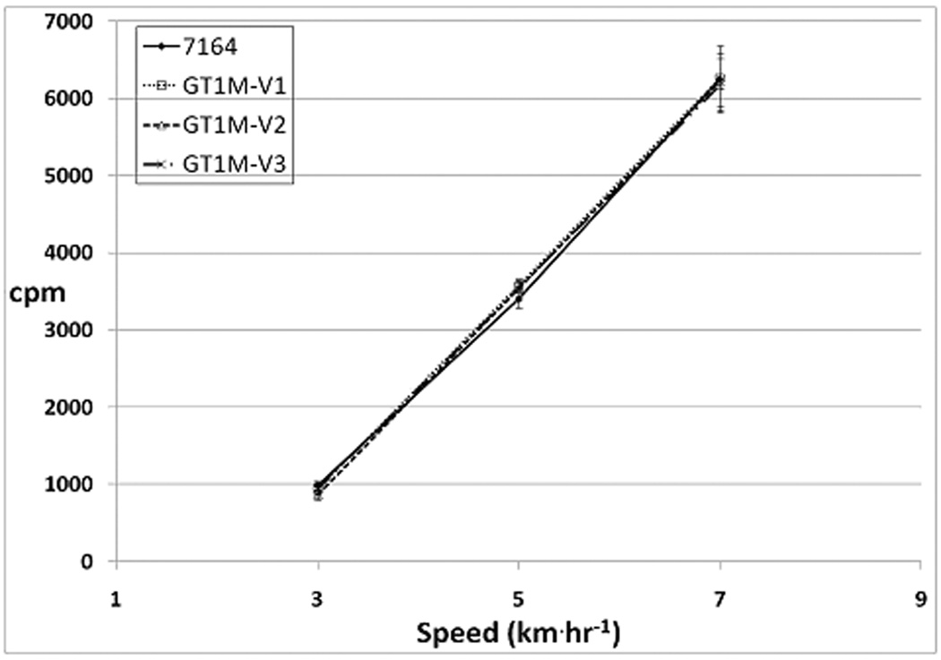
Two participants were unable to complete the exercise protocol during trial one. This resulted in two missing data points for the Actigraph 7164 and the GT1M-V3 at 20 km˙hr−1. Analysis of variance revealed significant inter- monitor differences between activity counts during walking at 3 and 5 km˙hr−1 only (p<0.05). However, these significant differences disappeared when Bonferroni adjustments were applied during the posthoc comparisons. Activity counts from all accelerometers increased linearly with increasing speed during the walking stages and are depicted in Figure 1. The maximum mean difference (138 ± 247 cpm) between any two accelerometers while walking was observed between the GT1M-V2 and the Actigraph 7164 at 5 km˙hr−1.
Figure 1.
Mean ± SE of activity counts during walking.
Figure 2 shows that during the running stages, activity counts from all the accelerometers rose curvilinearly, peaking at 14 km˙hr−1 (Actigraph 7164: 8974 ± 677 cpm; GT1M-V1 cpm: 9306 ± 1117 cpm; GT1M-V2: 9413 ± 982 cpm; GT1M-V3: 9170 ± 971 cpm) followed by a curvilinear decrease in activity counts. At all running speeds, activity counts from the Actigraph 7164 and the GT1M-V2 were lowest and highest, respectively. The maximum mean difference (680 ± 628 cpm) between any two accelerometers while running was observed between the Actigraph 7164 and GT1M-V2 at 18 km˙hr−1. With increasing running speeds, percent differences between activity counts from the Actigraph 7164 and the GT1M-V2 showed an increasing trend (2.5% at 8 km˙hr−1 to 8.3% at 20 km˙hr−1).
Figure 2.
Mean ± SE of activity counts during running.
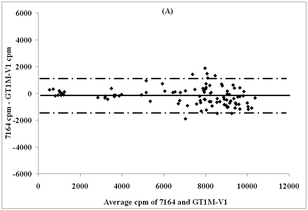
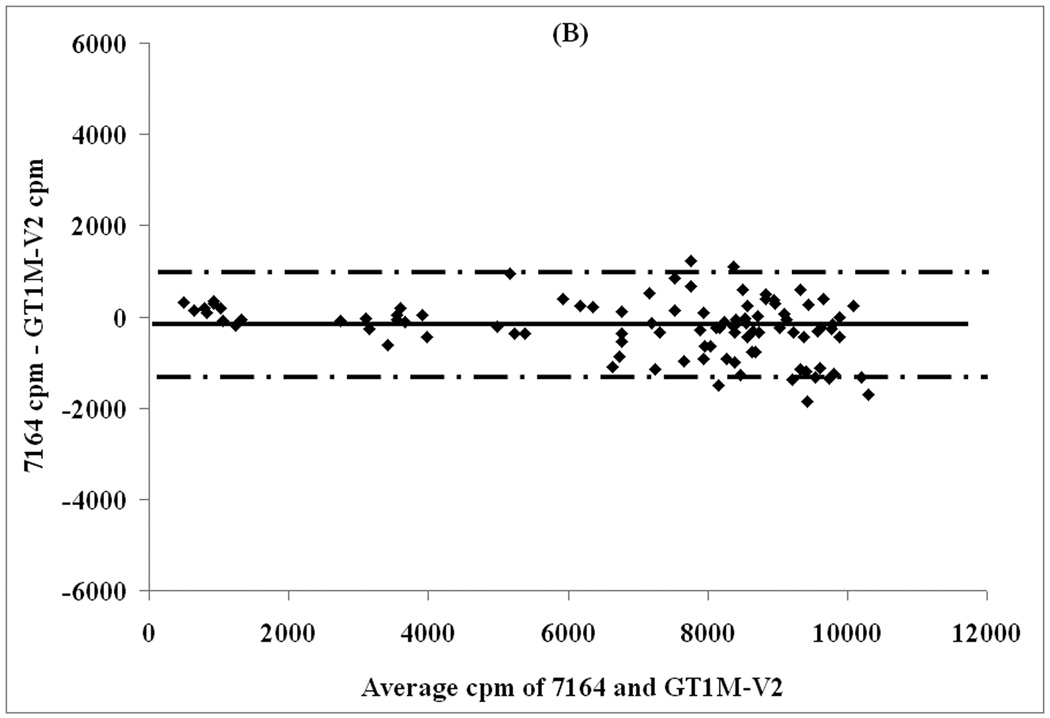
Figure 3 displays Bland-Altman plots that show the agreement between the Actigraph 7164 and the three GT1Ms. This analysis provides additional evidence to support the results of the ANOVA that there were no significant differences between the activity counts obtained from all four devices. It can be seen that the 95% limits of agreement between the Actigraph 7164 and all three GT1Ms are narrow and range between 724 to 1357 cpm.
Figure 3.
Bland-Altman plots assessing agreement with increasing activity counts between (A) Actigraph 7164 and GT1M-V1, (B) Actigraph 7164 and GT1M-V2, and (C) Actigraph 7164 and GT1M-V3. Solid line depicts mean activity count difference between devices and dashed lines depict ± 2 standard deviations of mean activity count difference.
Discussion
The major finding of this study was that the mean activity counts obtained from the Actigraph 7164, GT1M-V1, GT1M-V2, and GT1M-V3 were not significantly different from one another. The current study was the first to compare activity counts obtained from four Actigraph accelerometers at various walking and running speeds.
From our findings, it can be inferred that researchers interested in measuring physical activity or energy expenditure during running or walking could use any of the above mentioned Actigraph accelerometers in a single study. They could also draw direct comparisons with other investigations that used one or all of these devices. An example of this inference can be seen in table 2, where we quantified energy expenditure with the Freedson and Crouter regression equations during walking at 5 km˙hr−1 and running at 10 km˙hr−1 using mean activity counts from all four devices (7, 9). At a given speed, the predicted MET values using activity counts from all four accelerometers for a particular method (Freedson or Crouter) were within 0.2 METs of each other. Based on table 2, we can conclude that predicted energy expenditures from all four accelerometers used in our study are very close to each other.
Table 2.
Energy expenditure derived from accelerometer counts obtained at a walking (5 km˙hr−1) and running speed (10 km˙hr−1) using the Freedson and Crouter regression equations. Values are mean ± SD.
| Speed | Device | Freedson (METs) | Crouter (METs) |
|---|---|---|---|
| 5 km˙hr−1 | 7164 | 4.1 ± 0.3 | 6.7 ± 0.4 |
| V1 | 4.3 ± 0.3 | 6.8 ± 0.3 | |
| V2 | 4.1 ± 0.5 | 6.6 ± 0.6 | |
| V3 | 4.3 ± 0.3 | 6.8 ± 0.3 | |
| 10 km˙hr−1 | 7164 | 8.4 ± 0.6 | 9.8 ± 0.3 |
| V1 | 8.6 ± 0.8 | 9.8 ± 0.3 | |
| V2 | 8.6 ± 0.7 | 9.9 ± 0.3 | |
| V3 | 8.6 ± 0.8 | 9.8 ± 0.3 | |
Comparison With Previous Studies
Prior to the current study, only three studies have compared activity counts between Actigraph accelerometers (6, 10, 18). Among these, Rothney and colleagues used a mechanical shaker and Corder et al. examined activity counts in free living adolescents. Based on descriptions and photographs of the devices and via personal communication, we determined that Rothney et al. and Corder et al. compared the Actigraph 7164 to the GT1M-V1, while Fudge et al. compared the Actigraph 7164 to the GT1M-V2. This part of the discussion compares inter-model and GT1M inter-version peak activity counts from the studies of Rothney et al., Fudge et al., and the current study.
Similar to the study of Rothney et al., the current study did not find statistically significant differences in activity counts obtained from the Actigraph 7164 and all three versions of the GT1M (6, 18). Rothney and coworkers employed a mechanical orbital shaker to record activity counts from accelerometers placed at a fixed radius of 4.6 cm within the shaker while they were oscillated at increasing frequencies. This experiment is comparable to human locomotion at varying speeds because vertical displacement of the center of mass during walking lies between 2.7 ± 0.5 and 4.8 ± 0.9 cm (17). The Actigraph 7164 activity counts observed by Rothney et al, Fudge et al., and in the current study were relatively uniform, peaking between 9000 and 10,000 cpm (10, 18). On the other hand, GT1M activity counts in the current study and that of Rothney et al. peaked between 9200 and 9500 cpm, as compared to approximately 16,000 cpm in the study of Fudge et al. (10, 18). Fudge and colleagues observed very large differences (ranging from 6000 to 9000 cpm) between activity counts from the Actigraph 7164 and the GT1M at speeds of over 16 km˙hr−1. Although there was a similar trend in the current study, the differences were much smaller ranging between 237 to 612 cpm. Thus, the GT1M activity counts recorded by Fudge et al. differed greatly from both the Actigraph 7164 activity counts recorded in all three studies, and also from the GT1M activity counts recorded in our study and that of Rothney et al. (18).
Rothney et al. reported small inter-model discrepancies between the GT1M and the Actigraph 7164 that were of similar magnitude to those in the current study (18). Actigraph states that the GT1M records marginally higher counts than the Actigraph 7164 (especially at higher oscillation frequencies) due to a greater sampling rate than the Actigraph 7164 (30 Hz vs. 10 Hz) (Personal communication with John Schneider). However, it is interesting to note that unlike the current study and that of Fudge et al., Rothney et al. recorded lower activity counts using the GT1M as compared to the Actigraph 7164 (10, 18). Additionally Rothney et al. found that the GT1M is less sensitive at the lower end of the acceleration spectrum as compared to the Actigraph 7164 (18). This means that the GT1M may fail to record activity counts during certain very low intensity activities and thereby underestimate energy expenditure during these activities.
There were large differences (ranging from 638 cpm at 3 km˙hr−1 to 8494 cpm at 20 km˙hr−1) between average activity counts from the GT1M-V2 used by Fudge et al. and all three versions of the GT1M used in our study. Because the participants in both studies were highly trained runners, we consider it very unlikely that differences in running biomechanics caused the large discrepancy in GT1M activity counts between these studies. Thus, an alternative explanation would be inconsistencies between the two GT1M-V2 accelerometers used in these studies. We try to explore this possibility below.
All Actigraph GT1M accelerometers have a band pass filtering range of 0.25 to 2.5 Hz and can measure accelerations with a maximum amplitude of 2.0 g. According to Actigraph, accelerations at the hip during normal human motion mostly fall within these parameters and the band pass filter should help eliminate extraneous accelerations that are not due human movement (e.g., vibration artifacts). The peaking of GT1M activity counts at around 9300 cpm as observed by the current study and that of Rothney et al. can be attributed to the device’s amplitude and band pass filtering limitations (18) . However, the GT1M-V2 used by Fudge et al. seems to have had a wider band pass filtering and/or amplitude range, thereby recording much higher activity counts than in the current study and that of Rothney et al. If this discrepancy is due to the manufacturing process, it is possible that there are several flawed GT1Ms (all three versions) being used in research because unlike the Actigraph 7164, MEMS devices like the GT1M do not need user calibration and are mass produced using batch fabrication techniques.
When contacted, Actigraph was unable to provide an explanation for the high activity counts observed by Fudge et al. and the company stated that there were no manufacturing inconsistencies with regard to the GT1M-V2. This is contrary to findings of Fudge et al. Thus, when using the GT1M, investigators need to verify that their accelerometer provides similar activity counts to those reported in other studies.
‘Leveling-off/Plateau’ phenomenon
Actigraph accelerometers exhibit a phenomenon where activity counts reach a peak at a particular running speed or frequency (mechanical shaking) and do not increase in response to further speed/frequency increments (3, 10, 16, 18). Researchers have termed this phenomenon as a ‘leveling-off’ or ‘plateau’ and have attributed it to amplitude and band pass filtering limitations (1–3, 10, 12, 18, 19).
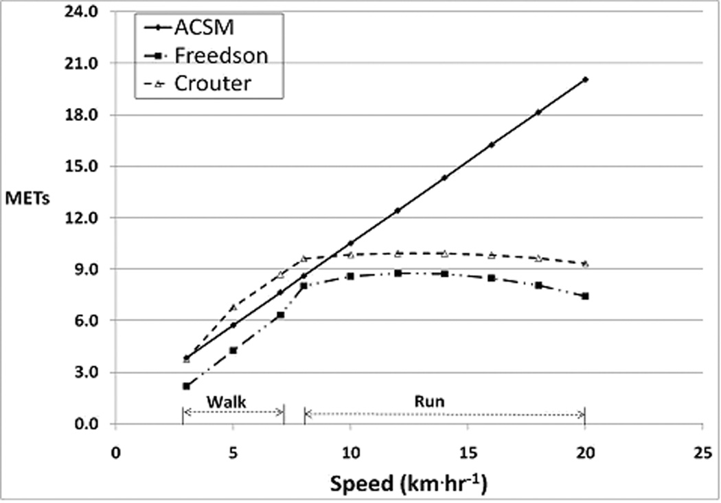
To accurately estimate energy expenditure, an accelerometer must demonstrate an increase (linear, curvilinear, exponential, etc.) in activity counts with increasing exercise intensity because energy expenditure regression equations are based on the theory that activity counts increase with increasing intensity of exercise. The practical implications of the undesirable leveling-off/plateau phenomenon can be seen in figure 4, which displays the calculated energy expenditure for all walking and running speeds using the ACSM prediction equation, the Crouter two-regression model, and the Freedson regression equation (7, 9). Since there were no significant differences between activity counts from all four accelerometers in our study, we calculated energy expenditure using data obtained from the GT1M-V3. It can be seen that both Freedson and Crouter regression equations under-predict METs at the higher running speeds as compared to the ACSM prediction equation. In fact, one would not be able to distinguish running at 8 km˙hr−1 from running at 18 km˙hr−1, due to the similarity of activity counts. Thus, investigators must be careful in using Actigraph accelerometers to estimate energy expenditure for running speeds above 10 km˙hr−1. Due to the absence of a true plateau or leveling off (activity counts peak and remain constant with subsequent speed increases), it may be more appropriate to call this behavior the ‘Inverted-U’ phenomenon.
Figure 4.
Walking and running energy expenditure estimated using the ACSM equation and predicted from mean GT1M-V3 activity counts using the Freedson, and Crouter regression equations.
To avoid early peaking of activity counts and the inverted-U phenomenon, Bouten et al. has recommended that accelerometers should have the ability to measure amplitudes and frequencies up to 6 g and 20 Hz, respectively (2). All versions of the GT1M fall short of this recommendation. Fudge et al. showed that vertical accelerations from the 3dNX (a triaxial accelerometer) did not completely plateau till running speed reached 16 km˙hr−1 and this can be attributed to the ability of the device to measure accelerations with amplitudes between 2.85 to 5.0 g (upper body acceleration range), at frequencies up to 10 Hz (10, 12, 13). In the same study, the Actiheart activity monitor, which has a uniaxial accelerometer with a wider band pass filtering width (1 to 7 Hz) and a higher upper limit for amplitude (2.5 g), compared to the GT1M, peaked at approximately 18 to 20 km˙hr−1.
Actigraph GT1M-V4 and GT3X
Actigraph released a new dual axis (vertical and horizontal) GT1M in 2008 and a triaxial activity monitor called the GT3X in early 2009. We are unsure of how these monitors would behave as compared to the Actigraph uniaxial accelerometers. Both new devices have a single solid-state accelerometer that records accelerations in more than just the vertical axis and they have a higher amplitude range (2.5 g). However, the band pass filtering width of these new monitors is the same as the previous uniaxial monitors. Another difference between the old uniaxial GT1M and the new monitors is that the newer ones have 12-bit A/D signal converters instead of 8-bit A/D signal converters. Additional research is needed to establish whether the newly introduced dual axis and triaxial Actigraph accelerometers are susceptible to the problem of premature peaking and the inverted-U phenomenon. Studies are also needed to examine the comparability of these new accelerometers to the older uniaxial devices.
Strengths and Limitations
Our study has several strengths and limitations. A strength of the study was that we conducted human trials to identify differences between accelerometers. Our protocol and participant characteristics were similar to previous research, which allowed us to draw direct comparisons with the same. Additionally, we compared multiple Actigraph models as compared to just two models in most other comparison studies. We used a single unit to represent each accelerometer model and version in order to eliminate possible errors attributable to inter-device variability. This could be viewed as a weakness if the Actigraph 7164 was found to not be in calibration at the end of the study or if the GT1Ms produced activity counts that differed from those seen in previous studies. However, this was not an issue because the Actigraph 7164 was found to be in calibration at the end of the study, and activity counts from the GT1Ms were comparable to those observed in other studies. In terms of weaknesses, we had a relatively small sample size (N=10). However, previous research studies have used comparable sample sizes to examine inter-model differences (3, 10). We collected data on only two accelerometers during one trial.
Conclusion
In summary, all four Actigraph accelerometers examined in this study behave similarly during walking and running and their activity counts are comparable to each other. Our study shows that physical activity researchers can use different models or versions of Actigraph accelerometers in the same study. However, researchers using the GT1Ms should ensure that their GT1M provides activity counts similar to those reported in other studies.
Although Actigraph accelerometers fail to record additional activity counts when running speed increases above 10 km˙hr−1, it must be kept in mind that most physical activity researchers do not use these devices to estimate minute by minute energy expenditure for short running bouts at speeds above 10 km˙hr−1. Rather, they typically only use them to record the total number of minutes spent in light (< 3 METs), moderate (3 to 6 METs), or vigorous intensity (> 6 METs) physical activity. Fortunately, Actigraph accelerometers correctly classify running as vigorous physical activity despite underestimating energy expenditure. These devices are widely used in physical activity research because they are easy to use, have low participant burden, allow extended periods of recording, and help identify the intensity and duration of activity.
Acknowledgements
Research supported by NIH R21CA122430.
The results of the present study do not constitute endorsement of the product by the authors or ACSM.
References
- 1.Bhattacharya A, McCutcheon EP, Shvartz E, Greenleaf JE. Body acceleration distribution and O2 uptake in humans during running and jumping. J Appl Physiol. 1980;49(5):881–887. doi: 10.1152/jappl.1980.49.5.881. [DOI] [PubMed] [Google Scholar]
- 2.Bouten CV, Koekkoek KT, Verduin M, Kodde R, Janssen JD. A triaxial accelerometer and portable data processing unit for the assessment of daily physical activity. IEEE Trans Biomed Eng. 1997;44(3):136–147. doi: 10.1109/10.554760. [DOI] [PubMed] [Google Scholar]
- 3.Brage S, Wedderkopp N, Franks PW, Andersen LB, Froberg K. Reexamination of validity and reliability of the CSA monitor in walking and running. Med Sci Sports Exerc. 2003;35(8):1447–1454. doi: 10.1249/01.MSS.0000079078.62035.EC. [DOI] [PubMed] [Google Scholar]
- 4.Cavagna G, Saibene F, Margaria R. A three-directional accelerometer for analyzing body movements. J Appl Physiol. 1961;16:191. doi: 10.1152/jappl.1961.16.1.191. [DOI] [PubMed] [Google Scholar]
- 5.Chen KY, Bassett DR., Jr. The technology of accelerometry-based activity monitors: current and future. Med Sci Sports Exerc. 2005;37(11 Suppl):S490–S500. doi: 10.1249/01.mss.0000185571.49104.82. [DOI] [PubMed] [Google Scholar]
- 6.Corder K, Brage S, Ramachandran A, Snehalatha C, Wareham N, Ekelund U. Comparison of two Actigraph models for assessing free-living physical activity in Indian adolescents. J Sports Sci. 2007;25(14):1607–1611. doi: 10.1080/02640410701283841. [DOI] [PubMed] [Google Scholar]
- 7.Crouter SE, Clowers KG, Bassett DR., Jr. A novel method for using accelerometer data to predict energy expenditure. J Appl Physiol. 2006;100(4):1324–1331. doi: 10.1152/japplphysiol.00818.2005. [DOI] [PubMed] [Google Scholar]
- 8.Daniels J. Daniels' Running Formula. Champaign (IL): Human Kinetics; 1998. p. 64. [Google Scholar]
- 9.Freedson PS, Melanson E, Sirard J. Calibration of the Computer Science and Applications, Inc. accelerometer. Med Sci Sports Exerc. 1998;30(5):777–781. doi: 10.1097/00005768-199805000-00021. [DOI] [PubMed] [Google Scholar]
- 10.Fudge BW, Wilson J, Easton C, Irwin L, Clark J, Haddow O, Kayser B, Pitsiladis YP. Estimation of oxygen uptake during fast running using accelerometry and heart rate. Med Sci Sports Exerc. 2007;39(1):192–198. doi: 10.1249/01.mss.0000235884.71487.21. [DOI] [PubMed] [Google Scholar]
- 11.Hendelman D, Miller K, Baggett C, Debold E, Freedson P. Validity of accelerometry for the assessment of moderate intensity physical activity in the field. Med Sci Sports Exerc. 2000;32(9 Suppl):S442–S449. doi: 10.1097/00005768-200009001-00002. [DOI] [PubMed] [Google Scholar]
- 12.Kram R, Griffin TM, Donelan JM, Chang YH. Force treadmill for measuring vertical and horizontal ground reaction forces. J Appl Physiol. 1998;85(2):764–769. doi: 10.1152/jappl.1998.85.2.764. [DOI] [PubMed] [Google Scholar]
- 13.Kyrolainen H, Belli A, Komi PV. Biomechanical factors affecting running economy. Med Sci Sports Exerc. 2001;33(8):1330–1337. doi: 10.1097/00005768-200108000-00014. [DOI] [PubMed] [Google Scholar]
- 14.Leenders NY, Nelson TE, Sherman WM. Ability of different physical activity monitors to detect movement during treadmill walking. Int J Sports Med. 2003;24(1):43–50. doi: 10.1055/s-2003-37196. [DOI] [PubMed] [Google Scholar]
- 15.Moeller NC, Korsholm L, Kristensen PL, Andersen LB, Wedderkopp N, Froberg K. Unit-specific calibration of Actigraph accelerometers in a mechanical setup - Is it worth the effort? The effect on random output variation caused by technical inter-instrument variability in the laboratory and in the field. [Cited 2009 March 20];BMC Medical Research Methodology [Internet] 2008 8(19) doi: 10.1186/1471-2288-8-19. Available from http://www.pubmedcentral.nih.gov/articlerender.fcgi?artid=2383921. [DOI] [PMC free article] [PubMed]
- 16.Nichols JF, Morgan CG, Chabot LE, Sallis JF, Calfas KJ. Assessment of physical activity with the Computer Science and Applications, Inc., accelerometer: laboratory versus field validation. Res Q Exerc Sport. 2000;71(1):36–43. doi: 10.1080/02701367.2000.10608878. [DOI] [PubMed] [Google Scholar]
- 17.Orendurff MS, Segal AD, Klute GK, Berge JS, Rohr ES, Kadel NJ. The effect of walking speed on center of mass displacement. J Rehabil Res Dev. 2004;41(6A):829–834. doi: 10.1682/jrrd.2003.10.0150. [DOI] [PubMed] [Google Scholar]
- 18.Rothney MP, Apker GA, Song Y, Chen KY. Comparing the performance of three generations of ActiGraph accelerometers. J Appl Physiol. 2008;105(4):1091–1097. doi: 10.1152/japplphysiol.90641.2008. [DOI] [PMC free article] [PubMed] [Google Scholar]
- 19.Rowlands AV, Stone MR, Eston RG. Influence of speed and step frequency during walking and running on motion sensor output. Med Sci Sports Exerc. 2007;39(4):716–727. doi: 10.1249/mss.0b013e318031126c. [DOI] [PubMed] [Google Scholar]
- 20.Swartz AM, Strath SJ, Bassett DR, Jr., O'Brien WL, King GA, Ainsworth BE. Estimation of energy expenditure using CSA accelerometers at hip and wrist sites. Med Sci Sports Exerc. 2000;32(9 Suppl):S450–S456. doi: 10.1097/00005768-200009001-00003. [DOI] [PubMed] [Google Scholar]
- 21.Troiano RP, Berrigan D, Dodd KW, Masse LC, Tilert T, McDowell M. Physical activity in the United States measured by accelerometer. Med Sci Sports Exerc. 2008;40(1):181–188. doi: 10.1249/mss.0b013e31815a51b3. [DOI] [PubMed] [Google Scholar]
- 22.Tryon WW. Fully Proportional Actigraphy: A new Instrument. Behavior research Methods, Instruments, and Computers. 1996;28(3):392–403. [Google Scholar]
- 23.Wong TC, Webster JG, Montoye HJ, Washburn R. Portable accelerometer device for measuring human energy expenditure. IEEE Trans Biomed Eng. 1981;28(6):467–471. doi: 10.1109/TBME.1981.324820. [DOI] [PubMed] [Google Scholar]
- 24.Yngve A, Nilsson A, Sjostrom M, Ekelund U. Effect of monitor placement and of activity setting on the MTI accelerometer output. Med Sci Sports Exerc. 2003;35(2):320–326. doi: 10.1249/01.MSS.0000048829.75758.A0. [DOI] [PubMed] [Google Scholar]
- 25.Yu J, Lan C. System modeling of microaccelerometer using piezoelectric thin films. Sensors & Actuators: A Physical. 2001;88(2):178–186. [Google Scholar]


