Abstract
Phosphatase inhibitor-1 (I-1) is a distal amplifier element of β-adrenergic signaling that functions by preventing dephosphorylation of downstream targets. I-1 is downregulated in human failing hearts, while overexpression of a constitutively active mutant form (I-1c) reverses contractile dysfunction in mouse failing hearts, suggesting that I-1c may be a candidate for gene therapy. We generated mice with conditional cardiomyocyte-restricted expression of I-1c (referred to herein as dTGI-1c mice) on an I-1–deficient background. Young adult dTGI-1c mice exhibited enhanced cardiac contractility but exaggerated contractile dysfunction and ventricular dilation upon catecholamine infusion. Telemetric ECG recordings revealed typical catecholamine-induced ventricular tachycardia and sudden death. Doxycycline feeding switched off expression of cardiomyocyte-restricted I-1c and reversed all abnormalities. Hearts from dTGI-1c mice showed hyperphosphorylation of phospholamban and the ryanodine receptor, and this was associated with an increased number of catecholamine-induced Ca2+ sparks in isolated myocytes. Aged dTGI-1c mice spontaneously developed a cardiomyopathic phenotype. These data were confirmed in a second independent transgenic mouse line, expressing a full-length I-1 mutant that could not be phosphorylated and thereby inactivated by PKC-α (I-1S67A). In conclusion, conditional expression of I-1c or I-1S67A enhanced steady-state phosphorylation of 2 key Ca2+-regulating sarcoplasmic reticulum enzymes. This was associated with increased contractile function in young animals but also with arrhythmias and cardiomyopathy after adrenergic stress and with aging. These data should be considered in the development of novel therapies for heart failure.
Introduction
Heart failure is among the most frequent causes of morbidity and mortality worldwide and is, despite improved treatment options, associated with poor prognosis. Current treatment with angiotensin-converting enzyme inhibitors, aldosterone receptor antagonists, and beta blockers is suboptimal, with the 5-year survival rate being less than 50%. New drug principles targeting neurohumoral activation mechanisms, such as antagonists of endothelin receptors, TNF-α or IL-6, and statins, failed to improve survival in clinical studies. Thus, new approaches are needed, and an attractive one is to target the abnormal function of cardiomyocytes in failing hearts directly (as opposed to the more indirect affection by neurohumoral blockade).
Two of the best studied alterations of failing myocyte function are (a) desensitization of the β-adrenergic signaling system (1, 2) and (b) alterations of intracellular Ca2+ handling (3, 4). The latter include decreased diastolic sarcoplasmic reticulum (SR) Ca2+ uptake via the SR Ca2+ ATPase (SERCA2a) and relatively increased diastolic sarcolemmal Ca2+ efflux through the Na+/Ca2+-exchanger (NCX), prolonged Ca2+ transients, and enhanced propensity for SR Ca2+ release (Ca2+ leak) via SR ryanodine receptor/Ca2+-release channel (RyR2) during diastole (5). These alterations aggravate contractile dysfunction particularly under exercise (less response to catecholamines, lower SR Ca2+ loading), and some of them (SR Ca2+ leak with subsequent Ca2+ efflux through NCX) contribute to electrical instability and arrhythmogenesis, which are further accelerated by reduced expression of repolarizing K+ channels (“acquired LQT syndrome”) (6). Indeed, sudden cardiac death, likely due to ventricular tachyarrhythmias, is responsible for half of all cardiac deaths in patients with heart failure.
On the other hand, some of the functional abnormalities of failing cardiomyocytes, particularly β-adrenergic desensitization, can also be interpreted as energy saving and at least partially protective adaptations (2). Accordingly, drugs intended to reverse or bypass β-adrenergic desensitization (PDE inhibitors, catecholamines, or other positive inotropic agents) caused symptomatic improvement but increased mortality in patients. Similarly, except for expression of adenylyl cyclase 6 and inhibitors of the G protein–coupled receptor kinase 2, transgenic overexpression of proximal elements of the β-adrenergic signaling pathway (receptors, G proteins, adenylyl cyclase 5, or PKA) caused short-term improvements in cardiac function but long-term cardiac pathology (2, 7). In contrast, transgenic or viral overexpression of SERCA2a, an important downstream target of β-adrenergic regulation of cardiac function (via phosphorylation of phospholamban [PLB]), improved diastolic and systolic function and the energetic state of failing hearts (8). Similar beneficial effects were seen after gene therapeutic knock down of the SERCA2a-inhibitor PLB (9). These studies validated the PLB/SERCA2a system and diastolic Ca2+ uptake into the SR as potential targets for effective heart failure therapy, and the first gene therapy trials in patients have been initiated (10).
An alternative target for treatment of heart failure is phosphatase inhibitor-1 (I-1), a small PKA substrate that in its PKA-phosphorylated form at Thr35 potently and specifically inhibits phosphatase-1 and thereby increases the phosphorylation state of PKA substrates such as PLB. As a consequence, I-1 amplifies β-adrenergic signals. In contrast, PKC-α phosphorylation at Ser67 attenuates I-1’s inhibitory activity toward phosphatase-1, and this regulation of I-1 has been associated with depressed cardiac function in PKC-α transgenic mice (11). Notably, protein levels and PKA phosphorylation of I-1 were reduced in human failing hearts and were associated with decreased phosphorylation of PLB (12). Thus, I-1 downregulation likely participates in decreased SR Ca2+ loading in failing myocytes. Conversely, overexpression of I-1 sensitized myocytes toward the positive inotropic effects of the β-adrenoceptor agonist isoprenaline (13), and overexpression of a truncated, constitutively active I-1 form (I-1c) reversed contractile dysfunction of failing myocytes (14) and, in transgenic mice, increased contractile function under basal conditions and in a model of pressure overload (15). These beneficial effects were associated with increased phosphorylation of PLB. Collectively, these data suggest that downregulation of I-1 may partially contribute to β-adrenergic desensitization in the failing heart and that normalization/overexpression of I-1 can increase contractile force and the response to catecholamines by increasing phosphorylation (and thus inactivation) of the SERCA2a-inhibitor PLB. Since phosphorylation of other PKA-targeted phosphoproteins like troponin I, myosin-binding protein C, and RyR2 were unaffected, I-1 was considered as a specific regulator of PLB. Based on this concept, I-1c was recently chosen as a new target for gene therapy in heart failure (15, 16).
On the other hand, I-1 knockout mice (Ppp1r1a KO mice) exhibited only mild reduction in sensitivity to catecholamines and were partially protected against acute and chronic toxicity of catecholamines (17). This protection was associated with reduced phosphorylation not only of PLB but also of RyR2, which calls into question the specificity of I-1 for PLB.
To further dissect the effects of I-1 in the heart and the role of its PKC-α–phosphorylation site at Ser67, we generated 2 strains of double-transgenic mice with conditional cardiac-restricted expression of I-1c (dTGI-1c) or the PKC-α phosphorylation-deficient mutant I-1S67A (dTGS67A) on a Ppp1r1a KO background, using the Tet-Off system. Given the potential of I-1c as a candidate for gene therapy in chronic heart failure, a disease of the elderly with increased risk for arrhythmias and sudden cardiac death, we studied consequences of I-1c expression and repression at rest, under catecholaminergic stress, and in aging and potential mechanisms. We found that expression of both variants improved cardiac contractility in young mice at rest but was deleterious and arrhythmogenic under catecholaminergic stress. All I-1–related abnormalities were reversed by shutting off transgene expression, indicating a direct causal relationship. Moreover, aged I-1 double-transgenic mice spontaneously developed a cardiomyopathic phenotype.
Results
Double-transgenic mice with conditional heart-specific expression of I-1c.
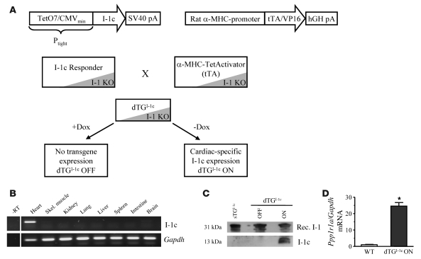
We generated a mouse model that expressed I-1c in a conditional and cardiomyocyte-restricted manner (α-MHC–regulated Tet-Off system; Figure 1A). All mice were backcrossed to C57BL/6J (5–6 generations) and crossed with Ppp1r1a KO mice until they were on a complete homozygote Ppp1r1a-null background. RT-PCR showed expression in heart only, with no expression in other tissues (Figure 1B). Western blots demonstrated robust I-1c expression in hearts from induced double-transgenic I-1c mice (dTGI-1c ON mice) and its absence (a) in noninduced double-transgenic I-1c mice (dTGI-1c OFF mice, i.e., mice that had been fed with doxycycline in utero and after birth) and (b) I-1c single transgenic mice (Figure 1C). These data demonstrate that doxycycline administration effectively suppressed I-1c transgene expression without significant constitutive promoter activity (leakiness). I-1c transcript concentrations in dTGI-1c ON mice were approximately 24-fold higher than endogenous Ppp1r1a mRNA concentrations in WT mice (Figure 1D).
Figure 1. In vivo cardiac expression of I-1c.
(A) Generation and crossing strategy of a conditional mouse model (Tet-Off system) to express I-1c on a complete Ppp1r1a KO background. Dox, doxycycline; Ptight, Tet-responsive promoter; SV40, simian virus 40; VP16, herpes simplex virus protein 16. (B) RT-PCR analysis for I-1c and Gapdh from various organs of induced I-1c double-transgenic mice shows cardiomyocyte-restricted expression. -RT, minus RT control; Skel., skeletal. (C) Western blots from cardiac extracts demonstrate doxycycline-dependent I-1c expression and lack of leakiness in the single transgenic I-1c responder (sTGI-1c). Rec., recombinant. (D) I-1c mRNA analysis in dTGI-1c ON hearts revealed a 24.5-fold expression compared with Ppp1r1a mRNA from WT mice (n ≥ 4 each). *P < 0.05 versus WT.
Enhanced contractility and phosphorylation of RyR2 and PLB in dTGI-1c ON mice.
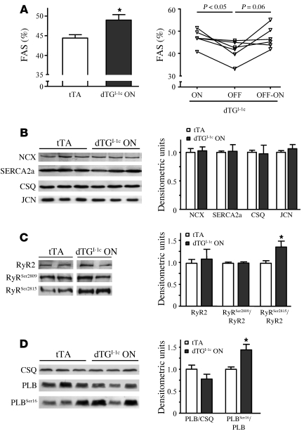
At 3 months of age, dTGI-1c ON mice exhibited a normal heart-to-body weight ratio (5.3 ± 0.1 mg/g vs. 5.4 ± 0.1 mg/g in tetracycline-controlled transactivator [tTA] mice; n = 6–8), atrial natriuretic peptide Anp mRNA levels (1.1 ± 0.3 vs. 1.0 ± 0.1 in tTA mice; n = 6), total β-adrenoceptor density (14.7 ± 1.6 fmol/mg vs. 14.0 ± 1.7 fmol/mg protein in tTA mice; n = 8), and phosphatase-1c protein levels (1.1 ± 0.1 vs. 1.0 ± 0.1 in tTA mice; n = 4) compared with tTA littermates. Echocardiographic examination revealed normal heart rate (481 ± 6 bpm vs. 488 ± 8 bpm in tTA mice; n = 13) and normal cardiac mass and volume (see Supplemental Table 1; supplemental material available online with this article; doi: 10.1172/JCI40545DS1). However, dTGI-1c ON mice showed higher fractional area shortening (FAS; Figure 2A, left) and calculated ejection fraction compared with tTA mice (see Supplemental Table 1). Hypercontractility in dTGI-1c ON mice could be fully reversed by 10 days doxycycline administration (dTGI-1c OFF) and reinduced by discontinuation of doxycycline in the drinking water for 7 weeks (dTGI-1c OFF-ON; Figure 2A, right). These data suggest that the higher contractility in dTGI-1c ON mice in vivo is a direct consequence of I-1c expression.
Figure 2. I-1c enhances basal contractility and is associated with higher phosphorylation of PLB and cardiac RyR2.
(A) Echocardiographic assessment of FAS in single transgenic tTA mice and dTGI-1c ON mice at the age of 3 months (n = 13 each; left panel). *P < 0.05 versus tTA. Reassessment of FAS after doxycycline feeding (dTGI-1c OFF) and doxycycline withdrawal (dTGI-1c OFF-ON) indicates temporally controllable I-1c effects (n = 6; age, 3 months; right panel). (B–D) Western blots and statistical analysis of total protein levels of NCX, SERCA2a, calsequestrin (CSQ), junctin (JCN), RyR2, and PLB and phosphorylation state of RyR2 (RyRSer2809 and RyRSer2815) and PLB (PLBSer16) in tTA and dTGI-1c ON hearts (n = 8 for each group). Samples were run on the same gel. *P < 0.05 versus tTA.
To identify cellular I-1c targets, we performed Western blots to determine total protein and phosphorylation levels of cardiac key regulatory proteins involved in Ca2+ homeostasis and the contractile machinery. The protein abundance of NCX, SERCA2a, calsequestrin, junctin, PLB, and RyR2 (Figure 2, B–D) and the abundance and phosphorylation level of the myofibrillar proteins troponin-I, myosin binding protein-C, and ventricular regulatory myosin light chain did not differ between dTGI-1c ON and tTA hearts (Supplemental Figure 1A). Similarly, the concentration of key proteins of hypertrophic pathways implicated in β-adrenergic signaling (p90 ribosomal S6 kinase, PKB, and mitogen activated kinases) did not differ between the groups (Supplemental Figure 1B). In contrast, phosphorylation levels of RyR2 at Ser2815 (Ca2+/calmodulin-activated protein kinase II [CaMKII] site, an increase of 38%; Figure 2C) and PLB at Ser16 (PKA site, an increase of 44%; Figure 2D) were consistently higher in dTGI-1c ON mice than in tTA mice. RyR2 phosphorylation at Ser2809 (PKA site) was not affected (Figure 2C).
Reversible ventricular arrhythmia in dTGI-1c ON mice.
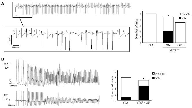
Previous work suggested a link between hyperphosphorylated RyR2, “leaky” RyR2 channels, delayed afterdepolarizations, and triggered activity/arrhythmias (18, 19). Therefore, we sought to determine whether I-1c mice are more susceptible to stress-induced cardiac arrhythmias. Telemetric ECG recordings in freely moving mice revealed normal resting heart rate in dTGI-1c ON (378 ± 13 bpm) versus tTA (414 ± 15 bpm; n = 9–10) mice. Using a stress protocol with 2 μg/g isoprenaline, followed by warm air-jet stress and a second injection of 2 μg/g isoprenaline, we detected ventricular tachycardia (VT) in 4 out of 9 dTGI-1c ON mice but in none of the tTA littermates (Figure 3A). Moreover, we documented a stress-induced lethal ventricular arrhythmia in a dTGI-1c ON mouse during an echocardiography exam (Supplemental Figure 2). Furthermore, 2 dTGI-1c ON mice equipped with the telemetry transmitter died suddenly, one of them exhibiting previous stress-induced arrhythmias during telemetry. In contrast, none of tTA telemetry littermates died, and none of I-1c double-transgenic mice died while on doxycycline (dTGI-1c OFF). Most importantly, doxycycline administration for 2 weeks completely suppressed VT induction in the very same (surviving) mice (Figure 3A), indicating casual relationship between I-1c expression and arrhythmogenesis. In isolated Langendorff-perfused hearts, ventricular arrhythmias developed spontaneously or with pacing in 5 out of 7 dTGI-1c ON hearts but in only 1 out of 8 tTA hearts (Figure 3B), confirming the in vivo results (Figure 3A). The findings indicate a higher susceptibility to triggered activity that may cause ventricular arrhythmias and sudden cardiac death in dTGI-1c ON mice.
Figure 3. Higher susceptibility to arrhythmia in I-1c double-transgenic mice.
(A) Original telemetric recording of VTs in a dTGI-1c ON mouse (top panel). Number of tTA and double-transgenic mice that developed VTs (black bars) after arrhythmia provocation in freely moving mice. VTs were completely reversible after doxycycline feeding (dTGI-1c OFF). *P < 0.05 by χ2 test. P-waves are denoted by §.(B) Original recordings of ventricular arrhythmias in isolated Langendorff-perfused hearts of dTGI-1c ON and tTA mice, elicited during high-frequency pacing, with 80 or 100 ms cycle length. Shown are left ventricular monophasic action potentials (MAPs) and a bipolar electrogram of the endocardial right ventricular octapolar electrophysiological pacing catheter (EP RV). *P < 0.05 by χ2 test.
Higher SR Ca2+ leak in dTGI-1c ON cardiomyocytes.
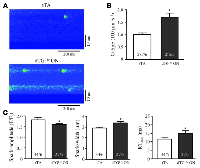
In order to investigate whether diastolic SR Ca2+ leak and increased incidence of SR Ca2+ release events contribute to arrhythmogenesis in dTGI-1c ON mice, isolated intact ventricular myocytes were loaded with the Ca2+-fluorescent dye Fluo-4 and electrically stimulated (1 Hz) in the presence of 10 nM isoprenaline. Representative confocal line-scan images of Ca2+ sparks are illustrated in Figure 4A. Myocytes from dTGI-1c ON mice showed approximately 70% higher Ca2+ spark frequency with unaltered Ca2+ spark amplitude but increased Ca2+ spark width and duration (Figure 4, B and C) compared with tTA. Consequently, the calculated total SR Ca2+ leak (frequency × amplitude × width × duration) was increased by approximately 155% in dTGI-1c ON (1.71 ± 0.24 milli–fluorescence units divided by diastolic baseline fluorescence [mF/F0]) versus tTA mice (0.67 ± 0.05 mF/F0; P < 0.05). SR Ca2+ content, as assessed by caffeine-induced SR Ca2+ release, was similar in dTGI-1c ON (7.98 ± 0.53 F/F0; n = 16) and tTA (7.35 ± 0.54 F/F0; n = 20) mice.
Figure 4. Isolated cardiomyocytes from dTGI-1c ON hearts show higher catecholamine-induced SR Ca2+ leak.
(A) Representative longitudinal line-scan images of tTA and dTGI-1c ON hearts. (B) Ca2+ spark frequency (CaSpF) in dTGI-1c ON versus tTA hearts (10 nM isoprenaline; *P < 0.05). (C) The Ca2+ spark characteristics, spark amplitude, spark width, and spark duration (time to 50% decay, RT50%), are shown. *P < 0.05 versus tTA. The numbers in the columns represent the numbers of myocytes and characterized sparks, respectively, from 5–6 hearts for each group.
Exaggerated toxicity of chronic catecholamine infusion in dTGI-1c ON mice.
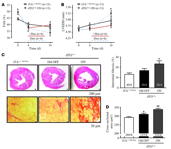
To study the consequences of I-1c overexpression in heart failure conditions, we next examined how dTGI-1c ON mice respond to prolonged adrenergic stress by subjecting dTGI-1c ON and tTA mice to a 14 day infusion with isoprenaline (30 μg/g per day) via mini-pumps. After 4 days, doxycycline was administered in half of I-1c double-transgenic mice (dTGI-1c 10d OFF) to turn off I-1c expression. Since tTA controls with and without doxycycline feeding did not differ in any of the investigated parameters, data were pooled and referred to as tTA+/–10d Dox (for details, see Supplemental Tables 2 and 3). In tTA+/–10d Dox, infusion of isoprenaline induced a moderate reduction in FAS and left ventricular dilation, which was stronger after 14 days than after 4 days (Figure 5). As expected, dTGI-1c ON mice exhibited a hypercontractile phenotype prior to infusion but an exaggerated decline in FAS (a decrease of 23% after 4 days and a decrease of 31% after 14 days; Figure 5A) and increase in left ventricular dilation (left ventricular end-diastolic diameter, an increase of 14% after 14 days; Figure 5B). Histological examination of the hearts revealed hypertrophy, increased interstitial fibrosis, and higher cardiomyocyte cross-sectional area in dTGI-1c ON versus tTA+/–10d Dox and/or dTGI-1c 10d OFF mice (Figure 5, C and D). Notably, the development of maladaptive cardiac phenotype between day 4 and 14 was partially prevented or reversed by doxycycline administration.
Figure 5. Accelerated morphometric and functional deterioration after chronic catecholaminergic stress in I-1c double-transgenic mice.
(A) Echocardiographically determined FAS in dTGI-1c ON and tTA mice before (day 0) and after (days 4 and 14) isoprenaline infusion, respectively. Exaggerated decline in FAS in I-1c double-transgenic mice was stopped in a subgroup of I-1c double-transgenic mice fed with doxycycline for 10 days (red line). *P < 0.05 versus tTA+/–10d Dox; #P < 0.05 dTGI-1c ON day 4 versus day 0. (B) Left ventricular end-diastolic diameter (LVEDD). The dashed line represents I-1c double-transgenic mice kept in the ON state, and the red line represents I-1c double-transgenic mice fed with doxycycline for 10 days. *P < 0.05 versus dTGI-1c 10d OFF. (C) H&E-stained paraffin sections (top row; representative of 4–8 hearts each) demonstrate dilation in dTGI-1c ON hearts. Sirius red–stained paraffin sections (bottom row) are representative of 4–8 hearts each and demonstrate fibrosis in dTGI-1c hearts, confirmed by quantitative analysis of interstitial fibrosis in dTGI-1c hearts. The first number in each column indicates the number of analyzed areas (20–40 areas), and the second number indicates the number of analyzed hearts. Scale bars: 200 μm (top row); 20 μm (bottom row). *P < 0.05 versus tTA+/–10d Dox. (D) Myocyte cross-sectional area in tTA+/–10d Dox, dTGI-1c 10d OFF, and dTGI-1c ON hearts treated with isoprenaline for 14 days (the first number in each column indicates the number of analyzed cardiomyocytes (≥132), and the second number indicates the number of analyzed hearts). *P < 0.05 versus tTA+/–10d Dox; #P < 0.05 versus dTGI-1c 10d OFF.
Progressive contractile dysfunction and dilation in aging dTGI-1c ON mice.
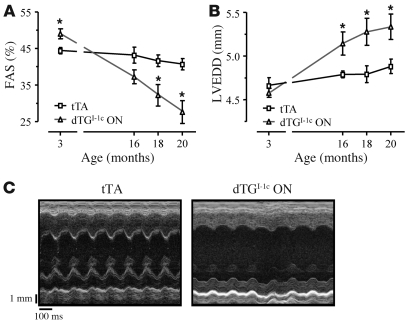
Serial echocardiography in aging dTGI-1c ON and tTA littermates (16, 18, and 20 months) showed a progressive decrease in contractile function and increase in left ventricular end-diastolic diameter, indicating a cardiomyopathic phenotype at rest with aging (Figure 6; for details, see Supplemental Table 4).
Figure 6. Aging I-1c double-transgenic ON littermates show contractile dysfunction and left ventricular dilation.
(A) FAS in tTA and dTGI-1c ON littermates at the age of 18 and 20 months (n = 5 each). *P < 0.05 versus tTA. (B) LVEDD (n = 5 each). *P < 0.05 versus tTA. (C) Representative M-mode views from tTA and dTGI-1c ON mice.
I-1S67A shows an identical phenotype.
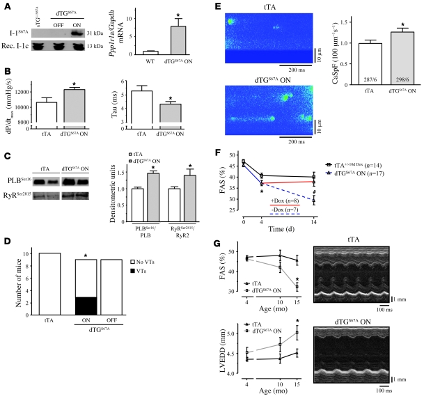
In parallel with I-1c a second double-transgenic line with conditional, heart-specific expression of a full-length mutant I-1 form, dTGS67A, was generated and brought on a Ppp1r1a-null background. Ser67 was replaced by the nonphosphorylatable Ala by PCR mutagenesis (see Supplemental Methods). Conserved PKA phosphorylation/activation and functionality of I-1S67A were confirmed by Western blot and phosphatase-1 activity assays (IC50, 18 ± 2 nM), respectively (Supplemental Figure 4, A–C). Western blots demonstrated I-1S67A protein only in dTGS67A ON hearts, with transcript concentrations being approximately 8-fold higher than endogenous Ppp1r1a mRNA in WT hearts (Figure 7A). Whereas echocardiography did not reveal significant differences (see Supplemental Table 5), the more sensitive cardiac catheterization technique demonstrated mild basal hypercontractility with enhanced systolic (maximum rate of rise in left ventricular pressure, dP/dtmax) and diastolic (time constant of isovolumetric relaxation, Tau) heart function at the age of 4 months (Figure 7B; for details, see Supplemental Table 6). Similar to the I-1c line, dTGS67A ON mice revealed (a) higher phosphorylation levels of PLB at PKA-Ser16 (an increase of 46% compared with tTA, n = 8; P < 0.05; Figure 7C) and RyR2 at CaMKII-Ser2815 (an increase of 41% compared with tTA, n = 6–8; P < 0.05; Figure 7C); (b) higher (and doxycycline-reversible) catecholamine-induced VTs in vivo (3 out of 9 mice, Figure 7D); (c) higher Ca2+ spark frequency after acute stimulation with 10 nM isoprenaline in isolated adult cardiomyocytes (an increase of 30% compared with tTA, Figure 7E; for details, see Supplemental Table 7); (d) an exaggerated adverse phenotype under chronic isoprenaline infusion (Figure 7F; for details, see Supplemental Table 8); and (e) a cardiomyopathic phenotype with aging (Figure 7G; for details, see Supplemental Table 9).
Figure 7. Characterization of I-1S67A–expressing mice.
(A) Doxycycline-dependent I-1S67A expression and a lack of leakiness in single transgenic I-1S67A responders (I-1S67A). I-1S67A mRNA amount in dTGS67A ON mice is 8-fold higher compared with Ppp1r1a mRNA from WT mice (n = 5 each). *P < 0.05 versus WT. (B) I-1S67A enhances cardiac contractility in vivo (n = 6 for each group). *P < 0.05 versus tTA. (C) Phosphorylation state of PLB (PLBSer16) and RyR2 (RyRSer2815) in tTA and dTGS67A ON hearts (n ≥ 6 for each group). Samples were run on the same gel. *P < 0.05 versus tTA. (D) Number of tTA and dTGS67A ON mice that developed VTs (black bar) after arrhythmia provocation in freely moving mice. Doxycycline application completely repressed the development of VTs (dTGS67A OFF). *P < 0.05 by χ2 test. (E) Representative longitudinal line-scan images of tTA and dTGS67A ON mice and Ca2+ spark frequency, respectively (10 nM isoprenaline). The numbers in the columns represent the number of myocytes and hearts from each group. *P < 0.05 versus tTA. (F) Isoprenaline-infusion accelerated the decrease in contractility in dTGS67A ON versus tTA mice after 4 and 14 days. Exaggerated decline of contractility in dTGS67A ON mice could be stopped by doxycycline feeding from day 4 to 14 (red line). The dashed line represents the I-1c double-transgenic mice kept on water at any time. *P < 0.05 tTA versus dTGS67A ON on day 4; #P < 0.05 dTGS67A ON versus dTGS67A 10d OFF and tTA. (G) Aging I-1S67A double-transgenic mice show an exaggerated decrease in FAS and increase in LVEDD (n ≥ 5 each) compared with tTA mice at the age of 15 months. *P < 0.05 versus tTA. Representative M-mode views from tTA and dTGS67A ON mice. Experiments were done in parallel to the I-1c double-transgenic mice (C–E).
Discussion
The exact role of I-1 in cardiac signaling remains incompletely understood. Studies overexpressing constitutively active I-1c reported improved contractility and resistance to cardiac overload and ischemia/reperfusion injury (15, 16). On the other hand, Ppp1r1a KO mice had essentially normal cardiac function and were partially protected from acute and chronic catecholamine toxicity (12). To dissect the specific roles of I-1 in normal and diseased conditions, we generated 2 mouse models that allowed investigation of the effects of conditional cardiac-specific expression of I-1c/I-1S67A on a Ppp1r1a KO background. Phenotypic evaluations were done in a strictly blinded fashion to prevent observer bias. The conditional, doxycycline-regulated mouse model allowed time-dependent, reversible expression of I-1c/I-1S67A, specifically in the heart. This allowed us to compare heart function in the absence of I-1 and in the (reversible) presence of the constitutively active form I-1c or I-1S67A. The strength of the Tet-Off system, namely reversible transgene expression, has been used in all cases in which a paired analysis (“before and after”) was possible (echocardiography under normal conditions and in isoprenaline-infused mice, telemetric analysis of rate and rhythm in isoprenaline-induced arrhythmias). Due to practical, ethical, and economic reasons, in all other cases tTA were used as controls. The following major results were obtained. (a) Young dTGI-1c/I-1S67A ON mice were apparently normal but exhibited hypercontractile heart function. (b) Hearts of young dTGI-1c/I-1S67A ON mice showed hyperphosphorylation of PLB and RyR2. (c) The latter was associated with higher susceptibility to catecholamine-induced VTs in vivo. Langendorff-perfused hearts from induced I-1c double-transgenic mice more frequently developed VTs, and isolated cardiomyocytes showed higher Ca2+ spark frequency. (d) dTGI-1c/I-1S67A ON mice developed exaggerated catecholamine-induced left ventricular dilation, contractile dysfunction, cardiomyocyte hypertrophy, and interstitial fibrosis. All abnormalities in both lines were prevented by doxycycline administration, underscoring a direct causal relationship. Finally, dTGI-1c/I-1S67A ON mice spontaneously developed a cardiomyopathic phenotype with aging. Taken together, expression of both I-1 variants in the heart was associated with enhanced heart function at the basal state but also with cardiac deterioration and increased propensity to arrhythmias after adrenergic stress and with aging. This phenotype is strikingly similar to that of mouse models overexpressing other key elements of the β-adrenergic signaling pathway (2, 7, 20). Overexpression of the β1-adrenergic receptor, β2-adrenergic receptor, or α-subunit of stimulatory G proteins leads initially to higher contractile performance but later in life causes cardiomyopathy and/or arrhythmia and premature death (21–23). Thus, this pattern is reminiscent of the well-known adverse effects of long-term adrenergic stimulation in experimental heart failure and positive inotropic agents in patients, raising a caveat with regard to the value of I-1c for gene therapy.
The present study confirms previous results, showing a hypercontractile phenotype in young I-1–overexpressing animals (15, 16). Overexpression of I-1c was associated with a robust stimulation of contractile function, both on a WT (15, 16) and a Ppp1r1a KO background (in this study), similar to what has been shown with adenoviral overexpression in cultured myocytes previously (13, 14). Here we extend these findings by showing fast reversibility of the phenotype by doxycycline. Switching on the gene by doxycycline withdrawal took several weeks, likely because of the complex pharmacokinetics of this drug (24). Collectively, the published data are all compatible with the idea that I-1 amplifies β-adrenergic signals by increasing the phosphorylation state of PLB with subsequent disinhibition of SERCA2a and greater SR Ca2+ uptake into the SR, enhanced SR Ca2+ loading, and systolic SR Ca2+ release. Hence, I-1c expression would be expected to mimic ablation of PLB (25). However, Plb KO mice, though also exhibiting a hypercontractile heart, remain healthy throughout their life span (25). Crossing mice into a Plb KO background even prevented the development of a cardiomyopathic phenotype in mice with muscle-LIM protein (Mlp) KO (26) or β1-adrenergic receptor overexpression (27). These findings point to critical differences between I-1c overexpression and Plb KO. A major difference between the 2 models, as identified in the present study, is that I-1c overexpression results in hyperphosphorylation of both PLB and RyR2. This finding was somewhat unexpected, because previous studies that have overexpressed I-1c failed to demonstrate changes in RyR2 phosphorylation at Ser2809 (PKA site), suggesting that I-1c specifically targets PLB (15, 16). We confirmed this finding with regard to RyR2 phosphorylation at Ser2809 but found higher phosphorylation at Ser2815 (CaMKII site), which to the best of our knowledge has not been studied in the context of I-1c expression yet. Ppp1r1a KO hearts exhibited the opposite alteration in RyR2-Ser2815 phosphorylation (17), indicating that I-1 and I-1c are similar in subcellular control of phosphatase-1. Further support for this interpretation comes from the observation of hyperphosphorylation of RyR2 at Ser2815 also in I-1S67A mice.
The mechanism of the selective hyperphosphorylation of RyR2 at the CaMKII site in I-1c/I-1S67A double-transgenic mice is unknown. One possibility is that phosphatase-1 is more active at the CaMKII site than at the PKA site of RyR2. Another one is that I-1 mediates a cross-talk between PKA and CaMKII, by inhibiting phosphatase-1–mediated dephosphorylation (therefore inactivation) of CaMKII at Thr286, thereby increasing CaMKII activity (28). However, neither dTGI-1c/I-1S67A ON (Supplemental Figure 3) nor Ppp1r1a KO hearts showed differences in CaMKII-Thr286 phosphorylation or activity (data not shown), and PLB-Thr17 phosphorylation at the CaMKII site was unchanged (data not shown), arguing against the relevance of this pathway in the heart. Thus, the exact reasons for preferential regulation of RyR2 at Ser2815 in dTGI-1c/I-1S67A ON hearts have yet to be determined.
Hyperphosphorylation of RyR2 at Ser2815 has previously been associated with increased Ca2+ spark frequency (18, 19), and Ca2+ spark frequency was indeed higher in dTGI-1c/I-1S67A ON than tTA mice (Figure 4), suggesting that the increased susceptibility to arrhythmia both in vivo and in Langendorff-perfused hearts in vitro is causally related to this alteration. In this model, hyperphosphorylation of RyR2 at Ser2815 increases the propensity toward spontaneous Ca2+ release from the SR, which, if it happened as an isolated event, would quickly lead to depletion of the SR and suspension of Ca2+ release events (29). However, simultaneous hyperphosphorylation of PLB would promote fast refilling of the SR, thereby supporting a continuous SR Ca2+ leak, which may lead to delayed afterdepolarizations, triggered activity, and ventricular arrhythmias. In fact, this mechanism has been proposed to explain the proarrhythmic effect of adrenergic stimulation in situations in which the open probability of RyR2 is increased, either experimentally by caffeine (30) or by mutations in RyR2 associated with catecholaminergic polymorphic VT (31). Our data suggest that I-1c and I-1S67A overexpression mimic this phenotype by producing 2 essential requirements for triggered activity and ventricular arrhythmogenesis: (a) hyperphosphorylation of PLB that indirectly enhances SR Ca2+ load by increasing SERCA2a activity and (b) hyperphosphorylation of RyR2, which increases diastolic SR Ca2+ leak by enhancing open probability of RyR2 channels.
Our data are not consistent with previous studies showing beneficial effects of I-1c expression against heart failure progression and postischemic injury (15, 16), with several possible explanations. Earlier studies on I-1c used mice in the FVB/N background instead of the C57Bl/6J background, and genetic backgrounds are known to alter morphology, function, and survival in transgenic mice. However, the FVB/N background is more susceptible to stress-induced arrhythmias than C57Bl/6J background (32), rendering background unlikely to explain the higher susceptibility to malignant arrhythmias in our model. Whereas previous studies expressed I-1c on top of endogenous I-1, this study expressed I-1c in the absence of endogenous I-1 for the following reasons: (a) it excludes confounding, e.g., competitive, effects between endogenous I-1 and I-1c, and (b) it resembles to some extent the “therapeutic” situation in human and experimental heart failure, in which I-1 is markedly downregulated and deactivated (12, 14, 33, 34). The present study used tTA littermates as control; another I-1c study used WT mice (16). Recent studies did also not address specifically the susceptibility to arrhythmias, which in this study was manifested under stress conditions only. Similarly, published work did not perform long-term studies with regular echocardiography for 20 months. Finally, previous reports neither analyzed the phosphorylation state of RyR2 at Ser2815 (CaMKII site), nor the propensity for diastolic SR Ca2+ leak in isolated cardiomyocytes.
One could argue that expression of I-1c, a constitutively active variant, at levels approximately 24 fold that of WT I-1 may give rise to artefacts, e.g., nonselective super stimulation of multiple pathways that then account for the pathology. However, the second mouse line expressing the nonconstitutively active (PKA-dependent), full-length mutant I-1S67A at approximately 8-fold WT levels showed essentially identical results. This mutant was constructed to be resistant to phosphorylation at Ser67 by PKC-α (11), because PKC-α phosphorylation at Ser67 has been associated with inactivation of I-1 and adverse effects of WT full-length I-1 on the heart (for discussion see refs. 2, 35). The fact that both lines showed almost superimposable results also argues against the concept that it is the PKC-α phosphorylation of I-1 that accounts for the adverse part of I-1 effects. Moreover, the data in 2 independent lines expressing different variants of I-1 provide more general evidence against the idea that Ppp1r1a gene transfer allows dissociation of a (beneficial) stimulation of force from the (adverse) consequences in terms of cardiac structure and arrhythmias. The risk is even more relevant since gene therapy with adeno-associated viruses will necessarily lead to inhomogeneous expression levels in the heart. Some cells will not be hit at all and others will be highly transduced. Thus, we think that our data should be considered when reevaluating the benefit/risk ratio of I-1c gene therapy in heart failure patients.
Methods
Generation of 2 double-transgenic mouse models with cardiac-specific and temporally regulated expression of I-1c and I-1S67A on a complete Ppp1r1a KO background.
I-1c and I-1S67A were both generated by PCR from full-length mouse I-1 by phosphomimetic site-directed mutagenesis. To generate I-1c, the full-length mouse I-1 was mutated at Thr35 (I-1-T35D) and subsequently truncated (amino acids 1–65). In I-1S67A, Ser67 was replaced by Ala (I-1-S67A). Functionality of the both I-1 forms (I-1c and I-1S67A) was tested and compared with PKA-phosphorylated full-length WT I-1 in vitro, by generating the corresponding recombinant proteins in bacteria and performing phosphatase-1 activity assays with 32P-phosphorylase-A as substrate. Successful WT I-1 as well as I-1S67A phosphorylation and loss of Thr35 in I-1c was confirmed by Western blot with an I-1 Thr35–phospho-specific antibody (Supplemental Figure 4A). As expected, PKA-phosphorylated WT I-1, I-1S67A, and I-1c inhibited phosphatase-1 activity potently, with IC50 values of 33 ± 2 nM, 18 ± 2 nM, and 151 ± 5 nM, respectively (Supplemental Figure 4B), demonstrating functional activity. The lower I-1c potency is consistent with previous reports (36).
Single transgenesis for I-1c and I-1S67A was achieved by pronucleus injection of a construct containing the Ptight promoter (Tet-responsive promoter, composed of 7 direct repeats of the tet-operator sequence [TetO7], followed by a CMV minimal promoter [CMVmin]) driving expression of the I-1c and I-1S67A cDNA, respectively (responder). Importantly, before injection these constructs were functionally tested in vitro in a HEK293 cell model: I-1c and I-1S67A expression was induced by CMV-driven tTA, and transactivation activity was completely abolished in the presence of doxycycline (1 μg/ml; Supplemental Figure 4C), demonstrating functionality of the Tet-Off system and indicating that the I-1c– as well as the I-1S67A–attenuated promoter did not exhibit leakage in vitro.
Consequently, both I-1 double-transgenic mice were backcrossed to C57BL/6J and genetically crossed with Ppp1r1a KO mice until they were on a complete homozygote Ppp1r1a-null background. The complex transgenic approach was continuously controlled by Southern blotting and PCR. The Tet-Off system requires a second transgenic line with a cardiac-specific promoter (α-MHC) driving expression of the tTA gene (37). This line was also bred into a homozygote Ppp1r1a-null background. Crossing both the I-1 responder lines with the tTA line revealed double-transgenic mice, which were expected to express I-1c and I-1S67A, respectively, only in the absence of doxycycline in drinking water. Animals were used and handled as approved by the governmental review board in Hamburg (G 21/1-42/05).
Telemetric ECG recordings.
Two weeks after implantation of telemetric transmitters (Data Sciences), ECGs were recorded in freely moving mice during a defined stress protocol, consisting of an intraperitoneal injection of isoprenaline (2 μg/g), followed by repetitive, intermittent warm air-jet stress to provoke mental stress for 15 minutes, followed by a second injection of isoprenaline (2 μg/g) (38). Telemetric ECGs were continuously recorded and analyzed for arrhythmias during stress tests and the following 2-hour recovery period. All stress test protocols were performed by blinded operators on matched pairs of mice.
Langendorff-perfused mouse hearts.
Isolated, beating Langendorff-perfused mouse hearts were studied using previously-published techniques (39). An octapolar murine electrophysiology catheter was inserted into the right atrium and right ventricle. Atrial and ventricular electrograms, a tissue bath ECG, and monophasic action potentials from left and right ventricle were simultaneously recorded. After instrumentation, the heart was subjected to a stabilization period of 10 minutes, followed by a period of spontaneous sinus rhythm of 5 minutes. Then, pacing was performed at constant rates at 100 and 80 ms cycle length using the octapolar catheter.
Ca2+ imaging using confocal microscopy.
Ca2+ sparks were recorded on a laser scanning confocal microscope (LSM 5 Pascal, Zeiss), using intact cardiomyocytes loaded with the Ca2+-fluorescent dye, Fluo-4 AM (10 μmol/l; Invitrogen). The dye was excited by an argon laser at 488 nm and emitted fluorescence was collected through a 505 nm long-pass emission filter. Fluorescence images were recorded in line-scan mode, during electrical stimulation at 1 Hz, and background fluorescence was compensated for. The amplitude of Ca2+ signals was inferred from fluorescence intensity divided by diastolic baseline fluorescence (F/F0). For each Ca2+ spark, spark amplitude (F/F0), duration (time to 50% decay, RT50%), and width were determined. Ca2+ spark frequency was normalized to scan-line length and scan duration. From this, total diastolic SR Ca2+ leak could be calculated as the product of frequency, amplitude, and duration. To assess SR Ca2+ content, we investigated Ca2+ transients induced by rapid application of 10 mM caffeine and evaluated F/F0 of these transients.
An expanded Supplemental Methods section can be found with the online data.
Statistics.
All measurements and analyses with the exception of protein biochemistry and mRNA quantification were performed by researchers blinded to genotype. All variables are reported as mean ± SEM. Unpaired Student t test (2-tailed) was used to compare means between 2 independent groups/samples. Paired t test (2-tailed) was used to compare values in mice before and after treatment. Fisher’s exact test and χ2 test were used to compare arrhythmia incidences between genotypes. P values of less than 0.05 were considered significant.
Supplementary Material
Acknowledgments
We thank Birgit Geertz, Jutta Starbatty, Sabine Kirsch, Annett Opitz, and Thomas Schulze for excellent technical assistance. This study was supported by the DFG (DFG EL 270/3-1, to A. El-Armouche and T. Eschenhagen; DFG MA 1982/2-1 and MA 1982/4-1, to L.S. Maier; DFG FA 413/3-1, to L. Fabritz; DFG Ki 713/1-1, to P. Kirchhof), the European Union (EUGene Heart to A. El-Armouche, T. Eschenhagen, and G. Hasenfuss), the German Heart Foundation/German Foundation of Heart Research (to A. El-Armouche), the German Federal Ministry of Education and Research through the Atrial Fibrillation Competence Network (grant 01Gi0204, to D. Dobrev and P. Kirchhof), by a grant from Fondation Leducq (07 CVD 03, to D. Dobrev and P. Kirchhof), and IZKF Münster (core unit CarTel, to P. Kirchhof).
Footnotes
Conflict of interest: The authors have declared that no conflict of interest exists.
Citation for this article: J Clin Invest. 2010;120(2):617–626. doi:10.1172/JCI40545.
References
- 1.Bristow MR. beta-adrenergic receptor blockade in chronic heart failure. Circulation. 2000;101(5):558–569. doi: 10.1161/01.cir.101.5.558. [DOI] [PubMed] [Google Scholar]
- 2.El-Armouche A, Eschenhagen T. beta-Adrenergic stimulation and myocardial function in the failing heart. Heart Fail Rev. 2008;14(4):225–241. doi: 10.1007/s10741-008-9132-8. [DOI] [PubMed] [Google Scholar]
- 3.Hasenfuss G, Pieske B. Calcium cycling in congestive heart failure. J Mol Cell Cardiol. 2002;34(8):951–969. doi: 10.1006/jmcc.2002.2037. [DOI] [PubMed] [Google Scholar]
- 4.Bers DM. Calcium cycling and signaling in cardiac myocytes. Annu Rev Physiol. 2008;70:23–49. doi: 10.1146/annurev.physiol.70.113006.100455. [DOI] [PubMed] [Google Scholar]
- 5.Venetucci LA, Trafford AW, O’Neill SC, Eisner DA. The sarcoplasmic reticulum and arrhythmogenic calcium release. Cardiovasc Res. 2008;77(2):285–292. doi: 10.1093/cvr/cvm009. [DOI] [PubMed] [Google Scholar]
- 6.Nabauer M, Kaab S. Potassium channel down-regulation in heart failure. Cardiovasc Res. 1998;37(2):324–334. doi: 10.1016/s0008-6363(97)00274-5. [DOI] [PubMed] [Google Scholar]
- 7.Dorn GW, II, Molkentin JD. Manipulating cardiac contractility in heart failure: data from mice and men. Circulation. 2004;109(2):150–158. doi: 10.1161/01.CIR.0000111581.15521.F5. [DOI] [PubMed] [Google Scholar]
- 8.Bukhari F, MacGillivray T, del Monte F, Hajjar RJ. Genetic maneuvers to ameliorate ventricular function in heart failure: therapeutic potential and future implications. Expert Rev Cardiovasc Ther. 2005;3(1):85–97. doi: 10.1586/14779072.3.1.85. [DOI] [PubMed] [Google Scholar]
- 9.Fechner H, et al. Highly efficient and specific modulation of cardiac calcium homeostasis by adenovector-derived short hairpin RNA targeting phospholamban. Gene Ther. 2007;14(3):211–218. doi: 10.1038/sj.gt.3302872. [DOI] [PubMed] [Google Scholar]
- 10.Hajjar RJ, et al. Design of a phase 1/2 trial of intracoronary administration of AAV1/SERCA2a in patients with heart failure. J Card Fail. 2008;14(5):355–367. doi: 10.1016/j.cardfail.2008.02.005. [DOI] [PubMed] [Google Scholar]
- 11.Braz JC, et al. PKC-alpha regulates cardiac contractility and propensity toward heart failure. Nat Med. 2004;10(3):248–254. doi: 10.1038/nm1000. [DOI] [PubMed] [Google Scholar]
- 12.El-Armouche A, Pamminger T, Ditz D, Zolk O, Eschenhagen T. Decreased protein and phosphorylation level of the protein phosphatase inhibitor-1 in failing human hearts. Cardiovasc Res. 2004;61(1):87–93. doi: 10.1016/j.cardiores.2003.11.005. [DOI] [PubMed] [Google Scholar]
- 13.El-Armouche A, et al. Evidence for protein phosphatase inhibitor-1 playing an amplifier role in beta-adrenergic signaling in cardiac myocytes. FASEB J. 2003;17(3):437–439. doi: 10.1096/fj.02-0057fje. [DOI] [PubMed] [Google Scholar]
- 14.Carr AN, et al. Type 1 phosphatase, a negative regulator of cardiac function. Mol Cell Biol. 2002;22(12):4124–4135. doi: 10.1128/MCB.22.12.4124-4135.2002. [DOI] [PMC free article] [PubMed] [Google Scholar]
- 15.Pathak A, et al. Enhancement of cardiac function and suppression of heart failure progression by inhibition of protein phosphatase 1. Circ Res. 2005;96(7):756–766. doi: 10.1161/01.RES.0000161256.85833.fa. [DOI] [PubMed] [Google Scholar]
- 16.Nicolaou P, et al. Inducible expression of active protein phosphatase-1 inhibitor-1 enhances basal cardiac function and protects against ischemia/reperfusion injury. Circ Res. 2009;104(8):1012–1020. doi: 10.1161/CIRCRESAHA.108.189811. [DOI] [PMC free article] [PubMed] [Google Scholar]
- 17.El-Armouche A, et al. Phosphatase inhibitor-1-deficient mice are protected from catecholamine-induced arrhythmias and myocardial hypertrophy. Cardiovasc Res. 2008;80(3):396–406. doi: 10.1093/cvr/cvn208. [DOI] [PubMed] [Google Scholar]
- 18.Curran J, Hinton MJ, Rios E, Bers DM, Shannon TR. Beta-adrenergic enhancement of sarcoplasmic reticulum calcium leak in cardiac myocytes is mediated by calcium/calmodulin-dependent protein kinase. Circ Res. 2007;100(3):391–398. doi: 10.1161/01.RES.0000258172.74570.e6. [DOI] [PubMed] [Google Scholar]
- 19.Wehrens XH, Lehnart SE, Reiken SR, Marks AR. Ca2+/calmodulin-dependent protein kinase II phosphorylation regulates the cardiac ryanodine receptor. Circ Res. 2004;94(6):e61–e70. doi: 10.1161/01.RES.0000125626.33738.E2. [DOI] [PubMed] [Google Scholar]
- 20.Lohse MJ, Engelhardt S, Eschenhagen T. What is the role of beta-adrenergic signaling in heart failure? Circ Res. 2003;93(10):896–906. doi: 10.1161/01.RES.0000102042.83024.CA. [DOI] [PubMed] [Google Scholar]
- 21.Engelhardt S, Hein L, Wiesmann F, Lohse MJ. Progressive hypertrophy and heart failure in beta1-adrenergic receptor transgenic mice. Proc Natl Acad Sci U S A. 1999;96(12):7059–7064. doi: 10.1073/pnas.96.12.7059. [DOI] [PMC free article] [PubMed] [Google Scholar]
- 22.Gaudin C, et al. Overexpression of Gs alpha protein in the hearts of transgenic mice. J Clin Invest. 1995;95(4):1676–1683. doi: 10.1172/JCI117843. [DOI] [PMC free article] [PubMed] [Google Scholar]
- 23.Liggett SB, et al. Early and delayed consequences of beta(2)-adrenergic receptor overexpression in mouse hearts: critical role for expression level. Circulation. 2000;101(14):1707–1714. doi: 10.1161/01.cir.101.14.1707. [DOI] [PubMed] [Google Scholar]
- 24.Kistner A, et al. Doxycycline-mediated quantitative and tissue-specific control of gene expression in transgenic mice. Proc Natl Acad Sci U S A. 1996;93(20):10933–10938. doi: 10.1073/pnas.93.20.10933. [DOI] [PMC free article] [PubMed] [Google Scholar]
- 25.Luo W, et al. Targeted ablation of the phospholamban gene is associated with markedly enhanced myocardial contractility and loss of beta-agonist stimulation. Circ Res. 1994;75(3):401–409. doi: 10.1161/01.res.75.3.401. [DOI] [PubMed] [Google Scholar]
- 26.Minamisawa S, et al. Chronic phospholamban-sarcoplasmic reticulum calcium ATPase interaction is the critical calcium cycling defect in dilated cardiomyopathy. Cell. 1999;99(3):313–322. doi: 10.1016/S0092-8674(00)81662-1. [DOI] [PubMed] [Google Scholar]
- 27.Engelhardt S, Hein L, Dyachenkow V, Kranias EG, Isenberg G, Lohse MJ. Altered calcium handling is critically involved in the cardiotoxic effects of chronic beta-adrenergic stimulation. Circulation. 2004;109(9):1154–1160. doi: 10.1161/01.CIR.0000117254.68497.39. [DOI] [PubMed] [Google Scholar]
- 28.Blitzer RD, et al. Gating of CaMKII by cAMP-regulated protein phosphatase activity during LTP. Science. 1998;280(5371):1940–1942. doi: 10.1126/science.280.5371.1940. [DOI] [PubMed] [Google Scholar]
- 29.Eisner DA, Kashimura T, O’Neill SC, Venetucci LA, Trafford AW. What role does modulation of the ryanodine receptor play in cardiac inotropy and arrhythmogenesis? J Mol Cell Cardiol. 2008;46(4):474–481. doi: 10.1016/j.yjmcc.2008.12.005. [DOI] [PubMed] [Google Scholar]
- 30.O’Neill SC, Miller L, Hinch R, Eisner DA. Interplay between SERCA and sarcolemmal Ca2+ efflux pathways controls spontaneous release of Ca2+ from the sarcoplasmic reticulum in rat ventricular myocytes. J Physiol. 2004;559(Pt 1):121–128. doi: 10.1113/jphysiol.2003.058917. [DOI] [PMC free article] [PubMed] [Google Scholar]
- 31.Marks AR, Priori S, Memmi M, Kontula K, Laitinen PJ. Involvement of the cardiac ryanodine receptor/calcium release channel in catecholaminergic polymorphic ventricular tachycardia. J Cell Physiol. 2002;190(1):1–6. doi: 10.1002/jcp.10031. [DOI] [PubMed] [Google Scholar]
- 32.Maguire CT, Wakimoto H, Patel VV, Hammer PE, Gauvreau K, Berul CI. Implications of ventricular arrhythmia vulnerability during murine electrophysiology studies. Physiol Genomics. 2003;15(1):84–91. doi: 10.1152/physiolgenomics.00034.2003. [DOI] [PubMed] [Google Scholar]
- 33.El-Armouche A, Gocht F, Jaeckel E, Wittkopper K, Peeck M, Eschenhagen T. Long-term beta-adrenergic stimulation leads to downregulation of protein phosphatase inhibitor-1 in the heart. Eur J Heart Fail. 2007;9(11):1077–1080. doi: 10.1016/j.ejheart.2007.09.006. [DOI] [PubMed] [Google Scholar]
- 34.Gupta RC, Mishra S, Rastogi S, Imai M, Habib O, Sabbah HN. Cardiac SR-coupled PP1 activity and expression are increased and inhibitor 1 protein expression is decreased in failing hearts. Am J Physiol Heart Circ Physiol. 2003;285(6):H2373–H2381. doi: 10.1152/ajpheart.00442.2003. [DOI] [PubMed] [Google Scholar]
- 35.Nicolaou P, Hajjar RJ, Kranias EG. Role of protein phosphatase-1 inhibitor-1 in cardiac physiology and pathophysiology. J Mol Cell Cardiol. 2009;47(3):365–371. doi: 10.1016/j.yjmcc.2009.05.010. [DOI] [PMC free article] [PubMed] [Google Scholar]
- 36.Endo S, Zhou X, Connor J, Wang B, Shenolikar S. Multiple structural elements define the specificity of recombinant human inhibitor-1 as a protein phosphatase-1 inhibitor. Biochemistry. 1996;35(16):5220–5228. doi: 10.1021/bi952940f. [DOI] [PubMed] [Google Scholar]
- 37.Passman RS, Fishman GI. Regulated expression of foreign genes in vivo after germline transfer. J Clin Invest. 1994;94(6):2421–2425. doi: 10.1172/JCI117609. [DOI] [PMC free article] [PubMed] [Google Scholar]
- 38.Kirchhof P, et al. Stress and high heart rate provoke ventricular tachycardia in mice expressing triadin. . J Mol Cell Cardiol. 2007;42(5):962–971. doi: 10.1016/j.yjmcc.2007.02.012. [DOI] [PubMed] [Google Scholar]
- 39.Fabritz L, et al. Effect of pacing and mexiletine on dispersion of repolarisation and arrhythmias in DeltaKPQ SCN5A (long QT3) mice. Cardiovasc Res. 20035741085–1093. . 10.1016/S0008-6363(02)00839-8 [DOI] [PubMed] [Google Scholar]
Associated Data
This section collects any data citations, data availability statements, or supplementary materials included in this article.


