
Figure 2.
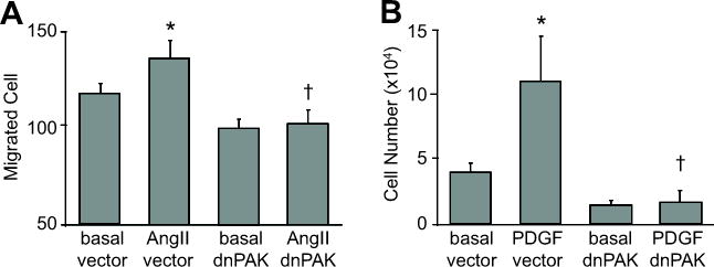
dnPAK1 inhibited migration and proliferation of VSMCs. A, Confluent VSMCs infected with adenovirus (100 moi) encoding dnPAK1 or the control GFP vector were scraped by a metal dental pick and stimulated with AngII for 24 hours in the presence of 5 mmol/L hydroxyurea to block cell proliferation completely. The nucleus was stained with Hoechst 33342 dye, and migrated VSMCs from the wound edge were counted in 4 independent view fields (100X). Data are mean±SEM of 4 experiments. B, VSMCs were infected with adenovirus (100 moi) encoding control GFP vector or dnPAK1 for 48 hours. The cells were then stimulated with 100 ng/mL PDGF-BB for 3 days and the cell number counted. Data are mean±SEM of 3 experiments. *p<0.05 compared to the basal control. †p<0.05 compared to the stimulated control.