Abstract
We present a case of thoracic splenosis in a 42-yr-old man with a medical history of abdominal surgery for a penetration injury with an iron bar of the left abdomen and back. He had been in good condition, but a chest radiograph taken during a regular checkup showed a multinodular left pleura-based mass. Computed tomography (CT) showed that the mass was well-enhanced and homogeneous, indicating a sclerosing hemangioma. Following its removal by video-assisted thoracoscopic surgery, the mass appeared similar to a hemangioma, with marked adhesion to the left side diaphragmatic pleura and lung parenchyma. Frozen section showed that the lesion was a solid mass consisted with abundant lymphoid cells, suggesting a low grade lymphoma. On permanent section, however, the mass was found to be composed of white pulp, red pulp, a thick capsule and trabeculae and was diagnosed as ectopic splenic tissue, or thoracic splenosis. Review of the patient's history and chest CT at admission revealed that the patient had undergone a splenectomy for the penetration injury 20 yr previously.
Keywords: Thoracic Cavity, Splenosis, Splenectomy
INTRODUCTION
Thoracic splenosis (TS) is a rare condition that usually follows simultaneous splenic and diaphragmatic injury, with autotransplantation of splenic tissue into the left hemithorax. TS typically presents on chest radiographs as an asymptomatic pulmonary mass, either solitary or multiple, and can mimic primary or metastatic lung cancer. Diagnosis is difficult without knowledge of previous splenic injury, leading to the use of biopsy, video-assisted thoracoscopic surgery (VATS) and even thoracotomy for diagnosis. Here, we describe a 42-yr-old Korean male diagnosed with TS 20 yr after splenectomy; this represents the first reported case of TS in Korea.
CASE REPORT
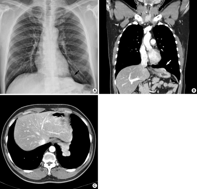
A 42-yr-old male was referred to Ulsan University Hospital for evaluation of a multinodular mass based on the left diaphragmatic pleura. This mass had been detected incidentally on a chest radiograph during the patient's annual medical checkup. The patient had been in good general condition, except for left inferior pleural thickening on chest radiographs taken from 1999 to 2004. A newly developed small nodule was detected in 2005, but the clinician regarded it as an inactive lesion. In 2007, the lesion had enlarged and formed a sizable mass, suggesting a malignancy (Fig. 1A). The patient had smoked one-half to one pack of cigarettes per day for 14 yr. On admission, all laboratory data were within normal ranges. Computed tomography (CT) revealed a homogeneously well-enhanced multinodular mass, measuring 4.8 cm in its greatest dimension, based on the diaphragm (Fig. 1B, C). The patient was suspected of having a tumor of pleural origin such as a sclerosing hemangioma. There was no evidence of a lung parenchymal mass or a primary malignancy of other organs. An excision by VATS was attempted.
Fig. 1.
Chest radiograph of the patient taken during an annual checkup (A) and coronal and transverse views of contrast enhanced computed tomographic (CT) images (B, C). A diaphragm-based mass (arrow) was observed on the chest radiograph (A). The homogeneously enhanced mass (arrow) abuts the left lateral segment of the liver and stomach (B, C). No spleen was detected in the left upper abdominal cavity (B, C).
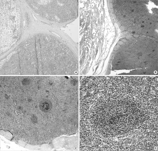
Intraoperatively, the mass had the gross appearance of a hemangioma containing blood. The mass, which was tightly attached to the diaphragm and adjacent lung tissue, was removed through a minimal thoracotomy. A piece of soft tissue labeled "diaphragm" was submitted for a frozen section, which revealed a multinodular solid mass containing abundant lymphoid cells in a diffuse or vaguely nodular arrangement, as well as a thick fibrous capsule and septae, suggesting it may be a low grade malignant lymphoma (Fig. 2A). The entire mass and the attached lung tissue were excised. Examination of a permanent section showed that the mass was ectopic splenic tissue, which included white and red pulp and central arteries (Fig. 2B-D).
Fig. 2.
Microscopic findings of the thoracic splenosis on frozen (A) and permanent (B-D) sections. Examination of the frozen section showed a thick capsule and trabeculae with abundant lymphoid tissue, but white and red pulp were indistinct (H&E stain, ×40) (A). The splenic tissue is tightly attached to the lung parenchyma without invasion (H&E stain, ×40) (B) and have normal splenic histology, with thick capsule and distinct white and red pulp (H&E stain, ×100 and ×200) (C, D).
A review of the patient's medical history showed that, 20 yr earlier, he had been pierced through the left abdomen and back by an iron bar. The patient, however, did not know what kind of operation had been performed. Review of his chest CT at admission revealed radiologic evidence that he had undergone a splenectomy.
DISCUSSION
Since the first report in 1937, a total of 57 cases of TS have been reported in the English language literature (1, 2). These patients range in age from 15 to 79 yr, and are predominantly male (47 of 57 cases) (2). The average interval between initial trauma and discovery of TS is 21 yr (3). Most patients are asymptomatic and discovered TS incidentally, but pleurisy or recurrent hemoptysis may occur (3, 4). TS presents on chest CT as multiple pleura-based nodules in 75% of patients and solitary pleura-based nodules in the remaining 25% (3). Differential diagnosis includes pleural metastases (most commonly arising from the lung, breast, or melanoma), lymphoma, localized fibrous tumor of the pleura, malignant mesothelioma, invasive thymoma and sclerosing hemangioma (4, 5).
TS used to be diagnosed at the time of surgery. However, if the diagnosis is confirmed preoperatively by appropriate radionuclide modalities in each patient with a new pulmonary mass and a history of thoraco-abdominal trauma, no matter how remote, thoracotomy can be avoided. Tc-99m sulfur colloid is sequestered in the reticulo-endothelial system and therefore can be used to detect all ectopic splenic tissue. Tc-99m heat-damaged erythrocytes and indium-111-labelled platelets are considered more sensitive and specific for the diagnosis of splenic sequestration and phagocytosis (6).
If a biopsy is performed without a preoperative suspicion of splenosis, the frozen section should be carefully examined for evidence of splenosis, although the quality of the frozen section may not be sufficient to assess the detailed structure of the splenic tissue. The pathologic differential diagnosis can include lymph nodes in any reactive condition, low grade lymphoma and thymoma. The microscopic feature of splenosis is almost identical with that of normal spleen, in that both have a thick capsule, trabeculae, and white and red pulp. In both, the subcapsular sinusoid or germinal center of the lymph node and epithelial cells of thymoma would not be present. Accessory spleens should be distinguished from splenosis, especially in patients with intra-abdominal splenosis. Accessory spleens are mainly located around the splenic hilum, and there are rarely more than three (7). Accessory spleens resemble the spleen in shape and have hilum where the arteries enter. Splenosis, however, has no hilum and the arterial supply can pass through any site in the capsule (7, 8).
Splenosis in the abdominal and pelvic cavities occurs more frequently than in the thoracic cavity and TS is commonly accompanied by abdominal splenosis (3, 9).
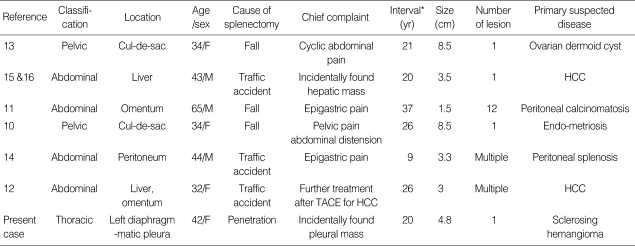
In Korea, four cases of abdominal and two cases of pelvic splenosis have been reported previously (10-16) (Table 1). All six patients had a history of splenic injury, due to falls or traffic accidents. In contrast to Western countries, in which 22 of 38 patients had splenic injuries due to gunshots, no patients with splenic injuries due to gunshots have been described in Korea (3).
Table 1.
Review of the clinical characteristics of seven patients of splenosis in Korea
*interval between splenic injury and diagnosis.
TACE, transcatheter arterial chemoembolization; HCC, hepatocellular carcinoma.
Surgical removal of the splenic tissue is generally inadvisable, except in symptomatic patients and those with hematological disease. The ectopic splenic tissue may protect against systemic encapsulated bacterial infection, which can be a major problem in asplenic patients (17-19).
In conclusion, a preoperative diagnosis of TS can prevent unnecessary biopsy or thoracotomy in patients with multiple, asymptomatic, left-side pleura-based lesions if the patient has an appropriate history of splenic injury. However, if a biopsy is performed without knowing the patient's history, the pathologists should consider the possibility of TS, even if the slide is offered for frozen diagnosis intra-operatively.
References
- 1.Shaw AF, Shafi A. Traumatic autoplastic transplantation of splenic tissue in a man with observation on the late results of splenectomy in six cases. J Pathol Bacteriol. 1937;45:215–235. [Google Scholar]
- 2.Khan AM, Manzoor K, Gordon D, Berman A. Thoracic splenosis: a diagnosis by history and imaging. Respirology. 2008;13:481–483. doi: 10.1111/j.1440-1843.2008.01272.x. [DOI] [PubMed] [Google Scholar]
- 3.Yammine JN, Yatim A, Barbari A. Radionuclide imaging in thoracic splenosis and a review of the literature. Clin Nucl Med. 2003;28:121–123. doi: 10.1097/01.RLU.0000048681.29894.BA. [DOI] [PubMed] [Google Scholar]
- 4.White CS, Meyer CA. General case of the day. Thoracic splenosis. Radiographics. 1998;18:255–257. doi: 10.1148/radiographics.18.1.9460132. [DOI] [PubMed] [Google Scholar]
- 5.Wold PB, Farrell MA. Pleural nodularity in a patient with pyrexia of unknown origin. Chest. 2002;122:718–720. doi: 10.1378/chest.122.2.718. [DOI] [PubMed] [Google Scholar]
- 6.Armas RR. Clinical studies with spleen-specific radiolabeled agents. Semin Nucl Med. 1985;15:260–275. doi: 10.1016/s0001-2998(85)80004-0. [DOI] [PubMed] [Google Scholar]
- 7.Darling JD, Flickinger FW. Splenosis mimicking neoplasm in the perirenal space: CT characteristics. J Comput Assist Tomogr. 1990;14:839–841. doi: 10.1097/00004728-199009000-00038. [DOI] [PubMed] [Google Scholar]
- 8.Fleming CR, Dickson ER, Harrison EG., Jr Splenosis: autotransplantation of splenic tissue. Am J Med. 1976;61:414–419. doi: 10.1016/0002-9343(76)90380-6. [DOI] [PubMed] [Google Scholar]
- 9.Normand JP, Rioux M, Dumont M, Bouchard G, Letourneau L. Thoracic splenosis after blunt trauma: frequency and imaging findings. AJR Am J Roentgenol. 1993;161:739–741. doi: 10.2214/ajr.161.4.8372748. [DOI] [PubMed] [Google Scholar]
- 10.Hoh JK, Kim KT, Cho SH, Hwang YY. A case of pelvic splenosis after splenectomy: a cause of pelvic pain. Korean J Obstet Gynecol. 2005;48:3004–3008. [Google Scholar]
- 11.Ryu SW, Kim IH, Sohn SS. Splenosis mimicking carcinomatosis peritonei in advanced gastric cancer. J Korean Surg Soc. 2005;68:61–64. [Google Scholar]
- 12.Choi GH, Ju MK, Kim JY, Kang CM, Kim KS, Choi JS, Han KH, Park MS, Park YN, Lee WJ, Kim BR. Hepatic splenosis preoperatively diagnosed as hepatocellular carcinoma in a patient with chronic hepatitis B: a case report. J Korean Med Sci. 2008;23:336–341. doi: 10.3346/jkms.2008.23.2.336. [DOI] [PMC free article] [PubMed] [Google Scholar]
- 13.Ha MK, Kwon OJ, Lee KS, Hwang YY, Hong EK. Splenosis with cyclic abdominal pain misdiagnosis as ovarian dermoid cyst. J Korean Surg Soc. 2002;62:450–452. [Google Scholar]
- 14.Yoon M, Hwang KH, Choe W. Multifocal peritoneal splenosis in Tc-99m-labeled heat denatured red blood cell scintigraphy. Nucl Med Mol Imaging. 2006;40:190–191. [Google Scholar]
- 15.Lee JB, Ryu KW, Song TJ, Suh SO, Kim YC, Koo BH, Choi SY. Hepatic splenosis diagnosed as hepatocellular carcinoma: report of a case. Surg Today. 2002;32:180–182. doi: 10.1007/s005950200016. [DOI] [PubMed] [Google Scholar]
- 16.Kim KA, Park CM, Kim CH, Choi SY, Park SW, Kang EY, Seol HY, Cha IH. An interesting hepatic mass: splenosis mimicking a hepatocellular carcinoma (2003:9b) Eur Radiol. 2003;13:2713–2715. doi: 10.1007/s00330-003-1978-5. [DOI] [PubMed] [Google Scholar]
- 17.Bordlee RP, Eshaghi N, Oz O. Thoracic splenosis: MR demonstration. J Thorac Imaging. 1995;10:146–149. doi: 10.1097/00005382-199521000-00014. [DOI] [PubMed] [Google Scholar]
- 18.Cordier JF, Gamondes JP, Marx P, Heinen I, Loire R. Thoracic splenosis presenting with hemoptysis. Chest. 1992;102:626–627. doi: 10.1378/chest.102.2.626. [DOI] [PubMed] [Google Scholar]
- 19.Pearson HA, Johnston D, Smith KA, Touloukian RJ. The born-again spleen. Return of splenic function after splenectomy for trauma. N Engl J Med. 1978;298:1389–1392. doi: 10.1056/NEJM197806222982504. [DOI] [PubMed] [Google Scholar]


