Abstract
Covalent conjugates of fullerene C60 and the highly effective anticancer drug doxorubicin (DOX) were prepared and studied. The conjugation was through the amide linkage to preserve the intrinsic properties of DOX and fullerene cage. As designed, the conjugates with hydrophilic ethylene glycol spacers exhibited much improved aqueous compatibility, with significant solubility in water-DMSO mixtures. The anti-neoplastic activities of DOX were apparently unaffected in the conjugates according to evaluations in vitro with a human breast cancer cell line.
Introduction
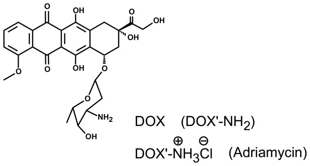
Fullerenes are extensively investigated and widely acknowledged for their unique properties as excellent electron acceptors, potent radiacal scavagers, and others.1–4 There has been much interest in exploiting these properties for potential biomedical applications of fullerenes.1,3,5 For example, fullerenes have been studied for their therapeutic effects on neurodegenerative diseases,6 as agents for DNA cleavage,7 and as nitric oxide synthase inhibitors.8 Among intersting recent investigations on potentially using fullerenes in medicine was the work by Wilson and coworkers, in which C60 was covalently conjugated with the widely used anticancer drug paclitaxel for in vitro pharmacological evaluations.9 Similar conjugates of fullerenes with other popular drugs, including the highly effective anticanecer drug doxorubicin (DOX, Scheme 1), have been explored or proposed for various medicinal purposes.10,11 In particular, there were many reports on using polyhydroxylated C60, often called fullerenols,12 to mitigate DOX-induced toxic side-effects.13 Beyond fullerenes, carbon nanotubes have been conjugated with DOX for the delivery of the drug to take advantage of the enhanced cellular uptake, selectivity to cancer cells, and pH regulated release.14,15
Scheme 1.
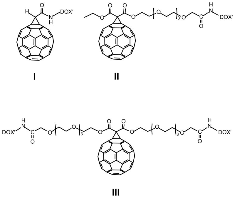
A major issue in the effort on using fullerene-DOX conjugates for improved drug formulation is on their aqueous solubility or compatibility. DOX in its natural form is marginally soluble in water,16 and the widely used commercial formulation is a salt with hydrochloric acid (under the trade name Adriamycin, Scheme 1).17 The fullerene cage is even less hydrophilic, hardly helpful to DOX in terms of compatibility with physiological media. Therefore, a special consideration is required in the conjugation of C60 with DOX to impart sufficient hydrophilicity into the conjugates for their desired bioavailability. Here we report the covalent conjugation of DOX with methano-C60 derivatives (Scheme 2), for which the molecular structures were determined unambiguously by using NMR and other techniques. As designed, the conjugates with hydrophilic linkages exhibited significantly improved aqueous compatibility, amenable to their targeted bioapplications. The anti-neoplastic activities of DOX were apparently preserved in the conjugates according to evaluations in vitro with human breast cancer cells. The results set the stage for further development of fullerene conjugates in the delievery of DOX for benefits similar to those found in the conjugation of fullerene with paclitaxel12 and those demonstrated with the use of fullerenols.13
Scheme 2.
Results and Discussion
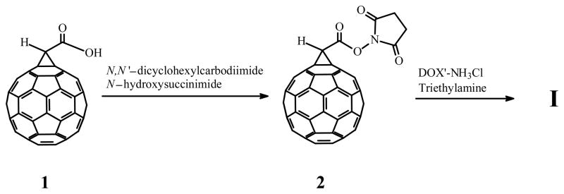
The conjugate I was synthesized to test the amidation of the carboxylic acid moiety in the methano-C60 derivative 1 by DOX (Scheme 3). The amide linkage was selected with the consideration of literature reports that the amidation of the primary amine in DOX had relatively minor effect on the anti-neoplastic activities of the drug.18 The synthesis was relatively straightforward, but the conjugate was found to be insoluble in water or aqueous solvent mixtures. This was hardly surprising since neat DOX (without the HCl salt in the commercial formulation) was known to have little solubility in water,16 even less for the fullerene. In order to increase the desired aqueous compatibility without inducing any major changes to the chemical structures (thus intrinsic properties) of the fullerene cage and/or DOX, hydrophilic moieties in the form of oligomeric ethylene glycol spacers were added to the conjugates (Scheme 2).
Scheme 3.
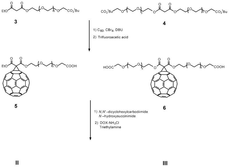
For the conjugate II, the methano-C60 adduct (with the unsymmetrical malonic ester 3) was obtained in the classical Bingel-Hirsch reaction (Scheme 4),19,20 with the observed yield (40%) comparable to those in other similar additions. Upon the selective hydrolysis of the terminal t-butyl ester, the resulting carboxylic acid was again activated by using the agent DCC (N,N′-dicyclohexylcarbodiimide) for amidation with DOX′-NH2 (Scheme 4).
Scheme 4.
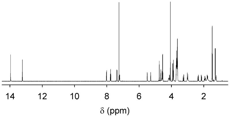
The molecular structure of II was confirmed by results from 1H- and 13C-NMR measurements and mass spectroscopy analyses. The 1H-NMR spectrum of the conjugate (Figure 1) was close to a superposition from those of the underlying fullerene and DOX. In the 13C-NMR spectrum of II, the 26 sp2 signals with a characteristic intensity distribution pattern for the fullerene cage suggested that the conjugate maintained the Cs symmetry (the same as that of 5 in Scheme 4).21 The signal for the two methano-fullerene bridgehead carbons (sp3) was at 71.5 ppm, and the other carbon of the cyclopropyl ring at 52.0 ppm. The matrix-assisted laser desorption ionization – time-of-flight (MALDI-TOF) MS pattern of II was compared with the prediction on the basis of isotopic populations (IsoPro 3.0). Shown in Figure 2 is an excellent match, with the expected parent peak mass of 1632.3 dalton (II+Na). This was further confirmed in the electrospray ionization (ESI) MS characterization, with both II+Na (1,632.32 vs 1,632.29 calculated) and II (1,610.21 vs 1,610.30 calculated) peaks observed.
Figure 1.
The 1H NMR spectrum of the conjugate II in deuterated chloroform.
Figure 2.
The observed MALDI-TOF MS trace of the conjugate II (in 2,5-dihydroxybenzoic acid matrix, bottom) is compared with that from calculation (IsoPro 3.0, top).
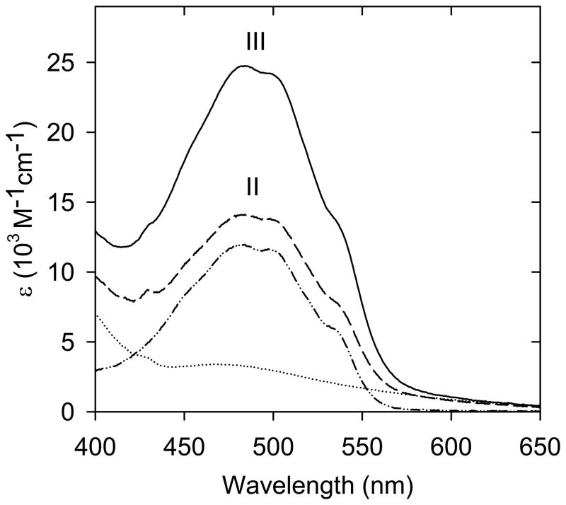
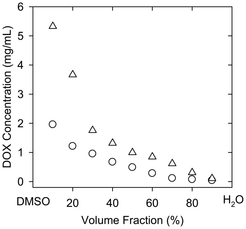
The observed UV/vis absorption spectrum of the conjugate II was also close to the superposition from those of the methano-C60 and DOX′-NH3Cl (Figure 3). Thus, the molar absorptivity at the spectral maximum of II could be approximated as the sum of those for the methano-C60 and DOX′-NH3Cl at the same wavelength, which allowed the use of observed absorbance to estimate the concentration of II in a solution (or the solubility of II in a saturated solution). The presence of the hydrophilic spacer in II made the conjugate more dispersible in water, but still practically insoluble. However, the conjugate was found to be soluble in some aqueous mixtures with a polar organic solvent, such as those with dimethyl sulfoxide (DMSO). The saturated solutions of II in water-DMSO mixtures of different compositions were prepared by pushing an excess amount of II into a given mixture, followed by vigorous high-speed centrifuging to keep the supernatant for absorption spectral measurements. The solubility results thus obtained are shown in Figure 4. In the 1:1 (v/v) water-DMSO mixture, for example, the solubility of II is ~1.5 mg/mL. The conjugate is obviously more soluble in mixtures with more DMSO (Figure 4).
Figure 3.
UV/vis spectra of III (solid line), II (dash line), DOX-NH3Cl (dash-dot line), and 5 (dot line) in DMSO solutions.
Figure 4.
The DOX-equivalent solubility of II and III as a function of the volume fraction in water-DMSO mixtures.
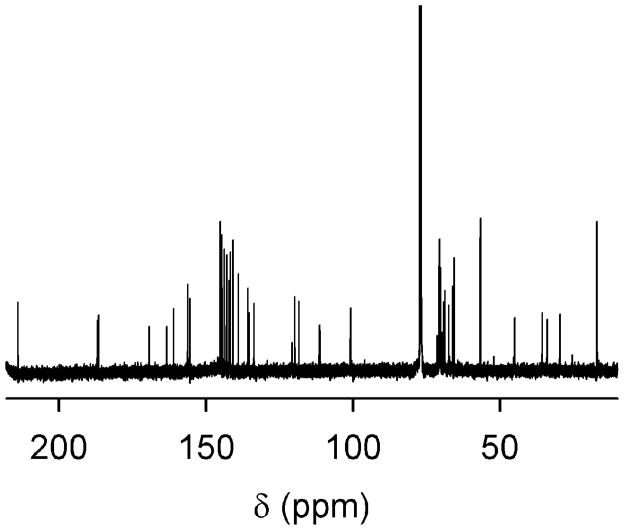
The conjugate III with two hydrophilic tethers and two DOX units sandwiching a C60 cage was designed for further improved aqueous compatibility, as well as the doubling in DOX loading per conjugate. As illustrated in Scheme 4, the malonic ester 4 was similarly added to C60 in the classical Bingel-Hirsch reaction, followed by the selective hydrolysis and then amidation reaction with DOX. The relatively more difficult coupling of the methano-C60 with two DOX molecules was reflected in the lower product yield, 42% in comparison with 65% found in the coupling for II. Again, the conjugate III was confirmed unambiguously in the characterization with 1H- and 13C-NMR techniques (Figure 5) and MS measurements (MALDI-TOF: 2,364.5; and ESI-MS/MS: 2,364.538, vs the expected 2,364.536 for III+Na).
Figure 5.
The 13C NMR spectrum of the conjugate III in deuterated chloroform.
Despite being only marginally soluble in water (less than 0.1 mg DOX-equivalent per mL), the conjugate III exhibited significantly improved aqueous compatibility. Again in the 1:1 (v/v) water-DMSO mixture, for example, the DOX-equivalent solubility was doubled (Figure 4). The improved aqueous compatibility of III over II was significant with respect to their bioevaluations. Specifically, both II and III were tested in experiments for their cytotoxicity evaluations, but those for II were very difficult and inconclusive (due a large part to significant aggregation of the conjugate in cell culture media). On the other hand, the same experiments for III were successful, enabling a comparison of anti-neoplastic activities between the conjugate and free DOX (the commercial formulation DOX′-NH3Cl).
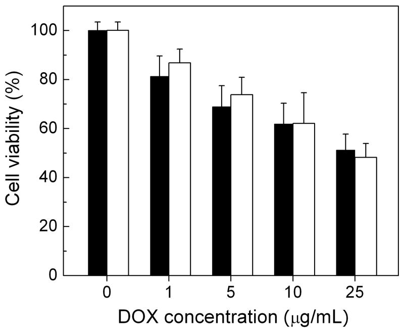
The viability of human breast cancer MCF-7 cells in the presence of III or free DOX was evaluated in the MTT assay (mitochondrial reduction of 3-(4,5-dimethylthiazol-2-yl)-2,5-diphenyltetrazolium bromide to formazan by succinic dehydrogenase22). A solution of III or free DOX in DMSO was diluted with the cell culture medium to the targeted concentration just prior to the cell exposure. The MCF-7 cells cultured in the free medium were taken as the control. The results of the MTT assay were obtained in terms of the relative cell viability (% of the control). As shown in Figure 6, the viability of MCF-7 cells exposed to the conjugate III decreased in a dose-dependent fashion, with about half of the cell viability inhibited at the DOX-equivalent concentration of 25 μg/mL. Between the conjugate III and free DOX (the commercial formulation DOX′-NH3Cl), the observed anti-neoplastic activities toward the breast cancer cell line were rather similar (Figure 6). This is interesting because a commonly observed phenomenon in the delivery of DOX by nanoscale carriers such as polymeric nanoparticles,23 carbon nanotubes,14 or nanodiamond,24 has been a decrease in the anti-neoplastic activities of the drug. Mechanistically, the anti-neoplastic function of DOX molecules has been attributed to their intercalation into DNA to inhibit the macromolecular biosynthesis, the inhibition of enzyme helicase to interfere with DNA unwinding, and/or the inhibition of topoisomerase II to induce DNA damage.25 The conjugate III is structurally flexible due to the ethylene glycol spacers, making the attached DOX units readily available for their biological functions. On the other hand, the extremely hydrophobic nature of the fullerene cage probably makes the wrapping of the cage by the hydrophilic spacers a favorable configuration, which thus keeps the conjugate compact in size, not to sterically hinder the activities of the DOX units. These structural characteristics might have contributed to the observed comparable in vitro performance of the conjugate to that of free DOX in the aqueous soluble formulation.
Figure 6.
The cell viability of MCF-7 cells after exposure to DOX′-NH3Cl (black) and the conjugate III (white) at various DOX-equivalent concentrations. Data presented as mean ± SD (n=4).
In summary, DOX could be covalently conjugated with methano-C60 through the amide linkage to preserve the intrinsic properties of DOX and fullerene cage. The improvement in aqueous compatibility could be accomplished by introducing hydrophilic ethylene glycol spacers into the conjugate structure, which imparted significant solubility of the conjugate in water-DMSO mixtures. The conjugate with two hydrophilic spacers was sufficiently aqueous compatible to enable bio-evaluations in vitro, from which the results suggested comparable anti-neoplastic activities of the conjugate with those of free DOX against the human breast cancer cells. The fullerene-spacer-DOX design apparently serves as a versatile platform for conjugates of desired properties, including availability of the drug, structural flexibility with hydrophilic and hydrophilic moieties for different cellular domains, etc. The platform may be further developed for conjugates of improved performances in both delivery and activities (including those for the mitigation of induced toxicities) of the drug. In particular, these conjugates are structurally uniquely defined, potentially offering pharmacological benefits that are not available in more complex and/or mixture-based systems such as those with fullerenols.13
Experimental Section
Materials
Fullerene C60 sample (>98%) from Bucky USA was purified on a silica gel column with toluene as eluent. N-hydroxysuccinimide (NHS), trifluoroacetic acid (TFA), triethylamine (TEA), and N,N′-dicyclohexylcarbodiimide (DCC) were purchased from Acros, carbon tetrabromide from Alfa Aesar, ethyl-malonyl chloride from Aldrich, 1,8-diazabicyclo[5.4.0]undec-7-ene (DBU) from Avocado Research Chemicals, and malonic dichloride from TCI America. Toluene, THF, and DMF were dried over molecular sieves, and then toluene and THF were freshly distilled over sodium and DMF distilled under reduced pressure before use. Other solvents were either spectrophotometry/HPLC grade or purified via simple distillation. Deuterated NMR solvents were obtained from Cambridge Isotope Laboratories.
Measurement
NMR spectra were measured on Bruker Avance 300 MHz and 500 MHz NMR spectrometers. MALDI-TOF MS analyses were performed on a Bruker AutoFlex system, and 2,5-dihydroxybenzoic acid was used as the matrix. Electrospray ionization MS results were obtained on a QSTAR XL hybrid LC/MS/MS system. UV/Vis absorption spectra were recorded on Shimadzu UV-2501 and UV-3100 spectrophotometers.
I
A solution of the methano-C60 1 (100 mg, 0.128 mmol)26 in CH2Cl2/dioxane (1:1 v/v, 100 mL) was prepared, and N-hydroxysuccinimide (30 mg, 0.26 mmol) was added. To the mixture was added dropwise (within ~10 min) a solution of DCC (60 mg, 0.29 mmol) in CH2Cl2/dioxane (1:1 v/v, 5 mL). After stirring for 12 h at room temperature, the reaction mixture was concentrated in vacuum and then separated by flash chromatography on silica gel with toluene as eluent to yield 2 as a brown solid (60 mg, 53% yield). Separately, a solution of DOX′-NH3Cl (14.5 mg, 0.025 mmol) in dry DMF (2 mL) was prepared, and to the solution was added TEA (4.14 μL, 0.03 mmol). Upon the mixture being stirred at room temperature for 2 min under argon, a solution of 2 (22 mg, 0.025 mmol) in dry DMF (2 mL) was added dropwise (within ~1 min). The resulting mixture was stirred in the dark at room temperature for 48 h. The subsequent separation of the reaction mixture by flash chromatography on silica gel with a chloroformmethanol mixture (95:5, v/v) as eluent yielded I as a reddish brown solid (16 mg, 49% yield). 1H NMR (CDCl3, 500 MHz, CDCl3): δ14.10 (s, 1H), 13.28 (s, 1H), 8.07 (d, J = 8 Hz, 1H), 7.81 (t, J = 8 Hz, 1H), 7.41 (d, J = 8 Hz, 1H), 7.29 (d, 1H), 5.63 (d, J = 3.5 Hz, 1H), 5.36 (s, 1H), 4.80 (t, J = 4.5 Hz, 2H), 4.64 (s, 1H), 4.50 (bs, 2H), 4.33 (q, J = 6.5 Hz, 1H), 4.10 (s, 3H), 3.91 (bs, 1H), 3.34 (d, J = 17.5 Hz, 1H), 3.09 (d, J = 19 Hz, 1H), 3.02 (t, 1H), 2.38 (d, 1H), 2.25 (m, 3H), 2.01 (m, 1H), 1.40 (d, J = 6.5 Hz, 3H) ppm; 13C NMR (75.46 MHz, CDCl3): δ 214.0, 188.0, 161.12, 156.23, 155.75, 148.44, 146.12, 145.62, 145.25, 145.15, 145.05, 144.68, 144.59, 144.53, 144.28, 143.94, 143.70, 143.32, 143.12, 142.96, 142.80, 142.42, 142.25, 142.09, 141.13, 140.96, 140.88, 140.20, 136.18, 135.62, 133.57, 133.41, 119.94, 118.50, 111.79, 100.86, 70.22, 69.59, 67.08, 65.55, 56.71, 46.19, 41.33, 35.81, 34.05, 29.68, 16.87 ppm. MALDI-TOF MS (M+Na)+: 1327.1 (1327.16 calculated).
II
A solution of C60 (600 mg, 0.83 mmol) in toluene (600 mL) was prepared, and to the solution was added the malonic ester 3 (300 mg, 0.71 mmol), CBr4 (240 mg, 0.72 mmol), and DBU (0.12 mL, 0.80 mmol). The resulting mixture was stirred at room temperature for 20 h, followed by the removal of solvent on a rotary evaporator. The crude reaction mixture was separated on a silica gel column with first toluene as eluent to remove unreacted C60 and then ethyl acetate to obtain the methano-C60 adduct. The sample was dissolved in CH2Cl2 (50 mL) to mix with TFA (50 mL). The mixture was stirred for 30 min, followed by solvent removal to yield the selectively hydrolyzed adduct 5 (270 mg, 36% yield).
A solution of 5 (100 mg, 0.092 mmol) in CH2Cl2 (40 mL) was prepared, and to the solution was added N-hydroxysuccinimide (11 mg, 0.097 mmol) and then dropwise a solution of DCC in CH2Cl2 (5 mL, 4 mg/mL). The mixture was stirred at room temperature for 12 h, filtered, concentrated, and then re-dissolved in dry DMF (2 mL). Separately, a solution of DOX′-NH3Cl (50 mg, 0.086 mmol) in dried DMF (20 mL) was prepared, and to the solution was added TEA (12.5 μL, 0.088 mmol). After brief stirring (about 2 min) at room temperature under argon, the DMF solution of the activated 5 above was added dropwise. After stirring in the dark at room temperature for 48 h, the reaction mixture was concentrated and separated on a silica gel column with chloroform-methanol (95:5, v/v) as eluent to obtain II as a reddish brown solid (96 mg, 65% yield). 1H NMR (500 MHz, CDCl3): δ 14.0 (s, 1H), 13.28 (s, 1H), 8.05 (d, J = 8 Hz, 1H), 7.80 (t, J = 7.5 Hz, 1H), 7.41 (d, J = 9Hz, 1H), 7.24 (d, J = 9 Hz, 1H), 5.54 (d, J = 4 Hz, 1H), 5.32 (s, 1H), 4.79 (d, J = 2 Hz, 2H), 4.70 – 4.68 (m, 2H), 4.59 (q, J = 14.5 Hz, 2H), 4.22 – 4.12 (m, 2H), 4.10 (s, 3H), 3.95 (d, J = 4.5 Hz, 2H), 3.91 (t, J = 4.5 Hz, 2 H), 3.75 – 3.67 (m, 14 Hz), 3.29 (d, J = 19 Hz, 1H), 3.04 (d, J = 18.5 Hz, 2H), 2.38 (d, J = 15 Hz, 2H), 2.18 (dd, J = 4 Hz, 14.5 Hz, 1H), 1.97 (dt, J = 4 Hz, 13.5 Hz, 1H), 1.85 (dd, J = 4 Hz, 13.25 Hz, 1H), 1.51 (t, J = 7 Hz, 3H), 1.31 (d, J = 6.5 Hz, 3H) ppm; 13C NMR (125.7 MHz, CDCl3): δ 213.9, 187.1, 186.7, 169.2, 163.6, 163.4, 161.07, 156.3, 155.7, 145.3, 145.27, 145.24, 145.18, 145.13, 144.88, 144.69, 144.66, 144.61, 144.57, 144.55, 143.88, 143.84, 143.09, 143.06, 143.02, 142.99, 142.93, 142.20, 142.16, 141.89, 141.82, 140.95, 140.89, 139.15, 138.92, 135.77, 135.57, 133.72, 119.89, 118.46, 111.64, 111.48, 100.9, 71.52, 70.92, 70.75, 70.63, 70.55, 70.50, 70.33, 70.20, 69.69, 69.17, 68.82, 67.46, 66.13, 65.60, 63.5, 56.7, 52.0, 44.9, 35.7, 34.0, 29.6, 17.0, 14.2 ppm.
III
C60 (230 mg, 0.32 mmol) was dissolved in toluene (500 mL), and to the solution was added the malonic ester 4 (200 mg, 0.29 mmol), CBr4 (97 mg, 0.29 mmol), and DBU (60 μL, 0.40 mmol). The mixture was stirred at room temperature for 30 min, followed by the solvent removal. The crude reaction mixture was separated on a silica gel column with first toluene as eluent and then ethyl acetate to obtain the methano-C60 adduct (190 mg, 46% yield). A portion of the sample (100 mg, 0.071 mmol) was dissolved in CH2Cl2 (50 mL) to mix with TFA (50 mL). The mixture was stirred for 30 min to yield the hydrolyzed adduct 6 quantitatively.
Similarly, 6 (100 mg, 0.77 mmol) was activated by N-hydroxysuccinimide (18 mg, 0.156 mmol) with DCC (33mg, 0.16 mmol), followed by the coupling with DOX′-NH3Cl (78 mg, 0.134 mmol) in dry DMF to obtain III as a reddish brown solid (75 mg, 42% yield).
1H NMR (500 MHz, CDCl3): δ 13.9 (s, 2H), 13.28 (s, 2H), 8.01 (d, J = 7.5 Hz, 2H), 7.80 (t, J = 7.5 Hz, 2H), 7.41 (d, J = 9 Hz, 2H), 7.24 (d, J = 9 Hz, 2H), 5.52 (d, J = 4 Hz, 2H), 5.27 (s, 2H), 4.77 (s, 4H), 4.71–4.64 (m, 4H), 4.62 (bs, 2H), 4.20–4.13 (m, 4H), 4.09 (s, 6H), 3.97–3.91 (m, 8H), 3.76–3.66 (m, 28H), 3.41 (bs, 2H), 3.19 (d, J = 19 Hz, 2H), 2.88 (d, J = 18.5 Hz, 2H), 2.36 (d, J = 15 Hz, 2H), 2.19 (dd, J = 4 Hz, 13.5 Hz, 2H), 1.97–1.92 (m, 2H), 1.79 (dd, J = 4 Hz, 13.25 Hz, 2H), 1.34 (d, J = 6.5 Hz, 6H) ppm; 13C-NMR (125.7 MHz, CDCl3): δ 213.9, 186.9, 186.5, 169.3, 163.4, 161.0, 156.2, 155.6, 145.3, 145.2, 145.1, 144.9, 144.6, 144.5, 143.8, 143.1, 143.0, 142.9, 142.2, 141.8, 140.9, 139.0, 135.7, 135.4, 133.8, 133.7, 119.8, 118.5, 111.5, 111.3, 100.9, 71.4, 70.9, 70.7, 70.6, 70.5, 70.3, 70.2, 69.7, 69.1, 68.8, 67.5, 66.2, 65.6, 56.7, 52.1, 44.9, 35.6, 33.9, 29.6, 17.0 ppm.
Bioassay
The human breast cancer MCF-7 cells (kindly provided by Dr. G. Huang in the Department of Biological Sciences at Clemson University) were cultured in Eagle’s Minimum Essential medium supplemented with 10% (v/v) fetal bovine serum and 1% of penicillin-streptomycin. The cells were cultivated in 75 cm2 flasks at 37 °C in a humidified atmosphere (5% CO2 and 95% air).
In the MTT assay on cell viability, DMSO solutions of the conjugate and free DOX were prepared with the same DOX-equivalent concentration of 1.1 mg/mL. The solutions, filtered with sterile syringe filters (Millipore, USA), were diluted with the cell culture medium to the targeted concentrations just prior to the cell exposure. MCF-7 cells were plated in 96-well plates (2×10⁴ cells per well) and incubated for 24 h. The conjugate and free DOX were introduced to the cells at DOX-equivalent concentrations of 1, 5, 10, and 25 μg/mL. Cells cultured in the free medium were taken as the control. After 24 h, the supernatants were discarded, and MTT (0.5 mg/mL in culture medium, 100 μL) was added to each well, followed by incubation for another 4 h. The formed formazan was dissolved by adding sodium dodecyl sulfate (SDS) solution (10%, also containing 5% isobutanol and 10 mM HCl, 100 μL). The optical density (OD) of formazan at 570 nm was recorded on a microplate reader (μQuant, Bio-Tek, USA). The relative cell viability was obtained as a percentage of ODTest/ODControl, where ODTest and ODControl are ODs of the exposed sample and the control, respectively.
Acknowledgments
Financial support from NSF and, in part, from NIH is gratefully acknowledged. S.L. was a participant of the summer undergraduate research program jointly sponsored by NSF and Clemson University.
References
- 1.(a) Prato M. J Mater Chem. 1997;7:1097. [Google Scholar]; (b) Bosi S, Da Ros T, Spalluto G, Prato M. Eur J Med Chem. 2003;38:913. doi: 10.1016/j.ejmech.2003.09.005. [DOI] [PubMed] [Google Scholar]
- 2.Sun Y-P. Photoexcited state and electron transfer properties of fullerenes and related materials. In: Shinar J, Vardeny ZV, Kafafi ZH, editors. Optical and Electronic Properties of Fullerenes and Fullerene-Based Materials. Marcel Dekker; New York: 1999. p. 43. [Google Scholar]
- 3.Hirsch A, Brettreich M. Fullerenes: chemistry and reactions. Wiley-VCH; Weinheim: 2005. [Google Scholar]
- 4.Guldi DM, Illescas BM, Atienza CM, Wielopolski M, Martín N. Chem Soc Rev. 2009;38:1587. doi: 10.1039/b900402p. [DOI] [PubMed] [Google Scholar]
- 5.Jensen AW, Wilson SR, Schuster DI. Bioorg Med Chem. 1996;4:767. doi: 10.1016/0968-0896(96)00081-8. [DOI] [PubMed] [Google Scholar]
- 6.Dugan LL, Turetsky DM, Du C, Lobner D, Wheeler M, Almli CR, Shen CKF, Luh TY, Choi DW, Lin TS. Proc Natl Acad Sci USA. 1997;94:9434. doi: 10.1073/pnas.94.17.9434. [DOI] [PMC free article] [PubMed] [Google Scholar]
- 7.(a) Ikeda A, Hatano H, Kawaguchi M, Suenaga H, Shinkai S. Chem Commun. 1999:1403. [Google Scholar]; (b) Ikeda A, Doi Y, Hashizume M, Kikuchi J, Konishi T. J Am Chem Soc. 2007;129:4140. doi: 10.1021/ja070243s. [DOI] [PubMed] [Google Scholar]
- 8.(a) Wolff DJ, Papoiu ADP, Mialkowski K, Richardson CF, Schuster DI, Wilson SR. Arch Biochem Biophys. 2000;378:216. doi: 10.1006/abbi.2000.1843. [DOI] [PubMed] [Google Scholar]; (b) Wolff DJ, Mialkowski K, Richardson CF, Wilson SR. Biochemistry. 2001;40:37. doi: 10.1021/bi0019444. [DOI] [PubMed] [Google Scholar]
- 9.Zakharian TY, Seryshev A, Sitharaman B, Gilbert BE, Knight V, Wilson LJ. J Am Chem Soc. 2005;127:12508. doi: 10.1021/ja0546525. [DOI] [PubMed] [Google Scholar]
- 10.(a) Hirsch A, Sagman U, Wilson SR. 7,070,810. US Patent. 2006; (b) Miwa N, Ito S, Matsubayashi K. 0,206,222. US Patent Application. 2008
- 11.Prasad GL. private communications. [Google Scholar]
- 12.Chiang LY, Swirczewski JW, Hsu CS, Chowdhury SK, Cameron S, Creegan K. Chem Commun. 1992:1791. [Google Scholar]
- 13.(a) Injac R, Strukej B. Technol Cancer Res Treat. 2008;7:497. doi: 10.1177/153303460800700611. [DOI] [PubMed] [Google Scholar]; (b) Injac R, Perse M, Obermajer N, Djordjevic-Milic V, Prijatelj M, Djordjevic A, Cerar A, Strukelj B. Biomaterials. 2008;29:3451. doi: 10.1016/j.biomaterials.2008.04.048. [DOI] [PubMed] [Google Scholar]; (c) Injac R, Perse M, Cerne M, Potocnik N, Radic N, Govedarica B, Djordjevic A, Cerar A, Strukelj B. Biomaterials. 2009;30:1184. doi: 10.1016/j.biomaterials.2008.10.060. [DOI] [PubMed] [Google Scholar]
- 14.Liu Z, Sun X, Nakayama-Ratchford N, Dai H. ACS Nano. 2007;1:50. doi: 10.1021/nn700040t. [DOI] [PubMed] [Google Scholar]
- 15.Ali-Boucetta H, Al-Jamal KT, McCarthy D, Prato M, Bianco A, Kostarelos K. Chem Commun. 2008:459. doi: 10.1039/b712350g. [DOI] [PubMed] [Google Scholar]
- 16.Fritze A, Hens F, Kimpfler A, Schubert R, Peschka-Süss R. Biochim Biophys Acta. 2006;1758:1633. doi: 10.1016/j.bbamem.2006.05.028. [DOI] [PubMed] [Google Scholar]
- 17.Blum RH, Carter SK. Ann Intern Med. 1974;80:249. doi: 10.7326/0003-4819-80-2-249. [DOI] [PubMed] [Google Scholar]
- 18.(a) Bakina E, Wu Z, Rosenblum M, Farquhar D. J Med Chem. 1997;40:4013. doi: 10.1021/jm970066d. [DOI] [PubMed] [Google Scholar]; (b) Farquhar D, Cherif A, Bakina E, Nelson JA. J Med Chem. 1998;41:965. doi: 10.1021/jm9706980. [DOI] [PubMed] [Google Scholar]; (c) Farquhar D, Newman RA, Zuckerman J, Andersson BS. J Med Chem. 1991;34:561. doi: 10.1021/jm00106a013. [DOI] [PubMed] [Google Scholar]
- 19.Bingel C. Chem Ber. 1993;126:1957. [Google Scholar]
- 20.(a) Hirsch A, Lamparth I, Karfunkel HR. Angew Chem, Int Ed. 1994;33:437. [Google Scholar]; (b) Hirsch A, Lamparth I, Grsser T, Karfunkel HR. J Am Chem Soc. 1994;116:9385. [Google Scholar]
- 21.Keshavarz MK, Knight B, Haddon RC, Wudl F. Tetrahedron. 1996;52:5149. [Google Scholar]
- 22.Carmichael J, DeGraff WG, Gazder AF, Minna JD, Mitchell JB. Cancer Res. 1987;47:936. [PubMed] [Google Scholar]
- 23.Zhang J, Chen X, Li Y, Liu C. Nanomedicine. 2007;3:258. doi: 10.1016/j.nano.2007.08.002. [DOI] [PubMed] [Google Scholar]
- 24.Huang H, Pierstorff E, Osawa E, Ho D. Nano Lett. 2007;7:3305. doi: 10.1021/nl071521o. [DOI] [PubMed] [Google Scholar]
- 25.Gewirtz DA. Biochem Pharmacol. 1999;57:727. doi: 10.1016/s0006-2952(98)00307-4. [DOI] [PubMed] [Google Scholar]
- 26.Isaacs L, Diederich F. Helv Chim Acta. 1993;76:2454. [Google Scholar]


