Abstract
In the present study, Staphylococcus (S.) hyicus strains isolated in Russia (n = 23) and Germany (n = 17) were investigated for the prevalence of the previously described genes sheta and shetb. Sheta was detected in 16 S. hyicus strains. Sheta-positive strains were mainly found among strains isolated from exudative epidermitis, and frequently together with the exfoliative toxin-encoding genes exhD and exhC. Partial sequencing of sheta in a single S. hyicus strain revealed an almost complete match with the sheta sequence obtained from GenBank. None of the S. hyicus strains displayed a positive reaction with the shetb-specific oligonucleotide primer used in the present study. According to the present results, the exotoxin encoding gene sheta seems to be distributed among S. hyicus strains in Russia and Germany. The toxigenic potential of this exotoxin, which does not have the classical structure of a staphylococcal exfoliative toxin, remains to be elucidated.
Keywords: exfoliative toxins, exudative epidermitis, sheta, shetb, Staphylococcus hyicus
Staphylococcus (S.) hyicus is a worldwide causative agent of exudative epidermitis in pigs, a generalized infection of the skin characterized by exudation, exfoliation, and vesicle formation [10]. S. hyicus isolated from exudative epidermitis generally produces exfoliation-inducing exotoxins, which show a close relation to comparable exfoliative toxins produced by Staphylococcus aureus isolated from Staphylococcal scaled skin syndrome infections in humans [7]. Exudative epidermitis-inducing exotoxins, originally described by Amtsberg [2], have been identified and purified from S. hyicus strains in Japan and Denmark and have been designated as SHETA and SHETB [8] and ExhA, ExhB, ExhC, and ExhD [1], respectively. The prevalence of the exfoliative toxin genes exhA, exhB, exhC, and exhD has been described for S. hyicus strains isolated from various countries [3-6]. However, little is known about the distribution of SHETA- and SHETB-encoding genes in S. hyicus or the combined occurrence of SHETA and SHETB and the exfoliative toxin-encoding genes described in Denmark. The present study was designed to investigate the distribution of sheta and shetb among previously characterized S. hyicus strains isolated in Russia and Germany.
A total of 40 S. hyicus strains, originally isolated in Russia and Germany, were investigated in the present study. The 40 S. hyicus strains and the reference strains S. hyicus S3588 (exhA), S. hyicus 1289D-88 (exhB), S. hyicus 842A-88 (exhC), S. hyicus A2869C (exhD), and S. hyicus DSM 20459 were identified and further characterized as described previously [6,9].
The sheta and shetb sequence data were obtained from GenBank (AB036768, AB036767). The design of the sheta- and shetb-specific oligonucleotide primers was performed using the computer program Oligo 4.0. The oligonucleotide primers used had the sequence 5'-GAACACGTTTTTCAGCCATATCTCC-3' and 5'-CGATTACAGTTGCCAATACCGTTTC-3' for sheta and 5'-GAGGCTTTACAGCCAAAATTATATGCTAG-3' and 5'-CAAATCGCTTCCTAGAGTATCTATTTTTTG-3' for shetb. Both oligonucleotide primers were synthesized by Operon (Germany).
The DNA preparation has been described previously [6]. The reaction mixture for sheta and shetb amplification contained 0.7 µl of each primer (10 pmol/µl), 0.8 µl of dNTP (10 mmol; Genecraft, Germany), 2.0 µl of 10 × Biotherm buffer with a final concentration of 1.5 mM MgCl2 (Genecraft, Germany), 0.2 µl of Biotherm polymerase (Genecraft, Germany) and 13 µl of H2O. The tubes were then subjected to thermal cycling (Gene Amp PCR System 2400; Perkin Elmer, Germany): 1 × 94℃ for 180 sec; 30 × (94℃ for 30 sec, 58℃ for 30 sec, 72℃ for 70 sec); and 1 × 72℃ for 300 sec. The presence of PCR products was determined by electrophoresis of 10 µl of reaction product on a 1.5% agarose gel (Gibco BRL, Germany) with Tris-acetate electrophoresis buffer (TAE, 4.0 mmol/l Tris-HCl, 1 mmol/l EDTA, pH 8.0) and visualized under UV light (Image Master VDS; Pharmacia Biotech, Germany).
For sequencing, the sheta amplicon of S. hyicus S3588 was eluted from the gel using QIAEX_II (Qiagen, Germany) according to the instructions of the manufacturer. The sequencing was performed using Sequence Genterprise (Mainz, Germany). A sequence comparison was carried out using the database of the National Centre for Biotechnology Information (NIH, USA). The toxin gene and protein sequences were compared with the exfoliative toxin gene and protein sequences of GenBank using a computer program, ClustalW (EBI, UK).
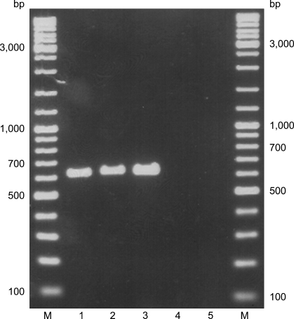
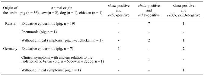
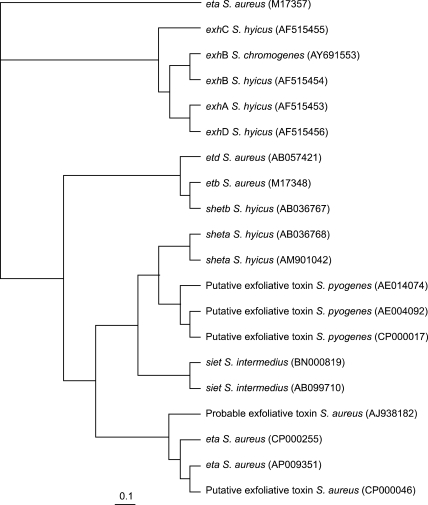
The S. hyicus strains investigated in the present study had been previously characterized and identified by phenotypic methods and by PCR-mediated amplification of S. hyicus-specific segments of the superoxide dismutase A-encoding gene sodA and by amplification of specific segments of the 16S-23S rDNA intergenic spacer region [9]. Screening of these strains for the exfoliative toxin genes exhA, exhB, exhC, and exhD by multiplex PCR revealed the presence of exhD in 17 of the S. hyicus strains isolated in Russia and the genes exhC and exhD for one and two S. hyicus strains, respectively, isolated in Germany [6]. Investigation of the S. hyicus strains for sheta and shetb yielded sheta positivity in 11 strains isolated in Russia and 5 strains isolated in Germany (Fig. 1). The origin of the strains and the PCR results are summarized in Table 1. Sequencing of the sheta gene from strain S. hyicus S3588 in the present study revealed an almost complete sequence match with the sheta sequence of GenBank. The sequencing results together with other available exfoliative toxin gene sequences are shown in Fig. 2. The presence of sheta in the present study occurred more frequently among strains isolated from exudative epidermitis, partly together with exhD and exhC. However, some of the sheta-positive strains were negative for exhC and exhD (Table 1). It was interesting to note that the exhA, exhB, exhC, and exhD reference strains and S. hyicus strain DSM 20459, which was also isolated from exudative epidermitis and was described as exhA-positive, were also sheta-positive.
Fig. 1.
Typical amplicons of sheta-positive Staphylococcus hyicus (1, 2, 3); sheta-negative Staphylococcus hyicus (4, 5).
Table 1.
Toxigenic properties of 23 Staphylococcus hyicus strains isolated in Russia and 17 Staphylococcus hyicus strains isolated in Germany
Fig. 2.
Dendrogram analysis of the sheta gene sequence of the present study and the published exfoliative toxin gene sequences of the genera Staphylococcus and Streptococcus. The dendrogram was prepared using the computer program ClustalW.
None of the strains was positive for shetb. The latter finding corresponded to the findings of Futagawa-Saito et al. [5]. These authors investigated 161 S. hyicus strains isolated from pigs with exudative epidermitis and 46 strains isolated from healthy pigs in Japan and could not detect plasmid-borne shetb. However, since no shetb reference strain is internationally available, the role SHETB plays in exudative epidermitis remains unclear.
According to Sato et al. [8] SHETA is under chromosomal control, and SHETB is plasmid-controlled. Strains investigated by Sato et al. [8] produced either SHETA or SHETB, or both. The typical signs of exudative epidermitis were observed in piglets inoculated with plasmid-carrying, SHETB-producing strains, as well as those inoculated with plasmidless SHETA-producing strains, indicating that both toxins seem to be involved in the clinical signs of exudative epidermitis. According to Ahrens and Andresen [1] and Yamaguchi et al. [11], SHETA does not posses the catalytic triad His-115, Asp-164, and Ser-239 of the S2 family of serine proteases, which is typical for the exfoliative toxins of S. aureus and S. hyicus, and also for SHETB. However, according to the findings of Sato et al. [8] and the results of the present study, SHETA seems to contribute to the clinical signs of exudative epidermitis, and PCR-amplification of sheta could additionally be used to detect virulent S. hyicus. At present, a target molecule or mode of action for SHETA has not been suggested.
References
- 1.Ahrens P, Andresen LO. Cloning and sequence analysis of genes encoding Staphylococcus hyicus exfoliative toxin types A, B, C, and D. J Bacteriol. 2004;186:1833–1837. doi: 10.1128/JB.186.6.1833-1837.2004. [DOI] [PMC free article] [PubMed] [Google Scholar]
- 2.Amtsberg G. Nachweis von Exofoliation auslösenden Substanzen in Kulturen von Staphylococcus hyicus des Schweines und Staphylococcus epidermidis Biotyp 2 des Rindes. Zentralbl Veterinarmed B. 1979;26:257–272. [PubMed] [Google Scholar]
- 3.Andresen LO. Production of exfoliative toxin by isolates of Staphylococcus hyicus from different countries. Vet Rec. 2005;157:376–378. doi: 10.1136/vr.157.13.376. [DOI] [PubMed] [Google Scholar]
- 4.Andresen LO, Ahrens P. A multiplex PCR for detection of genes encoding exfoliative toxins from Staphylococcus hyicus. J Appl Microbiol. 2004;96:1265–1270. doi: 10.1111/j.1365-2672.2004.02258.x. [DOI] [PubMed] [Google Scholar]
- 5.Futagawa-Saito K, Ba-Thein W, Higuchi T, Sakurai N, Fukuyasu T. Nationwide molecular surveillance of exfoliative toxigenic Staphylococcus hyicus on pig farms across Japan. Vet Microbiol. 2007;124:370–374. doi: 10.1016/j.vetmic.2007.04.036. [DOI] [PubMed] [Google Scholar]
- 6.Kanbar T, Voytenko AV, Alber J, Lämmler C, Weiss R, Zschöck M, Shilov IA, Dmitrenko OA, Gintsburg AL. Prevalence of genes encoding exfoliative toxins among Staphylococcus hyicus isolated in Russia and Germany. J Vet Med B Infect Dis Vet Public Health. 2006;53:429–433. doi: 10.1111/j.1439-0450.2006.00988.x. [DOI] [PubMed] [Google Scholar]
- 7.Prévost G, Couppié P, Monteil H. Staphylococcal epidermolysins. Curr Opin Infect Dis. 2003;16:71–76. doi: 10.1097/00001432-200304000-00002. [DOI] [PubMed] [Google Scholar]
- 8.Sato H, Watanabe T, Higuchi K, Teruya K, Ohtake A, Murata Y, Saito H, Aizawa C, Danbara H, Maehara N. Chromosomal and extrachromosomal synthesis of exfoliative toxin from Staphylococcus hyicus. J Bacteriol. 2000;182:4096–4100. doi: 10.1128/jb.182.14.4096-4100.2000. [DOI] [PMC free article] [PubMed] [Google Scholar]
- 9.Voytenko AV, Kanbar T, Alber J, Lämmler C, Weiss R, Prenger-Berninghoff E, Zschöck M, Akineden Ö, Hassan AA, Dmitrenko OA. Identification of Staphylococcus hyicus by polymerase chain reaction mediated amplification of species specific sequences of superoxide dismutase A encoding gene sodA. Vet Microbiol. 2006;116:211–216. doi: 10.1016/j.vetmic.2006.03.009. [DOI] [PubMed] [Google Scholar]
- 10.Wegener HC, Skov-Jensen EW. Exudative epidermitis. In: Straw BE, D'Allaire S, Mengeling WL, Taylor DJ, editors. Diseases of Swine. Ames: Iowa State University Press; 1999. pp. 469–474. [Google Scholar]
- 11.Yamaguchi T, Nishifuji K, Sasaki M, Fudaba Y, Aepfelbacher M, Takata T, Ohara M, Komatsuzawa H, Amagai M, Sugai M. Identification of the Staphylococcus aureus etd pathogenicity island which encodes a novel exfoliative toxin, ETD, and EDIN-B. Infect Immun. 2002;70:5835–5845. doi: 10.1128/IAI.70.10.5835-5845.2002. [DOI] [PMC free article] [PubMed] [Google Scholar]


