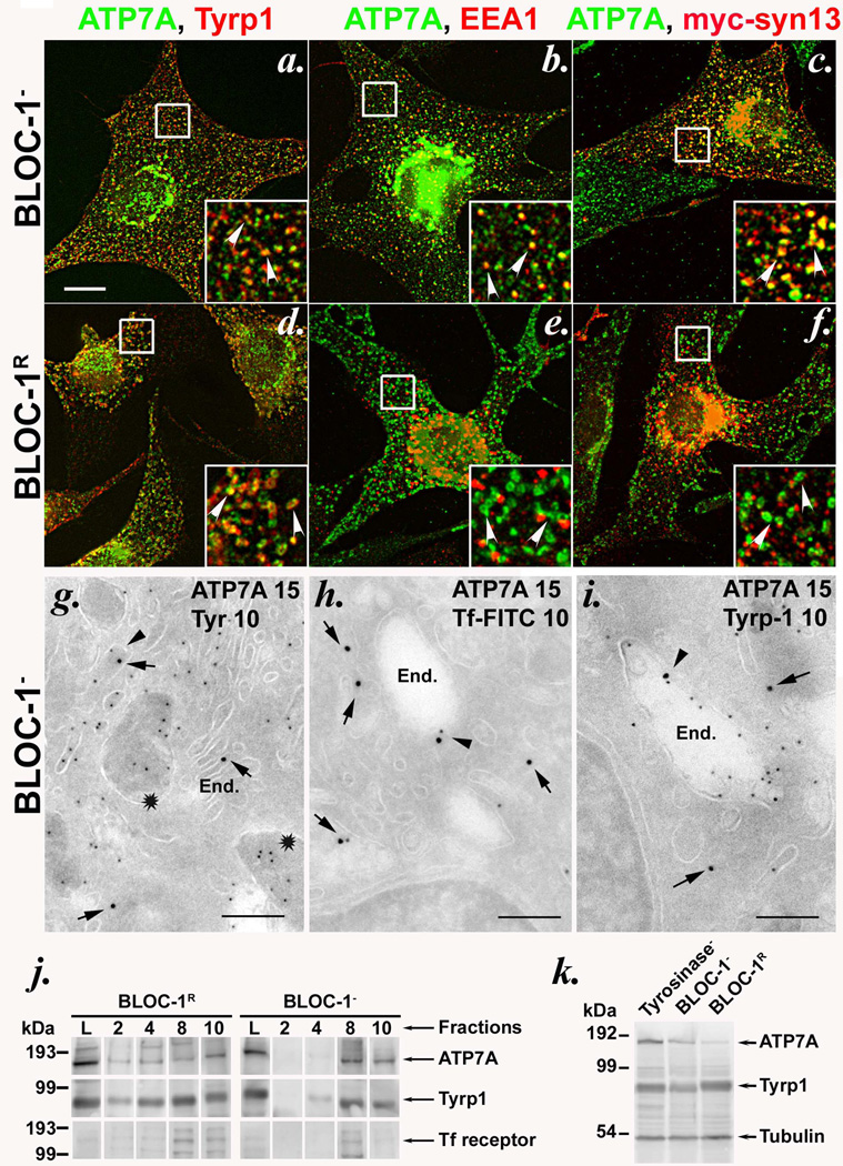
Figure 2. ATP7A is mislocalized to early endosomes in BLOC-1-deficient melanocytes.
(a–f) IFM analysis of BLOC-1− (melan-mu, a–c) and BLOC-1R (melan-mu: MuHA, d–f) melanocytes labeled with antibodies to ATP7A and either Tyrp1 (left), EEA1 (middle) or transiently expressed myc epitope-tagged Syntaxin 13 (myc-syn13, right). Insets, boxed regions magnified 3X. Arrowheads, ATP7A colocalized with the indicated marker. Bar, 10 µm. Note that Tyrp1 labels melanosomes in BLOC-1R cells but early endosomes in BLOC-1− cells15; EEA1 and syn13 label early endosomes in both cell types. (g–i) Ultrathin cryosections of BLOC-1− melan-mu cells were immunogold labeled with antibodies to ATP7A (15 nm gold) and tyrosinase (g), internalized Tf (h) or Tyrp1 (i)(10 nm gold) and analyzed by electron microscopy. Arrows indicate labeling of ATP7A and arrowheads indicate ATP7A with Tf or Tyrp1 on endosomes. Stars in (g) indicate striated melanosomes labeled for tyrosinase but not ATP7A. End., endosomes. Bars, 500 nm. See also Supplementary Figure S4. (j) Subcellular fractionation of BLOC-1− (melan-mu) and BLOC-1R (melan-mu:MuHA) cells on sucrose step gradients. Eluted fractions (2, 4, 8 and 10), collected from bottom to top, and lysates (L, input loading control) were probed by immunoblot with antibodies to ATP7A, Tyrp1 and Tf receptor (to label early endosomes). Left, molecular weight markers (in kDa). Arrows, relevant bands. Note the bands for ATP7A and Tyrp1, but not Tf receptor (middle band), in pigment-containing fractions 2 and 4 from melan-mu:MuHA but not melan-mu cells. (k) Whole cell lysates from tyrosinase-deficient (Tyrosinase-, melan-c), BLOC-1− (melan-mu) and BLOC-1R (melan-mu:MuHA) were fractionated by SDS-PAGE and immunoblotted with antibodies to the ATP7A, Tyrp1 and tubulin. Left, molecular weight markers (in kDa). Arrows, relevant bands.

