Abstract
DNA vaccines encoding different portions of the structural proteins of western equine encephalitis virus were tested for the efficacy of their protection in a 100% lethal mouse model of the virus. The 6K-E1 structural protein encoded by the DNA vaccine conferred complete protection against challenge with the homologous strain and limited protection against challenge with a heterologous strain.
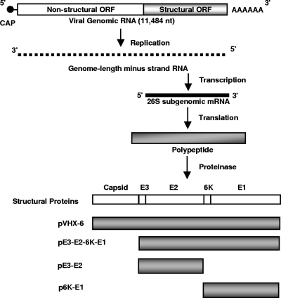
Western equine encephalitis (WEE) virus (WEEV) is an alphavirus that belongs to the family Togaviridae. The virus is a single-stranded, positive-sense, enveloped RNA virus with a genome of about 11.5 kb (11) (Fig. 1). The 5′ two-thirds of the genome encodes four nonstructural proteins, which form the replicase responsible for RNA replication and transcription. The 3′ one-third of the genome encodes structural proteins. The structural proteins are translated from a subgenomic 26S RNA, which is transcribed by the viral replicase from a promoter in the genome-length minus-strand RNA. Studies of other alphaviruses, such as Sindbis virus, Semliki Forest virus and Venezuelan equine encephalitis virus (VEEV), revealed that the structural proteins consist of a capsid protein, E3 (a signal peptide), E2 (an envelope glycoprotein responsible for receptor binding), 6K (a signal peptide and a possible viroporin), and E1 (an envelope glycoprotein responsible for the fusion of the viruses with cell membrane) (14). The E1 and E2 proteins are major determinants for the induction of neutralization antibodies against the alphaviruses. It is believed that the structural proteins of WEEV possess functions similar to those of other alphaviruses (11).
FIG. 1.
Schematic illustration of the synthesis of structural proteins of the alphaviruses and DNA plasmids constructed in this study.
WEEV is endemic in western North America, where the virus is transmitted via mosquitoes from its reservoir in wild birds to humans and horses. Infected individuals usually present with fever and headache before a gradual improvement. Severe cases can progress into coma and death, with the case mortality rate being 3 to 4% (4). Currently, no commercial vaccine and no antiviral drug are available for the prevention and treatment of WEEV infection. We previously developed a DNA vaccine (pVHX-6) encoding all the structural proteins (capsid-E3-E2-6K-E1) of the 71V-1658 strain of WEEV and demonstrated that the vaccine protected mice against challenge with a lethal dose of strain 71V-1658 (9). In this study, we further investigated which portion of the structural proteins encoded by the DNA vaccine is responsible for the protection. Three DNA vaccines encoding different portions of the 71V-1658 structural proteins were constructed (Fig. 1) and tested for their efficacies by intranasal challenge of mice in a lethal model of WEEV infection. We demonstrated that the DNA vaccine encoding the 6K-E1 protein was enough to provide complete protection against challenge with homologous strain 71V-1658. However, the DNA vaccine encoding 6K-E1 provided limited protection against challenge with a heterologous strain.
Construction and characterization of DNA vaccines.
The detailed construction of pVHX-6 was described previously (9). A DNA vaccine encoding E3-E2 (pE3-E2) was made by PCR amplification of the E3-E2 gene from pVHX-6 by using the following primer pair: WEE E3 start (5′-ATT CCG ATG GCA CTA GTT ACA GCG CTA TGC-3′) and WEE E2 end (5′-CTC TAG ATC ATT CGA AAG CGT TGG TTG GCC GAA TG-3′). The resulting PCR product was cloned into pCRII-TOPO (Invitrogen, Carlsbad, CA) to generate pCRII-TOPO-E3-E2. The E3-E2 gene was then isolated from pCRII-TOPO-E3-E2 and inserted into the mammalian expression vector pVAX (Invitrogen) to produce pE3-E2. A DNA vaccine encoding 6K-E1 (p6K-E1) was made in a similar fashion. The 6K-E1 gene was amplified from pVHX-6 by using primers WEE 6K st2 (5′-ATT CCG ATG GAA ACA TTT GGA GAA AC-3′) and WEE E1 end (5′-CTC TAG ATC ATT CGA ATC TAC GTG TGT TTA TAA GC-3′). The PCR product was cloned into pCRII-TOPO and inserted into pVAX to generate p6K-E1. A DNA vaccine encoding E3-E2-6K-E1 (pE3-E2-6K-E1) was constructed by deletion of the capsid-coding region of pVHX-6 (9). To do this, a HindIII-BstEII fragment was isolated from pE3-E2 and ligated into HindIII-BstEII-digested pVHX-6. The coding sequences in the DNA vaccines were verified by DNA sequencing. Plasmid DNA was purified with a Wizard Plus SV minipreps DNA purification system (Promega, Madison, WI), which does not have the step for endotoxin removal.
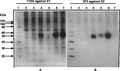
The expression of proteins E1 and E2 from the DNA vaccines was determined by Western blotting. Human embryonic kidney 293S cells seeded in six-well plates were transfected with pE3-E2-6K-E1, pE3-E2, and p6K-E1 by using Lipofectamine 2000 (Invitrogen). Controls include mock-, pVAX vector-, and pVHX-6-transfected cells. A total of 200 μl of cell lysates was collected at 24 h after transfection, and 1 μl of the cell lysate from each sample was loaded onto 12.5% SDS-polyacrylamide gels prior to transfer to a nitrocellulose membrane. The total protein in each cell lysate sample was not normalized. The E1 and E2 proteins in the lysates were probed with monoclonal antibodies 11D2 and 3F3, respectively (8). As shown in Fig. 2, both the E1 and the E2 proteins, each with a molecular mass of 50 kDa, were detected in cells transfected with pE3-E2-6K-E1 (Fig. 2A and B, lanes 6). The E1 protein was seen in p6K-E1-transfected cells (Fig. 2A, lane 7), and the E2 protein was seen in pE3-E2-transfected cells (Fig. 2B, lane 5). No 50-kDa protein band was shown in either mock- or pVAX-transfected cells (Fig. 2A and B, lanes 2 and 3), suggesting that the expression of E1 and E2 is specific for the DNA vaccines.
FIG. 2.
Expression of E1 and E2 proteins from DNA vaccines. Proteins from 293S cells transfected with the DNA vaccine were separated by electrophoresis through 12.5% polyacrylamide gels, transferred to nitrocellulose membranes, probed with an anti-E1 (A) or an anti-E2 (B) monoclonal antibody, and incubated with goat anti-mouse IgG conjugated with horseradish peroxidase (Sigma-Aldrich, St. Louis, MO). The E1 and E2 proteins were visualized by use of an Amersham ECL Plus Western blotting detection kit (GE Healthcare, Buckinghamshire, United Kingdom). Lane 1, molecular mass standard (Invitrogen); lane 2, mock-transfected cells (negative control); lane 3, pVAX-transfected cells (vector control); lane 4, pVHX-6-transfected cells; lane 5, pE3-E2-transfected cells; lane 6, pE3-E2-6K-E1-transfected cells; and lane 7, p6K-E1-transfected cells.
Figure 2B, lane 5, shows that the molecular mass of the E2 protein from the pE3-E2-transfected cells was slightly larger than the molecular masses of the E2 proteins from pVHX-6-transfected (lane 4) and pE3-E2-6K-E1-transfected (lane 6) cells. The size of the larger-molecular-mass protein is consistent with that of the uncleaved E3-E2 protein. It has previously been shown for VEEV that E2 is poorly processed in the absence of E1 (5).
Challenge and protection studies with mice.
Having confirmed that the E1 and E2 proteins were produced from DNA vaccines, we next evaluated the efficacies of these DNA vaccines in a mouse lethal challenge model of WEEV previously developed in our laboratory (10). Female BALB/c mice (weight, 17 to 25 g) were obtained from the mouse breeding colony at the Defense Research and Development Canada (DRDC)—Suffield. The original breeding pairs were purchased from Charles River Canada (St. Constant, Quebec, Canada). The protocols for the mouse experiments were reviewed and approved by the Animal Care Committee of DRDC—Suffield. The guidelines of the Canadian Council on Animal Care were followed for the caring and the handling of the mice. Groups of eight mice each were vaccinated with one of the following DNA vaccines: pE3-E2-6K-E1, pE3-E2, p6K-E1, pVHX-6, or pVAX (vector control). Inactivated WEE vaccine (9) was used as a positive control. Three doses of the DNA vaccine containing 2 μg of plasmid DNA in each vaccine were administered 14 days apart by using a Helios gene gun (Bio-Rad Laboratories, Mississauga, Ontario, Canada). Delivery of the DNA vaccine by use of a gene gun has been shown to improve immune protection through the induction of a predominant Th2-type immune response. Studies have shown that the improved immune protection is related to the increased transfection efficacy of antigen-presenting cells and the bombardment procedure of gene gun vaccination (2). Three vaccinations of a 50-μl dose of the WEE inactivated vaccine (the Salk WEE inactivated vaccine) were given to each mouse by the intramuscular route 14 days apart. At 14 days after the third vaccination, the mice were challenged intranasally with 1,500 PFU (25 50% lethal doses [LD50s]) of the homologous 71V-1658 strain of WEEV, 1,500 PFU (25 LD50s) of the heterologous Fleming strain, or 1,500 PFU of the heterologous CBA87 strain. The challenged mice were observed for 14 days for survival and the severity of the infection.
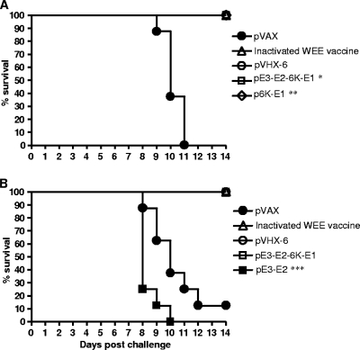
As shown in Fig. 3A, all the mice vaccinated with p6K-E1 survived by day 14 after challenge with the homologous 71V-1658 strain. The surviving mice did not show any signs of infection throughout the 14-day observation period (data not shown), demonstrating that the DNA vaccine encoding 6K-E1 conferred complete protection against the challenge with the homologous strain. This finding is similar to that obtained from the vaccine study of VEEV, in which a recombinant baculovirus expressing 6K-E1 conferred complete protection against challenge with the homologous strain of the virus (5). A previous study by Das et al. found that the recombinant WEEV E1 protein expressed by Escherichia coli did not provide any protection against the lethal challenge with the homologous WEEV strain (1). This may be because the E1 protein expressed by E. coli was not glycosylated, resulting in the misfolding of the E1 protein. In addition, immunization with the recombinant E1 protein without adjuvant could result in a poor immune response. The result from our study suggests that DNA vaccination with the 6K-E1 gene is a better approach for WEEV vaccine development.
FIG. 3.
Kaplan-Meier survival curves for the mice (eight mice per group) vaccinated with pE3-E2-6K-E1, p6K-E1, or pE3-E2 and challenged with a lethal dose of the homologous strain of WEEV. The vaccinated mice were challenged intranasally with strain 71V-1658 and monitored for 14 days. The log rank test was used to assess the differences in the survival rates of the challenged mice. *, P < 0.01 for pE3-E2-6K-E1 versus pVAX; **, P < 0.01 for p6K-E1 versus pVAX; ***, P > 0.05 for pE3-E2 versus pVAX.
Figure 3 also shows that pE3-E2-6K-E1 lacking the capsid-coding region conferred the same protection as pVHX-6 (capsid-E3-E2-6K-E1). All the mice vaccinated with pE3-E2-6K-E1 survived up to day 14 postchallenge. The mice did not show any signs of infection throughout the observation period, suggesting that the protection provided by pE3-E2-6K-E1 was complete. This result demonstrates that the capsid is not required for protection by DNA vaccination.
The pE3-E2 DNA vaccine containing the E3-E2-coding region did not provide any protection (Fig. 3B). This finding is different from that obtained in studies of VEEV (12, 13), in which the E3-E2-6K-coding region of VEEV gave good protection against challenge with the homologous strain. The presence of the 6K gene, which might help with the folding and the processing of E3-E2, may account for this difference. The E3-E2-6K gene in the VEEV studies was also delivered by an adenovirus vector instead of plasmid DNA, which can significantly enhance delivery (6). Figure 3B shows that about 10% (one of eight) of the mice given the pVAX control survived after challenge. We speculate that the surviving mouse might not have taken enough challenge virus during the intranasal inoculation.
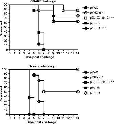
Having identified that 6K-E1 is enough to confer complete protection against challenge with homologous WEEV strain 71V-1658, we investigated whether 6K-E1 would also protect mice against challenge with a heterologous strain. Pathogenesis studies with different WEEV strains revealed that the strains can be grouped into high- and low-virulence strains on the basis of the percent mortality and the mean time to death (7, 10). We tested the efficacy of the DNA vaccine encoding 6K-E1 against CBA87, a low-virulence strain. As shown in Fig. 4, 6K-E1 alone was able to protect the majority of the mice from the challenge; however, 6K-E1 was unable to provide protection against the highly virulent Fleming strain of WEEV. Additionally, we found that pE3-E2-6K-E1 gave better protection against the Fleming strain than pVHX-6 did (100% and 50%, respectively; P < 0.05, log rank test). The reason for this is unclear. A previous study found that the capsid protein of VEEV inhibits the transcription and translation of cellular proteins (3). Therefore, the presence of the capsid-coding region in pVHX-6 might reduce the amount of E1 and E2 proteins produced in vivo, which could result in the induction of weaker immune protection.
FIG. 4.
Kaplan-Meier survival curves for the mice (eight mice per group) vaccinated with pE3-E2-6K-E1, p6K-E1, or pE3-E2 and challenged with a lethal dose of a heterologous strain of WEEV. The vaccinated mice were challenged intranasally with low-virulence strain CBA87 or high-virulence strain Fleming and were monitored for 14 days. The log rank test was used to assess the differences in the survival rates of the challenged mice. *, P < 0.01 for pVHX-6 versus pVAX; **, P < 0.01 for pE3-E2-6K-E1 versus pVAX; ***, P < 0.01 for p6K-E1 versus pVAX; #, P < 0.01 for pVHX-6 versus pVAX; ##, P < 0.01 for pE3-E2-6K-E1 versus pVAX.
In summary, we found that the DNA vaccine encoding the 6K-E1 protein conferred complete protection against challenge with the homologous strain of WEEV and limited protection against challenge with a heterologous strain. We also found that the DNA vaccine encoding the E3-E2 protein provided no protection and that deletion of the capsid-coding region from the DNA vaccine appears to improve the efficacy of the vaccine against WEEV challenge. Further studies will be carried out to determine the minimum region in 6K-E1 required for protection. The results from those studies will aid with the development of an improved vaccine for WEEV.
Acknowledgments
We thank staff from the animal care unit at DRDC—Suffield for providing mice and caring for the vaccinated mice during the weekend.
This study was partially supported by the Chemical, Biological, Radiological or Nuclear Research and Technology Initiative (CRTI) of Canada and the Technology Investment Fund (TIF) of DRDC.
Footnotes
Published ahead of print on 18 November 2009.
REFERENCES
- 1.Das, D., S. L. Gares, L. P. Nagata, and M. R. Suresh. 2004. Evaluation of a western equine encephalitis recombinant E1 protein for protective immunity and diagnostics. Antivir. Res. 64:85-92. [DOI] [PubMed] [Google Scholar]
- 2.Doria-Rose, N. A., and N. L. Haigwood. 2003. DNA vaccine strategies: candidates for immune modulation and immunization regimens. Methods 31:207-216. [DOI] [PubMed] [Google Scholar]
- 3.Garmashova, N., S. Atasheva, W. Kang, S. C. Weaver, E. Frolova, and I. Frolov. 2007. Analysis of Venezuelan equine encephalitis virus capsid protein function in the inhibition of cellular transcription. J. Virol. 81:13552-13565. [DOI] [PMC free article] [PubMed] [Google Scholar]
- 4.Griffin, D. E. 2007. Alphaviruses, p. 1023-1067. In D. M. Knipe and P. M. Howley (ed.), Fields virology, vol. 1, 5th ed. Lippincott Williams & Wilkins, Philadelphia, PA. [Google Scholar]
- 5.Hodgson, L. A., G. V. Ludwig, and J. F. Smith. 1999. Expression, processing, and immunogenicity of the structural proteins of Venezuelan equine encephalitis virus from recombinant baculovirus vectors. Vaccine 17:1151-1160. [DOI] [PubMed] [Google Scholar]
- 6.Lasaro, M. O., and H. C. Ertl. 2009. New insights on adenovirus as vaccine vectors. Mol. Ther. 17:1333-1339. [DOI] [PMC free article] [PubMed] [Google Scholar]
- 7.Logue, C. H., C. F. Bosio, T. Welte, K. M. Keene, J. P. Ledermann, A. Phillips, B. J. Sheahan, D. J. Pierro, N. Marlenee, A. C. Brault, C. M. Bosio, A. J. Singh, A. M. Powers, and K. E. Olson. 2009. Virulence variation among isolates of western equine encephalitis virus in an outbred mouse model. J. Gen. Virol. 90:1848-1858. [DOI] [PMC free article] [PubMed] [Google Scholar]
- 8.Long, M. C., L. P. Nagata, G. V. Ludwig, A. Z. Alvi, J. D. Conley, A. R. Bhatti, M. R. Suresh, and R. E. Fulton. 2000. Construction and characterization of monoclonal antibodies against western equine encephalitis virus. Hybridoma 19:121-127. [DOI] [PubMed] [Google Scholar]
- 9.Nagata, L. P., W. G. Hu, S. A. Masri, G. A. Rayner, F. L. Schmaltz, D. Das, J. Wu, M. C. Long, C. Chan, D. Proll, S. Jager, L. Jebailey, M. R. Suresh, and J. P. Wong. 2005. Efficacy of DNA vaccination against western equine encephalitis virus infection. Vaccine 23:2280-2283. [DOI] [PubMed] [Google Scholar]
- 10.Nagata, L. P., W. G. Hu, M. Parker, D. Chau, G. A. Rayner, F. L. Schmaltz, and J. P. Wong. 2006. Infectivity variation and genetic diversity among strains of Western equine encephalitis virus. J. Gen. Virol. 87:2353-2361. [DOI] [PubMed] [Google Scholar]
- 11.Netolitzky, D. J., F. L. Schmaltz, M. D. Parker, G. A. Rayner, G. R. Fisher, D. W. Trent, D. E. Bader, and L. P. Nagata. 2000. Complete genomic RNA sequence of western equine encephalitis virus and expression of the structural genes. J. Gen. Virol. 81:151-159. [DOI] [PubMed] [Google Scholar]
- 12.Perkins, S. D., L. M. O'Brien, and R. J. Phillpotts. 2006. Boosting with an adenovirus-based vaccine improves protective efficacy against Venezuelan equine encephalitis virus following DNA vaccination. Vaccine 24:3440-3445. [DOI] [PubMed] [Google Scholar]
- 13.Phillpotts, R. J., L. O'Brien, R. E. Appleton, S. Carr, and A. Bennett. 2005. Intranasal immunisation with defective adenovirus serotype 5 expressing the Venezuelan equine encephalitis virus E2 glycoprotein protects against airborne challenge with virulent virus. Vaccine 23:1615-1623. [DOI] [PubMed] [Google Scholar]
- 14.Richard, J. K. 2007. Togaviridae: the viruses and their replication, p. 1000-1022. In D. M. Knipe and P. M. Howley (ed.), Fields virology, vol. 1, 5th ed. Lippincott Williams & Wilkins, Philadelphia, PA. [Google Scholar]


