Abstract
To improve the diagnosis efficiency of human strongyloidiasis by using formalin-ether concentration technique (FECT), the effects of various factors on the recovery rates of Strongyloides stercoralis larvae were comparatively evaluated. Fresh stool and a short time exposure of larvae to formalin yielded significantly higher numbers of larvae than preserved stool and 10-min exposure. Likewise, straining through wire mesh yielded a significantly higher number of larvae recovered than straining through gauze did. In addition, centrifugation for 5 min for separation of larvae from debris yielded a significantly greater number of larvae recovered than centrifugation for 2 min did. The efficacies of the five versions of FECT with different factors-FECT 1, FECT 2, FECT 3, FECT 4, and FECT 5-were then compared. It was found that FECT 5 was 1.8, 2.0, 1.9, and 1.4 times more effective than FECT 1, FECT 2, FECT 3, and FECT 4, respectively. FECT 5 is a modified FECT method, whose modifications included using fresh stool without a preservative substance; a short-time rather than 10-min formalin exposure; and the use of wire mesh instead of gauze.
Strongyloidiasis caused by Strongyloides stercoralis is a harmful infectious disease for immunosuppressed patients (5). The most accurate method for diagnosis of strongyloidiasis, which is widely used in areas where this disease is endemic, is to detect S. stercoralis larvae in stool, whereas detection by indirect methods, especially immunological techniques, are not as effective (11). Agar plate culture (APC) has consistently been found to be 1.6 to 6.0 times more effective than formalin-ether concentration technique (FECT) (1, 8). However, APC has many disadvantages: it is time-consuming and costly, has a tendency of risk of larval infection (7, 8), and demonstrates a particular inability to detect 10% (6) to 20% (12) of positive samples detected by FECT. Thus, if the efficacy of FECT can be improved, a more precise diagnosis of strongyloidiasis will be obtained. Many laboratory personnel believe that variations in the skill and experience of examiners caused the effectiveness of APC shown to be 1.6 to 6.0 times greater than FECT (1). However, there could possibly be some concealed factors causing the lower sensitivity of FECT.
A pilot study was thus conducted using a large number of S. stercoralis larvae in diluted stool samples from patients with strongylodiasis (kept for 2 months in 10% formalin) for the capability of larval counting. FECT was performed, after which four layers were formed: ether, plug of debris, formalin and sediment. The results showed that 62.5% of larvae were in the debris plug, 32.5% floated in the formalin layer, and only 5% settled in the sediment. The diluted stool samples were also comparatively filtered either through two layers of wet gauze or two layers of wire mesh (0.7- by 0.7-mm and 1.2- by 1.2-mm pore sizes). The results revealed that a significantly lower number of larvae were recovered in the experiment using gauze than with the two wire meshes. The fine wire mesh also yielded a significantly lower number of larvae than the coarse one did. This work therefore aims to compare various factors affecting the number of larvae dwelling in fresh stool that may affect the recovery rate of S. stercoralis larvae and to determine suitable modification conditions of the FECT method to obtain a more precise diagnosis of strongyloidiasis.
MATERIALS AND METHODS
Samples.
For comparison of factors affecting the recovery of S. stercoralis larvae, 20 stool samples were collected from villagers living in Moklan in Nakhon Si Thammarat province, Thailand, 7 km away from the laboratory unit in Walailak University. These samples were taken from villagers with previously known cases of asymptomatic strongyloidiasis, and the samples were received from other research projects and diagnosed by the agar plate culture method (1, 8). The specimens were used only for comparing the factors affecting S. stercoralis larvae recovery (four comparisons, namely, comparison of fresh versus preserved stool samples, comparison of a short-time versus 10-min exposure of larvae to formalin before adding diethyl ether, comparison of gauze versus wire mesh, and comparison of 2 min versus 5 min of centrifugation at 700 × g) (see below). For comparison purposes, 100 samples were collected by random sampling from primary schoolchildren whose disease status was unknown; the schoolchildren attended Wat Moklan primary school, Thasala, Nakhon Si Thammarat, Thailand. Each stool sample was kept in a box and sent to the laboratory for testing within 4 h after defecation. For confirmation of diagnosis of strongyloidiasis, a 3-g portion of individual stool sample was determined by the agar plate culture method (1, 8). Of 100 samples, 29 were positive by the agar plate culture method. These samples are used in a blind manner in comparative efficacy studies of five versions of FECT with various factors (see below). This research was approved by the Ethical Clearance Committee on Human Rights Related to Research Involving Human Subjects, Walailak University.
Formalin-ether concentration technique.
FECT was performed as described previously (2). Briefly, 2 g of either preserved or fresh stool sample was suspended in a tube containing 10 ml of 0.85% saline. The suspension was strained through two layers of wet gauze into another centrifuge tube. The strained suspension was centrifuged at 700 × g for 5 min, and the supernatant was then decanted. After that, the volume was adjusted to 7 ml with 10% formalin, mixed well, and allowed to stand for 10 min (for fresh stool samples). Later, 3 ml of diethyl ether was added. The tube was closed, vigorously shaken by hand for 1 min, and centrifuged at 700 × g for 5 min. The debris plug was loosened, and the top three layers were discarded. One milliliter of 10% formalin was then added to the sediment. The number of larvae per gram (lpg) of stool was counted for quantitation of the comparative studies.
Comparison of fresh versus preserved stool samples.
Fresh stool (4 g) was suspended in a beaker containing 8 ml of 0.85% saline and then divided equally into two tubes: one contained 6 ml of 0.85% saline and was promptly tested, while the other contained 6 ml of 10% formalin and was left for 30 min. FECT was performed as described above, except that wire mesh was used instead of gauze, and a shorter exposure time (rather than 10 min) of larvae to formalin was employed. The plug of debris was poured off into a plastic petri dish for examination of trapped larvae.
Comparison of a short-time versus 10-min exposure of larvae to formalin before adding diethyl ether.
Fresh stool (4 g) was suspended in a beaker containing 20 ml of 0.85% saline and then divided equally into two centrifuge tubes. After the suspension was strained through gauze, the tubes were centrifuged and the supernatant was discarded. A 10% formalin solution was added to one of the tubes, followed promptly by the addition of 3 ml diethyl ether, while the other tube had 10% formalin added, which was then mixed and allowed to stand for 10 min before the addition of diethyl ether. The FECT process was continued as described above. The plug of debris was also poured off for examination of trapped larvae.
Comparison of gauze versus wire mesh.
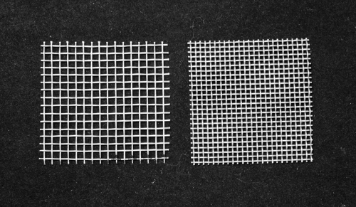
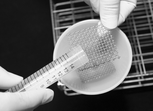
Fresh stool (4 g) was suspended and stirred well in a beaker containing 20 ml of 0.85% saline and then divided equally into two centrifuge tubes. The contents of one tube was strained through two layers of wet gauze, while the contents of the other tube was strained through two pieces of 4-cm wire mesh (the finer wire mesh with 1.2- by 1.2-mm holes was on a funnel, and the second mesh with 2- by 2-mm holes was held by hand or forceps) (Fig. 1 and 2). The trapped fecal material on both gauze and wire mesh was washed with 3 ml of 0.85% saline. The FECT process was continued as described above.
FIG. 1.
Coarse and fine wire meshes.
FIG. 2.
Straining method through two wire meshes.
Comparison of 2 min versus 5 min of centrifugation at 700 × g for separation of larvae from debris.
Fresh stool (4 g) was suspended and stirred well in a beaker containing 20 ml of 0.85% saline and then divided equally into two centrifuge tubes. After the contents of the tubes were strained through gauze and centrifuged at 700 × g for 5 min, the larvae were exposed for a short time to formalin. After diethyl ether was added to the tube and vigorously shaken for 1 min, one tube was centrifuged at 700 × g for 2 min, while the other tube was centrifuged at the same speed for 5 min. The FECT process was continued as described above.
Comparative efficacies of five versions of FECT with various factors.
Fresh stool (10 g) was suspended in a beaker containing 20 ml of 0.85% saline and then divided equally into five tubes. Three tubes each had 6 ml of 0.85% saline added and were then promptly tested (fresh stools), while the other two tubes each had 6 ml of 10% formalin added and were then kept for 30 min (preserved stools). Five versions of FECT with the following factors were compared: using fresh stool, gauze, and 10-min exposure of larvae to formalin (FECT 1); using preserved stool, gauze, and a short time exposure of larvae to formalin (FECT 2); using preserved stool, wire mesh, and a short time exposure of larvae to formalin (FECT 3); using fresh stool, wire mesh, and 10-min exposure of larvae to formalin (FECT 4); and using fresh stool, wire mesh, and a short time exposure of larvae to formalin (FECT 5).
Statistical analysis.
A paired t test was used to test the difference between 20 pairs in four quantitative comparisons. A nonparametric McNemar test was used for qualitative comparisons of various factors among the 5 versions of FECT; a P value of <0.05 was considered statistically significant. All statistical analysis was performed by SPSS 13.0 for Windows.
RESULTS
The 10-min exposure of larvae in stool samples to formalin before adding diethyl ether, as well as 30-min stool preservation before straining, caused approximately 40% of the larvae to be trapped in the lower part of the debris plug (data not shown). Comparatively, approximately 32% of larvae in fresh stool samples were trapped in gauze rather than in wire mesh (data not shown). Likewise, centrifugation at 700 × g for 2 min for larval separation from debris resulted in a decrease of 30% of larval numbers compared to centrifugation for 5 min. However, there was no observational data regarding whether the lost larvae were floating in the 10% formalin layer (Table 1) .
TABLE 1.
Comparative studies of four factors affecting the recovery of S. stercoralis larvae
| Comparison and factor | No. of larvae per g of stool (mean ± SD) | Pa | Larvae in the plug of debris |
|---|---|---|---|
| Fresh vs preserved stool | |||
| Fresh stool | 462.6 ± 349.7 | <0.001 | Not found |
| Preserved stool | 279.9 ± 195.0 | Numerous | |
| Short vs long time exposure | |||
| Short larva-formalin time exposure | 313.0 ± 276.0 | 0.001 | Not found |
| Long larva-formalin time exposure | 193.3 ± 154.9 | Numerous | |
| Wire mesh vs gauze | |||
| Wire mesh | 268.8 ± 253.0 | <0.001 | Not done |
| Gauze | 184.7 ± 174.1 | Not done | |
| 5-min vs 2-min centrifugation | |||
| 5-min centrifugation | 327.2 ± 467.2 | 0.017 | Not done |
| 2-min centrifugation | 229.2 ± 301.4 | Not done |
The P values comparing the values (number of larvae per g of stool) for the two factors in a comparison (e.g., comparing fresh stool to preserved stool).
The efficacies of the five different FECT procedures were compared. Out of 100 stool samples, 28 were found to be positive by at least one of the five methods. The positive detection values for FECT 1, FECT 2, FECT 3, FECT 4, and FECT 5 were 16%, 14%, 15%, 20%, and 28%, respectively. The positive rate by FECT 5 was significantly different from the positive rate by FECT 1 (P < 0.001), FECT 2 (P < 0.001), FECT 3 (P < 0.001), and FECT 4 (P = 0.008). The positive rate by FECT 4 was not significantly different from FECT 1 (P = 0.125) or FECT 3 (P = 0.063) but was significantly different from FECT 2 (P = 0.031). FECT 1 showed no significant difference from FECT 2 (P = 0.500) or FECT 3 (P = 1.000). FECT 5 was 1.8, 2.0, 1.9, and 1.4 times more effective than FECT 1, FECT 2, FECT 3, and FECT 4, respectively.
For detection of other parasites in 100 stool samples from individuals with unknown status re strongyloidiasis, no protozoa were found, but there were positive results for Ascaris lumbricoides, Trichuris trichiura, and hookworm eggs. When determined by FECT 1 to FECT 5 methods, the positive detection of the three worms had the same values: 6%, 20%, and 52% were found to be positive (A. lumbricoides, T. trichiura, and hookworm eggs, respectively). Mixed infections were as follows: S. stercoralis and hookworm, 18%; T. trichiura and S. stercoralis, 3%; A. lumbricoides, S. stercoralis, and hookworm, 2%; and A. lumbricoides, T. trichiura, hookworm, and S. stercoralis, 2%.
DISCUSSION
The formalin solution had biochemical and physical effects on S. stercoralis larvae. It caused the larval bodies to float in the fecal water solution, consequently becoming trapped in the lower part of the debris plug and then later being discarded during the FECT process. Previous parasitological laboratory procedures have revealed (2) that preserving the stool for at least 30 min is recommended prior to performing the FECT procedure. In addition, even though fresh stool is used (4), the larvae must still be preserved for 10 min during the FECT process; otherwise, the larvae are trapped in the plug of debris.
Our unpublished study showed that the preserved larvae moderately decreased (from 100% to approximately 50%) during the course of exposure to formalin from a few minutes to 2 h. However, they rapidly decreased from 2 h to 24 h of exposure and then gradually decreased after 24 h of preservation. Nevertheless, there may be variation on a case-by-case basis. In a comparative observation using fresh stool samples (tested within 4 h after defecation) and 24-h-preserved stool samples, the result revealed that larval intensity in fresh stool was 1,500 lpg of stool, while that in the 24-h-preserved stool was only 50 lpg. We also observed a larval intensity of 300 lpg in a fresh stool experiment, contrasting with 2 lpg in 60-day-preserved stool. The plug of debris was examined, and a large number of larvae were found in both cases. Thus, consideration should be given to the beneficial factors of avoiding long-time exposure of larvae to formalin, as well as the advantages of performing the FECT procedure by the FECT 5 method, which used fresh stool and a short time exposure of larvae to formalin. Moreover, the use of 1 g or 2 g of stool for processing during FECT yielded no significant differences in the number of larvae (data not shown). Furthermore, formalin also affected the recovery of hookworm eggs and Taenia eggs but had little effect on Opisthorchis viverrini eggs (data not shown). However, stool samples preserved by sodium acetate, acetic acid, and formalin yielded a higher recovery rate of intestinal protozoa (9), probably because formalin has an effect on larvae and some helminth eggs rather than on protozoon cysts and trophozoites.
The use of wire mesh was superior to gauze in yielding a higher number of larvae. The size of the holes in the gauze, varying in pore size from 0.6 mm to 2.0 mm (10), was not much different from the finer wire mesh (1.2 mm) used in our study. However, the cotton gauze was folded into two layers, and the gauze is also composed of fragmented cotton fibers that might make the holes smaller, thus more easily trapping the larvae. Previously, a comparative evaluation of five stool concentration systems revealed that devices with large filter pore sizes produced better results with large parasite forms (10).
The centrifugation time for separation of larvae from the debris is also important. The speed of 700 × g for 2 min was not enough to allow separation of floating larvae from the formalin layer. Unfortunately, we have no further information on this issue except from the pilot study, which used diluted stool samples that revealed 32.5% of larvae floating in the formalin layer. However, when the preserved larvae in a diluted strongylodiasis stool sample were added to the preserved stool sample from a healthy subject, the recovery rate (number of larvae in the sediment) revealed an increase in the larval numbers from 5% to 20%, possibly because heavy stool particles enhanced the larvae settling down in the sediment. Thus, centrifugation at 700 × g for 5 min is sufficient to separate the floating larvae from the formalin layer. In addition, centrifugation for 5 and 10 min yielded no significance differences in the number of larvae recovered (data not shown). Similarly, the increase in centrifugation time and acceleration of gravity resulted in the detection of a higher number of Cryptosporidium oocysts and positive samples (3).
FECT 1 and FECT 2 are the traditional FECT methods widely used in general laboratory sections (2, 4). FECT 3, FECT 4, and FECT 5 are performed as modified versions. The combination of the use of fresh stool, a short time exposure of larvae to formalin, and wire mesh—as demonstrated in the FECT 5 procedure—provided the highest efficacy compared to FECT procedures 1 to 4. FECT 5 is a modified FECT method that was effectively superior to conventional FECT (FECT 1 and FECT 2) by 1.8 and 2.0 times, respectively. However, the efficacy should be further compared with other sensitive methods, including APC and the Baermann method, by using a large number of samples.
When examining other helminthic detection in samples by the FECT 1 to FECT 5 methods, the positive detection rates of A. lumbricoides, T. trichiura, and hookworm eggs all had the same values, regardless of method. This result is possibly due to the stool samples having been collected from areas where infection is endemic, resulting in the lack of effect on the recovery rates of other parasites.
Acknowledgments
We thank Pawilai Dermlim, Marisa Thongrod, and Siraporn Innimit for technical assistance.
P.M.I. and W.M. were supported by a Khon Kaen University grant.
Footnotes
Published ahead of print on 18 November 2009.
REFERENCES
- 1.Arakaki, T., M. Iwanaga, F. Kinjo, A. Saito, R. Asato, and T. Ikeshiro. 1990. Efficacy of agar plate culture in detection of Strongyloides stercoralis infection. J. Parasitol. 76:425-428. [PubMed] [Google Scholar]
- 2.Beaver, P. C., R. C. Jung, and E. W. Cupp. 1984. Examination of specimens for parasites, p. 733-758. In P. C. Beaver, R. C. Jung, and E. W. Cupp (ed.). Clinical parasitology, 9th ed. Lea & Febiger, Philadelphia, PA.
- 3.Clavel, A., A. Arnal, E. Sanchez, M. Varea, J. Quilez, I. Ramirez, and F. J. Castillo. 1996. Comparison of 2 centrifugation procedures in the formalin-ethyl acetate stool concentration technique for the detection of Cryptosporidium oocysts. Int. J. Parasitol. 26:671-672. [DOI] [PubMed] [Google Scholar]
- 4.Grove, D. I. (ed.). 1989. Diagnosis, p. 175-197. In D. I. Grove (ed.), Strongyloidiasis: a major roundworm infection of man. Taylor & Francis, Philadelphia, PA.
- 5.Grove, D. I. 1996. Human strongyloidiasis. Adv. Parasitol. 38:251-309. [DOI] [PubMed] [Google Scholar]
- 6.Intapan, P. M., W. Maleewong, T. Wongsaroj, S. Singthong, and N. Morakote. 2005. Comparison of the quantitative formalin ethyl acetate concentration technique and agar plate culture for diagnosis of human strongyloidiasis. J. Clin. Microbiol. 43:1932-1933. [DOI] [PMC free article] [PubMed] [Google Scholar]
- 7.Kaminsky, R. G. 1993. Evaluation of three methods for laboratory diagnosis of Strongyloides stercoralis infection. J. Parasitol. 79:277-280. [PubMed] [Google Scholar]
- 8.Koga, K., S. Kasuya, C. Khamboonruang, K. Sukhavat, Y. Nakamura, S. Tani, M. Ieda, K. Tomita, S. Tomita, N. Hattan, M. Mori, and S. Makino. 1990. An evaluation of the agar plate method for the detection of Strongyloides stercoralis in northern Thailand. J. Trop. Med. Hyg. 93:183-188. [PubMed] [Google Scholar]
- 9.Mank, T. G., J. O. M. Zaat, J. Blotkamp, and A. M. Polderman. 1995. Comparison of fresh versus sodium acetate acetic acid formalin preserved stool specimens for diagnosis of intestinal protozoal infections. Eur. J. Clin. Microbiol. Infect. Dis. 14:1076-1081. [DOI] [PubMed] [Google Scholar]
- 10.Perry, J. L., J. S. Matthews, and G. R. Miller. 1990. Parasite detection efficiencies of five stool concentration systems. J. Clin. Microbiol. 28:1094-1097. [DOI] [PMC free article] [PubMed] [Google Scholar]
- 11.Siddiqui, A. A., and S. L. Berk. 2001. Diagnosis of Strongyloides stercoralis infection. Clin. Infect. Dis. 33:1040-1047. [DOI] [PubMed] [Google Scholar]
- 12.Sukhavat, K., N. Morakote, P. Chaiwong, and S. Piangjai. 1994. Comparative efficacy of four methods for the detection of Strongyloides stercoralis in human stool specimens. Ann. Trop. Med. Parasitol. 88:95-96. [DOI] [PubMed] [Google Scholar]


