Abstract
Bacterial spores remain dormant and highly resistant to environmental stress until they germinate. Completion of germination requires the degradation of spore cortex peptidoglycan by germination-specific lytic enzymes (GSLEs). Bacillus anthracis has four GSLEs: CwlJ1, CwlJ2, SleB, and SleL. In this study, the cooperative action of all four GSLEs in vivo was investigated by combining in-frame deletion mutations to generate all possible double, triple, and quadruple GSLE mutant strains. Analyses of mutant strains during spore germination and outgrowth combined observations of optical density loss, colony-producing ability, and quantitative identification of spore cortex fragments. The lytic transglycosylase SleB alone can facilitate enough digestion to allow full spore viability and generates a variety of small and large cortex fragments. CwlJ1 is also sufficient to allow completion of nutrient-triggered germination independently and is a major factor in Ca2+-dipicolinic acid (DPA)-triggered germination, but its enzymatic activity remains unidentified because its products are large and not readily released from the spore's integuments. CwlJ2 contributes the least to overall cortex digestion but plays a subsidiary role in Ca2+-DPA-induced germination. SleL is an N-acetylglucosaminidase that plays the major role in hydrolyzing the large products of other GSLEs into small, rapidly released muropeptides. As the roles of these enzymes in cortex degradation become clearer, they will be targets for methods to stimulate premature germination of B. anthracis spores, greatly simplifying decontamination measures.
The Gram-positive bacterium Bacillus anthracis is the etiologic agent of cutaneous, gastrointestinal, and inhalational anthrax (24). An anthrax infection begins when the host is infected with highly resistant, quiescent B. anthracis spores (1, 24). Within the host, the spore's sensory mechanism recognizes chemical signals, known as germinants, and triggers germination, which leads to the resumption of metabolism (36). Spores that have differentiated into vegetative cells produce a protective capsule and deadly toxins. These virulence factors allow the bacteria to evade the host's immune system and establish an infection resulting in septicemia, toxemia, and frequently death (24). Although vegetative cells produce virulence factors that are potentially fatal, these cells cannot initiate infections and are much more susceptible to antimicrobial treatments than spores (24). Therefore, efficient triggering of spore germination may enhance current decontamination methods.
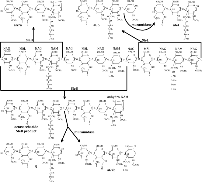
Spores are highly resistant to many environmental insults because the spore core (cytoplasm) is dehydrated, dormant, and surrounded by multiple protective layers, including a modified layer of peptidoglycan (PG) known as the cortex (36). The cortex functions to maintain dormancy and heat resistance by preventing core rehydration (9). It is composed of alternating N-acetylglucosamine (NAG) and N-acetylmuramic acid (NAM) sugars (Fig. 1). Peptide side chains on the NAM residues are either involved in interstrand cross-linking, cleaved to single l-alanine side chains, or fully removed with accompanying formation of muramic-δ-lactam (2, 31, 38). After germination is initiated by either nutrient or nonnutrient germinants, the cortex is depolymerized, resulting in complete core rehydration, resumption of metabolic activity, and outgrowth (33, 36).
FIG. 1.
Spore PG structure and hydrolysis. The central structure shows a representative spore PG strand with alternating NAG and NAM or muramic-δ-lactam (MδL) residues and with tetrapeptide or l-Ala side chains on the NAM residues. Forked arrows originate at sites of hydrolysis by the indicated enzymes and point to muropeptide products. The indicated “aG” muropeptide names are as previously published (7, 11). SleB lytic transglycosylase activity produces muropeptides terminating in anhydro-NAM. Cleavage at adjacent NAM residues produces the tetrasaccharide aG7a or aG7b, while cleavage further apart can produce octasaccharides or larger fragments. These can be further cleaved by muramidase treatment, resulting in the production of tetrasaccharide N, which terminates in NAM. The N-acetylglucosaminidase activity of SleL produces tetrasaccharides terminating in NAG, which can be further cleaved by muramidase to trisaccharides terminating in NAM.
Cortex hydrolysis is driven by autolysins called germination-specific cortex lytic enzymes (GSLEs) that recognize the cortex-specific muramic-δ-lactam residues (2, 4, 21, 32). GSLEs fall into two classes: spore cortex lytic enzymes (SCLEs), which are thought to depolymerize intact cortical PG, and cortical fragment lytic enzymes (CFLEs), which further degrade partially hydrolyzed cortex (21). Both SCLEs and CFLEs have been identified in a variety of spore-forming species, including B. anthracis (11, 18, 19), Bacillus cereus (4, 20, 26), Bacillus megaterium (8, 34), Bacillus subtilis (13, 16, 25), Bacillus thuringiensis (12), and Clostridium perfringens (5, 23). Of the four GSLEs identified in B. anthracis, CwlJ1, CwlJ2, and SleB are predicted to be SCLEs (11), whereas SleL is thought to be a CFLE (18).
Recently, independent studies showed that CwlJ1 and the lytic transglycosylase SleB (Fig. 1) play partially redundant roles and that either is sufficient for spore germination and outgrowth (10, 11). However, these same studies report conflicting results concerning the role of CwlJ2 during germination. Heffron et al. found no effect of CwlJ2 on the biochemistry of cortex hydrolysis or on colony-forming efficiency of spores (11). Giebel et al. reported that loss of CwlJ2 caused a minor defect in germination kinetics and that in the absence of SleB and CwlJ1, further loss of CwlJ2 had a major effect on colony forming efficiency (10). SleL in Bacillus anthracis is proposed to be an N-acetylglucosaminidase (Fig. 1) whose role is to further degrade cortex fragments resulting from SCLE hydrolysis (18). SleL is not essential for the completion of germination but does promote the release of small muropeptides to the spore's surrounding environment (18).
This study reports the effects of multiple deletion mutations affecting GSLEs on spore germination efficiency and kinetics of cortex hydrolysis. The data confirm the dominant roles played by CwlJ1 and SleB in the initiation of cortex hydrolysis and the major role of SleL in release of small cortex fragments. A minor role of CwlJ2 in nutrient-triggered germination and the contributions of CwlJ1 and CwlJ2 to Ca2+-dipicolinic acid (DPA)-triggered germination were revealed.
MATERIALS AND METHODS
Bacterial strains and growth conditions.
B. anthracis strains and plasmids used are listed in Table 1. Escherichia coli strains used to propagate plasmids were grown in LB with 500 μg/ml erythromycin (Fisher) or 100 μg/ml ampicillin (Jersey Lab Supply) and incubated at 37°C. B. anthracis strains were grown in brain heart infusion (BHI; Difco) medium containing 5 μg/ml erythromycin or 10 μg/ml tetracycline (Jersey Lab Supply) and incubated at 22°C prior to allelic exchange or 37°C afterwards.
TABLE 1.
B. anthracis strains and plasmids
| Strain or plasmid | Relevant genotypea | Constructionb | Source or reference |
|---|---|---|---|
| Strains | |||
| Sterne 34F2 | pXO1+, pXO2− | P. Hanna | |
| DPBa35 | ΔsleL | pDPV351→34F2 | 18 |
| DPBa38 | ΔsleB | pDPV383→34F2 | 11 |
| DPBa55 | ΔsleB ΔsleL | pDPV383→DPBa35 | This study |
| DPBa60 | ΔcwlJ2 | pDPV344→34F2 | This study |
| DPBa61 | ΔcwlJ1 | pDPV347→34F2 | 11 |
| DPBa66 | ΔcwlJ2 ΔsleL | pDPV344→DPBa35 | This study |
| DPBa69 | ΔcwlJ1 ΔsleL | pDPV347→DPBa35 | This study |
| DPBa72 | ΔcwlJ1 ΔsleB ΔsleL | pDPV347→DPBa55 | This study |
| DPBa73 | ΔcwlJ2 ΔsleB | pDPV383→DPBa60 | This study |
| DPBa74 | ΔcwlJ1 ΔsleB | pDPV383→DPBa61 | This study |
| DPBa78 | ΔcwlJ1 ΔcwlJ2 | pDPV347→DPBa60 | This study |
| DPBa82 | ΔcwlJ2 ΔsleB ΔsleL | pDPV344→DPBa55 | This study |
| DPBa83 | ΔcwlJ1 ΔcwlJ2 ΔsleL | pDPV344→DPBa69 | This study |
| DPBa84 | ΔcwlJ1 ΔcwlJ2 ΔsleB ΔsleL | pDPV344→DPBa72 | This study |
| DPBa85 | ΔcwlJ1 ΔcwlJ2 ΔsleB | pDPV347→DPBa73 | This study |
| Plasmids | |||
| pBKJ223 | Tetr, Pamy-I-SceI | 14 | |
| pBKJ236 | Err orits | 14 | |
| pDPV344 | ΔcwlJ2 | pBKJ236 | This study |
| pDPV347 | ΔcwlJ1 | pBKJ236 | 11 |
| pDPV351 | ΔsleL | pBKJ236 | 18 |
| pDPV383 | ΔsleB | pBKJ236 | 11 |
Tetr, tetracycline resistance; Err, erythromycin resistance; orits, temperature-sensitive origin of replication.
Strains were constructed by conjugation followed by the published series of steps required for recombination of the deletion mutation into the chromosome. The designation preceding the arrow is the plasmid, and the designation following the arrow is the recipient strain. Single plasmid designations indicate the vector in which the deletion construct was created.
In-frame deletion mutagenesis of cwlJ1, cwlJ2, sleB, and sleL has previously been published (11, 18). Each deletion allele encoded only 6 to 9 codons of the original gene. In-frame deletions were integrated into the B. anthracis chromosome by using markerless gene replacement as previously described (14). The deletion mutations were verified in each new strain by PCR amplification of each locus and sequencing the regions including >250 bp both up- and downstream.
Spore preparation.
B. anthracis strains were incubated at 37°C with shaking in modified G broth (15) for 3 days. Spores were harvested by repeated centrifugation and water washing. Vegetative cells were heat killed at 65°C for 30 min. Spores were purified by centrifugation through 50% sodium diatrizoate (Sigma) (27). Purified spores were ∼99% free of vegetative cells and were stored in deionized water at 4°C until analysis.
Spores at an optical density at 600 nm (OD) of 2 were decoated at 70°C for 30 min in 0.1 N NaOH, 0.1 M NaCl, 1% (wt/vol) sodium dodecyl sulfate, and 0.1 M dithiothreitol. After decoating, spores were washed five times in sterile deionized water. To evaluate the effects of decoating on viability, decoated and intact spores were serially diluted and plated onto BHI plates.
Spore germination assays.
To evaluate colony formation efficiency, intact or decoated spores were resuspended at an OD of 0.2. After heat activation at 70°C for 30 min, the suspensions were serially diluted, spotted on BHI plateswith or without 1 μg/ml lysozyme (Sigma), and incubated at 30°C overnight.
Spore germination and outgrowth in liquid BHI were assayed by monitoring OD as previously described, except incubation was done at 37°C (11, 18). Ca2+-DPA treatment to trigger germination was carried out essentially as previously described (28). Spores were suspended at an OD of 1 in water or 50 mM Ca2+-DPA solution and incubated at 25°C for 60 min. Spore suspensions were then heated at 70°C for 20 min, serially diluted in water, spotted onto BHI plates, and incubated at 37°C overnight to determine heat-resistant titers.
Biochemical analyses of PG hydrolysis and release.
After heat activation, spores were induced to germinate in a buffered solution containing 10 mM l-alanine and 1 mM inosine as previously described (18). Spore-associated (pellet) and exudate (supernatant) fractions collected throughout germination were assayed for muramic acid and diaminopimelic acid (Dpm) content as described previously (22). PG was purified and prepared for reverse-phase high-pressure liquid chromatography (RP-HPLC) analysis as previously described (7). Briefly, the germinated spore suspension was separated into pellet and supernatant fractions. Lytic enzymes were inactivated with heat (supernatant) or with heat and detergent (pellet), and PG was purified. The PG material from the pellets and half of each exudate fraction was then digested with the muramidase mutanolysin (Sigma). All fractions were reduced with NaBH4 prior to HPLC separation.
RESULTS
Effects of GSLEs on nutrient-triggered germination.
Wild-type and mutant spores were allowed to germinate and produce colonies on BHI plates overnight. All strains containing either CwlJ1 or SleB produced the same number of colonies per OD unit as did the wild-type strain (Table 2 and data not shown). However, all strains lacking both CwlJ1 and SleB exhibited a >103-fold decrease in colony formation. There was no further decrease in colony formation when other GSLEs, CwlJ2 and/or SleL, were also absent. Colony formation efficiency was restored to near wild-type levels in strains lacking both CwlJ1 and SleB when decoated spores were plated onto BHI plates supplemented with 1 μg/ml of lysozyme (Table 2). Spores were decoated to facilitate the penetration of lysozyme to the cortex, but this treatment did not affect colony formation, and lysozyme did not affect colony formation by nondecoated spores (data not shown).
TABLE 2.
Spore plating efficiency and lysozyme recovery
| Strain | Genotype | CFU/ml/OD ona: |
|
|---|---|---|---|
| BHI | BHI + lysob | ||
| 34F2 | Wild type | 1.5 × 10⁸ | 1.1 × 10⁸ |
| DPBa72 | ΔcwlJ1 ΔsleB ΔsleL | 3.7 × 10⁴c | 1.2 × 10⁸ |
| DPBa74 | ΔcwlJ1 ΔsleB | 4.3 × 10⁴c | 1.5 × 10⁸ |
| DPBa82 | ΔcwlJ2 ΔsleB ΔsleL | 9.9 × 10⁷ | ND |
| DPBa83 | ΔcwlJ1 ΔcwlJ2 ΔsleL | 9.3 × 10⁷ | ND |
| DPBa84 | ΔcwlJ1 ΔcwlJ2 ΔsleB ΔsleL | 2.9 × 10⁴c | 7.9 × 10⁷ |
| DPBa85 | ΔcwlJ1 ΔcwlJ2 ΔsleB | 1.3 × 10⁵c | 6.1 × 10⁷ |
Values are averages for three independent spore preparations.
Spores were decoated and plated onto BHI medium containing 1 μg/ml of lysozyme. ND, not determined.
Value that is significantly different from that for wild-type spores with a P value of ≤0.005 as determined by a two-tailed analysis of variance.
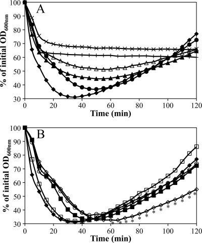
Spores lacking GSLEs in various combinations were germinated in liquid BHI and monitored by measuring the change in OD. Germinating spores rapidly lose approximately 50% of their OD in the first few minutes, and the OD then increases as the population continues into vegetative growth. As previously demonstrated (11, 18), spores of the wild-type strain and strains lacking any single GSLE were successful at synchronous germination, and those strains without functional CwlJ1, SleB, or SleL had delays in loss of OD during germination, though these did not always translate to a delay in the initiation of outgrowth (Fig. 2A and B). When both cwlJ1 and sleB were deleted, spores were capable of initiating germination and lost OD for only 10 min, but at that point, germination was arrested and no further changes were observed (Fig. 2A) (11). This was the most dramatic phenotype, and adding any number of additional GSLE-eliminating mutations to the ΔcwlJ1 ΔsleB background did not produce further significant changes (P > 0.05, as determined by a Tukey-Kramer honestly significant difference [HSD] analysis) (Fig. 2A and data not shown).
FIG. 2.
Germination and outgrowth of spores in BHI medium. Spores were heat activated in water and germinated in BHI medium at 37°C, and OD was monitored. Data shown are averages of the results for three independent spore preparations; error bars are omitted for clarity. (A) Germination of wild-type (⧫), ΔsleB (▴), ΔsleL (•), ΔsleB ΔsleL (Δ), ΔcwlJ1 ΔsleB (+), and ΔcwlJ1 ΔcwlJ2 ΔsleB ΔsleL (×) spores. (B) Germination of wild-type (⧫), ΔcwlJ1 (▪), ΔcwlJ2 (□), ΔcwlJ1 ΔcwlJ2 (⋄), and ΔcwlJ1 ΔsleL (○) spores. Asterisks indicate those time points at which ΔcwlJ1 ΔcwlJ2 spores were significantly different (P ≤ 0.01) from ΔcwlJ1 spores.
Adding a ΔsleL mutation to ΔcwlJ1, ΔsleB, ΔcwlJ1 ΔcwlJ2, and ΔsleB ΔcwlJ2 strains generated spores with increased delays in OD loss during germination that were evidenced by shallower curves (Fig. 2A and 2B and data not shown). Despite this consistent trend, these differences were not statistically significant (P > 0.05, as determined by a Tukey-Kramer HSD analysis), which is likely the result of variability inherent in assay of multiple independent spore preparations. Placing a ΔcwlJ2 deletion into any other GSLE mutant strains, whether single, double, or triple, did not impact the preexisting germination phenotypes except in the case of ΔcwlJ1 ΔcwlJ2 spores. This strain exhibited a slowed OD increase compared to ΔcwlJ1 spores with statistically significant delays from 60 min onward (P ≤ 0.01, as determined by a Tukey-Kramer HSD analysis) (Fig. 2B).
Effects of GSLEs on Ca2+-DPA-triggered germination.
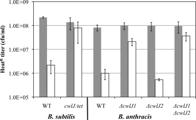
It was previously demonstrated that B. subtilis and B. megaterium spores that lack CwlJ are extremely unresponsive to the nonnutrient germinant Ca2+-DPA (28, 34). Spores that germinate in response to Ca2+-DPA become heat sensitive. For example, after treatment with Ca2+-DPA, our control wild-type B. subtilis spores lost a significant level of heat resistance, indicating that 99% of the spores had germinated, while B. subtilis spores carrying a cwlJ mutation were not affected (Fig. 3).
FIG. 3.
Germination of spores in response to Ca2+-DPA. Spores from B. subtilis strains PS832 (wild-type) and FB111 (ΔcwlJ::tet) (28), and B. anthracis strains DPBa2 (wild-type), DPBa61 (ΔcwlJ1), DPBa60 (ΔcwlJ2), and DPBa78 (ΔcwlJ1 ΔcwlJ2) were incubated in water (gray bars) or 50 mM Ca2+-DPA (pH 8.0) (white bars) for 60 min at 25°C before being heated at 70°C for 20 min, serially diluted, and plated onto BHI medium. Values are averages of at least three independent spore preparations. Error bars represent 1 standard deviation of the mean. WT, wild type.
Wild-type B. anthracis spores incubated with exogenous Ca2+-DPA exhibited a 99% decrease in heat-resistant CFU (Fig. 3). Surprisingly, ΔcwlJ1 spores were also significantly heat sensitive after Ca2+-DPA exposure, suffering a 79%-lowered titer. While ΔcwlJ2 spores performed similarly to those of the wild-type strain, spores lacking both cwlJ1 and cwlJ2 had no significant change in heat resistance after Ca2+-DPA treatment (Fig. 3). Phase-contrast microscopy of spores during their Ca2+-DPA incubation was also carried out, since germinating spores transition from phase bright to phase dark. Consistent with the heat resistance assay, 98% of wild-type and ΔcwlJ2 spores became phase dark after 60 min and 60% of ΔcwlJ1 spores transitioned, but only 6% of spores carrying both cwlJ1 and cwlJ2 deletions became phase dark (data not shown). Together, these observations show that, in B. anthracis spores, both CwlJ1 and CwlJ2 independently contribute to a Ca2+-DPA germination response and that CwlJ1 is responsible for the majority of the activity. Deletions of either sleB or sleL had no impact on the spore response to Ca2+-DPA (data not shown).
Release of cortex fragments from germinating spores.
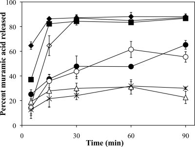
During germination, spores release cortex fragments that can be quantified based upon muramic acid and Dpm content. Data are presented only for muramic acid release, but in all cases, Dpm release was analyzed and paralleled the release of muramic acid (data not shown). Wild-type B. anthracis spores released nearly 90% of their muramic acid within 15 min of germination initiation (Fig. 4). The ΔcwlJ1 spores had an early delay in muramic acid release, but these spores still released nearly as much muramic acid as did those of the wild-type strain after 15 min (Fig. 4) (11). While the ΔcwlJ2 mutant was indistinguishable from the wild-type strain (11 and data not shown), ΔcwlJ1 ΔcwlJ2 spores exhibited an additive decrease in muramic acid release which was significant during the first 15 min of germination in comparison to both the wild-type and ΔcwlJ1 strains (Fig. 4). However, by 30 min, the double mutant was still able to release a normal amount of cortex fragments.
FIG. 4.
Release of cortex fragments from germinating spores. Dormant wild-type (⧫), ΔcwlJ1 (▪), ΔsleL (•), ΔcwlJ1 ΔsleL (○), ΔsleB ΔsleL (Δ), ΔcwlJ1 ΔcwlJ2 (⋄), and ΔcwlJ1 ΔcwlJ2 ΔsleB ΔsleL (×) spores in buffer were sampled for assay of the release of muramic acid following exposure to the germinants l-alanine and inosine. Error bars represent 1 standard deviation of the mean of the results for three independent spore preparations. All points have error bars, but in some cases, these are too small to be visible.
The ΔsleL mutation caused dramatic effects on muramic acid release (Fig. 4) (18). These spores released ≤40% of their muramic acid within 15 min, and even after 90 min expelled a maximum of only 65% of this cortex component. The ΔsleL ΔcwlJ1 double mutant spores displayed kinetics similar to those displayed by the ΔsleL single mutant over the course of the assay. However, ΔsleB ΔsleL spores were more severely impaired at discharging cortical fragments. While ΔsleB spores have only a minor delay in cortex release (11), ΔsleB ΔsleL double mutant spores released considerably less muramic acid throughout the assay than did those of the single mutants (Fig. 4) (11). In fact, the ΔsleB ΔsleL spores released muramic acid with kinetic similar to those of the quadruple mutant lacking all four GSLEs. These two strains released a maximum of ∼30% of their muramic acid; however, the ΔsleB ΔsleL spore accomplished this within 15 min, whereas the quadruple mutant required 60 min (Fig. 4).
GSLE effects on cortex hydrolysis.
Cortex structural changes during germination of spores lacking multiple GSLEs were analyzed using an RP-HPLC technique that was previously employed to identify the cortex lytic activities of SleB and SleL in B. anthracis spores (11, 18). In these earlier studies, spores lacking CwlJ2 were indistinguishable from those of the wild type. Spores of strains carrying mutations in cwlJ1, sleB, or sleL released smaller amounts of muropeptides, and in the cases of sleB and sleL, specific muropeptides indicative of those gene products' activities were lacking. It was shown that ΔcwlJ1 ΔsleB spores released essentially no measurable muropeptides, consistent with their germination block. For the present study, spores with a combination of cwlJ1 and sleB deletions were not investigated for muropeptide release due to this severe phenotype.
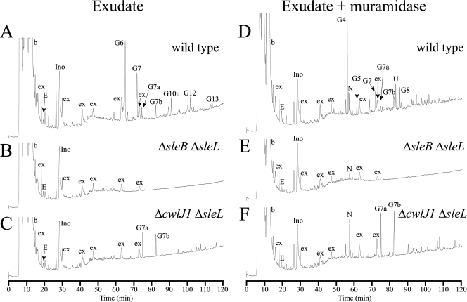
The muropeptides released from ΔcwlJ1 ΔsleL spores were anticipated to be the consequence of SleB's lytic transglycosylase activity. After germinating for 2 h, this strain released more than 60% of cortex PG into the exudate and, as expected, generated the lytic transglycosylase products aG7a and aG7b (Fig. 1 and 5B and Table 3) (11). Further digestion of the exudate with the muramidase mutanolysin increased the relative amounts of aG7a and aG7b by 44% and 114%, respectively, and generated a large amount of muropeptide N (Fig. 1 and 5E). Muropeptide N is a tetrasaccharide that is the predominant product produced by muramidase digestion of intact cortex strands (Table 3) (7). Based on the sizes of peaks aG7a, aG7b, and N released from ΔcwlJ1 ΔsleL spores, we calculate that 42% of SleB products are anhydrotetrasaccharides (aG7a and aG7b) and that the remaining 58% are larger fragments. Also, after muramidase digestion, the increase in anhydrotetrasaccharides compared to that in muropeptide N is in a ratio of nearly 1:1, which suggests that the average large muropeptide after SleB digestion is eight sugars in length (Fig. 1). Muramidase digestion of the PG retained in the germinated ΔcwlJ1 ΔsleL spore pellet released a small amount of aG7a and aG7b, demonstrating that a few SleB products remained too large to be released from the spore (data not shown).
FIG. 5.
RP-HPLC separation of muropeptides released from germinating spores. PG was prepared from germinating spore suspensions as described in Materials and Methods. Samples were collected after spores were allowed to germinate for 120 min. Fifty percent of each exudate (D to F) sample was digested with muramidase, reduced, and separated as previously described (22). The other 50% of each exudate (A to C) was reduced and separated without muramidase digestion. Peaks are numbered as in reference 11 and Table 3, but the initial “a” in the germination-specific peak names were omitted for space considerations. Early-eluting peaks labeled “b” are buffer components present in blank samples. Peaks labeled “ex” are spore exudate components that are not derived from PG. Peaks labeled “Ino” are from the inosine used as germinant.
TABLE 3.
Muropeptide peak identification
| Namea | Structureb | Enzymatic origin |
|---|---|---|
| N | TS-TP | Muramidase |
| U | HS-TP-Ac | Muramidase |
| aG4 | TriS-TP | N-acetylglucosaminidase + muramidase |
| aG5 | TriS-Ala | N-acetylglucosaminidase + muramidase |
| aG6 | TS-TP NAGr | N-acetylglucosaminidase |
| aG7 | TS-Ala NAGr | N-acetylglucosaminidase |
| aG7a | TS-TP anhydro | Lytic transglycosylase |
| aG7b | TS-Ala anhydro | Lytic transglycosylase |
| aG8 | PS-TP-Ac | N-acetylglucosaminidase + muramidase |
| aG10u | HS-TP-Ac NAGr | N-acetylglucosaminidase |
| aG12 | HS-Ala-Ac NAGr | N-acetylglucosaminidase |
| aG13 | HS-TP NAGr | Cortex, N-acetylglucosaminidase |
Muropeptide names are as previously published (7, 11). Muropeptide names preceded by “a” indicate those generated by B. anthracis in order to differentiate them from those generated by other species.
TS, tetrasaccharide (NAG-MδL-NAG-NAM); HS, hexasaccharide (NAG-MδL-NAG-MδL-NAG-NAM); TriS, trisaccharide (MδL-NAG-NAM); PS, pentasaccharide (MδL-NAG-MδL-NAG-NAM); TP, tetrapeptide (Ala-Glu-Dpm-Ala); Ac, deacetylated glucosamine; NAGr, NAG at the reducing end. “Anhydro” indicates that the NAM at the reducing end is in the anhydro form.
The chromatograms of spore germination exudates from ΔsleB ΔsleL (Fig. 5C) and ΔcwlJ2 ΔsleB ΔsleL strains (data not shown) were indistinguishable; any cortex fragments released from these strains are expected to be products of CwlJ1 activity. These spores released no detectable small muropeptides (Fig. 5C); however, treatment of the exudates with muramidase produced a small quantity of muropeptide N (Fig. 5F). This suggests that depolymerization of cortex catalyzed by CwlJ1 alone produced large muropeptides that are not resolved with this method. In fact, assays of released cortex fragments (Fig. 4) and of cortex released from the germinated spore pellet by muramidase (data not shown) indicate that ≥75% of CwlJ1-generated cortex fragments are too large to be released from the spore.
DISCUSSION
In the simplest measure of spore germination kinetics, the tracking of OD loss, slight delays in germination are observed in single mutants lacking cwlJ1, sleB, or sleL, and combinations of these mutations result in additive effects. In the cases of sleB and sleL mutants, germination is indistinguishable from that of the wild-type strain during the first 10 min, and delays are obvious only after that time. A cwlJ1 mutation affects OD loss even earlier, though it has the least effect on the overall kinetics of cortex hydrolysis and release, suggesting that this mutation has an additional effect on some other aspect of the germination process. A combination of cwlJ1 and sleB mutations renders spores essentially incapable of completing germination (10, 11); cortex hydrolysis is completely blocked, and OD decrease ceases after 10 min. This indicates that OD decrease during the first 10 min of germination is primarily associated with release of spore solutes and uptake of water, with accompanying loss of spore refractility and decrease in spore core density. Studies of other species demonstrated that cwlJ mutations slowed the process of Ca2+-DPA release (13, 30, 34), and we suggest that the early delay in OD loss in a cwlJ1 mutant is due to a slowed process of spore solute exchange. How might a cortex lytic enzyme affect solute movement into and out of the spore? We propose that CwlJ1 (and CwlJ2) acts initially on the outer layers of the cortex, due to their apparent location at or near the cortex/coat interface (3, 6, 11, 17). Outer cortex layers are more highly cross-linked than the inner layers (22) and thus may produce a slightly greater diffusion barrier and/or might exert a greater influence on the ability of the spore core to expand and take up water in the first moments of germination. Such water uptake may be critical for solubilization and movement of Ca2+-DPA, for dissociation of SASP proteins from the spore DNA, and for activation of a germination-specific protease in the core (35, 36).
Giebel et al. found that spores of a cwlJ2 mutant lost OD slightly more slowly than did those of the wild-type strain during germination, but with an overall effect that was less dramatic than that seen with those of cwlJ1 and sleB mutants (10). While our analyses with single mutants have never demonstrated a significant phenotypic change due to a single cwlJ2 mutation (11), we and Giebel et al. (10) did observe that a cwlJ2 mutation had additive effects to those of a cwlJ1 mutation. In particular, we find that cwlJ1 cwlJ2 spores exhibit a delay in outgrowth that is significantly greater than that which might be expected based on the relative delays in OD loss and cortex fragment release among all the mutant strains analyzed. We also found that cwlJ2 plays a role in Ca2+-DPA-stimulated germination. Studies of other species have demonstrated roles of CwlJ proteins in response to this nonnutrient germinant, and release of Ca2+-DPA from the germinating spore is apparently the mechanism by which this lytic enzyme is normally activated (28). In B. anthracis, Ca2+-DPA-triggered germination utilizes CwlJ1 and CwlJ2 working independently and with partial redundancy, since loss of both enzymes is required to completely eliminate the response. Low expression of cwlJ2 (11) may produce spores with an insufficient amount of CwlJ2 to respond to Ca2+-DPA in the absence of CwlJ1. We assert that CwlJ1 and CwlJ2 carry out identical functions given, as follows: (i) the high level of sequence identity (58%) between the proteins; (ii) that deletion of the former is needed in order to demonstrate activity for the latter; and (iii) the requirement of both enzymes for maximum response to Ca2+-DPA.
Colony formation is unaffected in B. anthracis spores that contain at least SleB or CwlJ1 (10, 11), supporting the idea that either one of these SCLEs is sufficient to degrade the cortex to a great enough degree that metabolic activity resumes and the cell can grow out of its integuments. When both SleB and CwlJ1 are absent from spores, in strains with or without other GSLEs present, we observe a 1,000-fold decrease in colony-forming efficiency (Table 2) (11). In each case, colony-forming ability can be rescued by the addition of lysozyme, indicating that the block to germination is due to incomplete cortex hydrolysis. Contrary to our results, a recent report indicated that the additional loss of CwlJ2 from a ΔcwlJ1 ΔsleB double mutant resulted in a much more profound loss of colony-forming efficiency (10). To explain this difference, we must ask why 0.1% of spores that lack all known GSLEs are able to produce colonies in our studies, but not in those of Giebel et al. One potential explanation is that some vegetative cell-derived lytic enzyme copurified with our spores and might be hydrolyzing the cortex PG enough to allow a small percentage of spores to complete germination. A contribution of vegetative lytic activities to germination of defective C. perfringens spores has been postulated (29). However, our decoating procedure, which should inactivate spore surface-associated enzymes, did not significantly reduce the colony-forming ability of ΔcwlJ1 ΔsleB spores (data not shown). An alternative explanation is that small amounts of exogenous lytic enzymes in our growth media might be hydrolyzing the cortex PG, despite the fact that we have used BHI medium from the same manufacturer as that used by Giebel et al. (10). The potential for exogenous lytic enzymes to allow germination of GSLE-deficient spores was also cited as an explanation for the virulence of these spores at a level greater than that which would be expected from their colony-forming efficiency on laboratory medium (10).
Cortex fragment release from germinating B. anthracis spores follows three different kinetic paths, depending on which proteins are actively digesting the cortex PG. Maximum total release occurs when both SleB and SleL are functional, and CwlJ1 and CwlJ2 affect only the initial rate of release. SleB digests the PG into fragments of various sizes, some of which are released, but the majority of which are initially retained within the spore. SleL then acts on these fragments, thus increasing the proportion that is small enough for rapid release. In the absence of SleL, muropeptides found in the exudates are significantly reduced in quantity. Given enough time, SleB alone can digest the PG to fragments small enough, primarily tetra- and octasaccharides, for release of >50% of the cortex from the spore. In the absence of both SleB and SleL, cortex degradation can still be accomplished by CwlJ1 and CwlJ2 to allow outgrowth. However, the cortex fragments are apparently so large that they are not released, and muramic acid release by sleB sleL spores is indistinguishable from that of spores lacking all four GSLEs.
The fact that the products of CwlJ1 activity are so large has prevented determination of the site of PG cleavage by this enzyme. Muramidase digestion of germinated cwlJ2 sleB sleL spore PG, which presumably has been cleaved only by CwlJ1, has yielded only muramidase products (data not shown). This suggests either that CwlJ1 is a muramidase or that its cleavage sites are so few as to be undetectable by our current methods. The possibility of CwlJ1 muramidase activity is consistent with the significant sequence homology between CwlJ and SleB lytic transglycosylase proteins. Lytic transglycosylases and muramidases cleave the same bond in PG, but differ in the chemistry of their products, the former producing anhydro-N-acetylmuramic acid and the latter producing N-acetylmuramic acid. Similar protein folds can result in these two enzymatic activities (37). Ongoing efforts to produce significant CwlJ1 activity in vitro may allow cleavage of a purified substrate to an extent sufficient to directly identify the enzymatic products.
While the roles played by the B. anthracis GSLEs and their requirements for germination both in and out of the host (10, 11, 18) are becoming clear, many questions remain concerning the mechanisms by which they are held inactive in the dormant spore and activated during germination. Future studies of the localization, processing, and interaction partners of these enzymes may answer these questions. A strategy for efficient external activation of GSLEs, and therefore, initiation of germination, will allow the development of simpler methods for decontamination of sites of spore release.
Acknowledgments
This research was supported by Public Health Service grant AI060726 from the National Institute of Allergy and Infectious Disease.
We thank P. Setlow for providing strains. We thank Mark Seiss and Chongrui Yu at the VT Laboratory for Interdisciplinary Statistical Analysis for their advice.
Footnotes
Published ahead of print on 4 December 2009.
REFERENCES
- 1.Albrink, W. S. 1961. Pathogenesis of inhalation anthrax. Bacteriol. Rev. 25:268-273. [DOI] [PMC free article] [PubMed] [Google Scholar]
- 2.Atrih, A., P. Zollner, G. Allmaier, and S. J. Foster. 1996. Structural analysis of Bacillus subtilis 168 endospore peptidoglycan and its role during differentiation. J. Bacteriol. 178:6173-6183. [DOI] [PMC free article] [PubMed] [Google Scholar]
- 3.Bagyan, I., and P. Setlow. 2002. Localization of the cortex lytic enzyme CwlJ in spores of Bacillus subtilis. J. Bacteriol. 184:1219-1224. [DOI] [PMC free article] [PubMed] [Google Scholar]
- 4.Chen, Y., S. Fukuoka, and S. Makino. 2000. A novel spore peptidoglycan hydrolase of Bacillus cereus: biochemical characterization and nucleotide sequence of the corresponding gene, sleL. J. Bacteriol. 182:1499-1506. [DOI] [PMC free article] [PubMed] [Google Scholar]
- 5.Chen, Y., S. Miyata, S. Makino, and R. Moriyama. 1997. Molecular characterization of a germination-specific muramidase from Clostridium perfringens S40 spores and nucleotide sequence of the corresponding gene. J. Bacteriol. 179:3181-3187. [DOI] [PMC free article] [PubMed] [Google Scholar]
- 6.Chirakkal, H., M. O'Rourke, A. Atrih, S. J. Foster, and A. Moir. 2002. Analysis of spore cortex lytic enzymes and related proteins in Bacillus subtilis endospore germination. Microbiology 148:2383-2392. [DOI] [PubMed] [Google Scholar]
- 7.Dowd, M. M., B. Orsburn, and D. L. Popham. 2008. Cortex peptidoglycan lytic activity in germinating Bacillus anthracis spores. J. Bacteriol. 190:4541-4548. [DOI] [PMC free article] [PubMed] [Google Scholar]
- 8.Foster, S. J., and K. Johnstone. 1987. Purification and properties of a germination-specific cortex-lytic enzyme from spores of Bacillus megaterium KM. Biochem. J. 242:573-579. [DOI] [PMC free article] [PubMed] [Google Scholar]
- 9.Gerhardt, P., and R. E. Marquis. 1989. Spore thermoresistance mechanisms, p. 43-63. In I. Smith, R. A. Slepecky, and P. Setlow (ed.), Regulation of prokaryotic development. American Society for Microbiology, Washington, DC.
- 10.Giebel, J. D., K. A. Carr, E. C. Anderson, and P. C. Hanna. 2009. Germination-specific lytic enzymes SleB, CwlJ1, and CwlJ2 each contribute to Bacillus anthracis spore germination and virulence. J. Bacteriol. 191:5569-5576. [DOI] [PMC free article] [PubMed] [Google Scholar]
- 11.Heffron, J. D., B. Orsburn, and D. L. Popham. 2009. Roles of germination-specific lytic enzymes CwlJ and SleB in Bacillus anthracis. J. Bacteriol. 191:2237-2247. [DOI] [PMC free article] [PubMed] [Google Scholar]
- 12.Hu, K., H. Yang, G. Liu, and H. Tan. 2007. Cloning and identification of a gene encoding spore cortex-lytic enzyme in Bacillus thuringiensis. Curr. Microbiol. 54:292-295. [DOI] [PubMed] [Google Scholar]
- 13.Ishikawa, S., K. Yamane, and J. Sekiguchi. 1998. Regulation and characterization of a newly deduced cell wall hydrolase gene (cwlJ) which affects germination of Bacillus subtilis spores. J. Bacteriol. 180:1375-1380. [DOI] [PMC free article] [PubMed] [Google Scholar]
- 14.Janes, B. K., and S. Stibitz. 2006. Routine markerless gene replacement in Bacillus anthracis. Infect. Immun. 74:1949-1953. [DOI] [PMC free article] [PubMed] [Google Scholar]
- 15.Kim, H. U., and J. M. Goepfert. 1974. A sporulation medium for Bacillus anthracis. J. Appl. Bacteriol. 37:265-267. [DOI] [PubMed] [Google Scholar]
- 16.Kodama, T., H. Takamatsu, K. Asai, K. Kobayashi, N. Ogasawara, and K. Watabe. 1999. The Bacillus subtilis yaaH gene is transcribed by SigE RNA polymerase during sporulation, and its product is involved in germination of spores. J. Bacteriol. 181:4584-4591. [DOI] [PMC free article] [PubMed] [Google Scholar]
- 17.Lai, E. M., N. D. Phadke, M. T. Kachman, R. Giorno, S. Vazquez, J. A. Vazquez, J. R. Maddock, and A. Driks. 2003. Proteomic analysis of the spore coats of Bacillus subtilis and Bacillus anthracis. J. Bacteriol. 185:1443-1454. [DOI] [PMC free article] [PubMed] [Google Scholar]
- 18.Lambert, E. A., and D. L. Popham. 2008. The Bacillus anthracis SleL (YaaH) protein is an N-acetylglucosaminidase involved in spore cortex depolymerization. J. Bacteriol. 190:7601-7607. [DOI] [PMC free article] [PubMed] [Google Scholar]
- 19.Liu, H., N. H. Bergman, B. Thomason, S. Shallom, A. Hazen, J. Crossno, D. A. Rasko, J. Ravel, T. D. Read, S. N. Peterson, J. Yates III, and P. C. Hanna. 2004. Formation and composition of the Bacillus anthracis endospore. J. Bacteriol. 186:164-178. [DOI] [PMC free article] [PubMed] [Google Scholar]
- 20.Makino, S., N. Ito, T. Inoue, S. Miyata, and R. Moriyama. 1994. A spore-lytic enzyme released from Bacillus cereus spores during germination. Microbiology 140:1403-1410. [DOI] [PubMed] [Google Scholar]
- 21.Makino, S., and R. Moriyama. 2002. Hydrolysis of cortex peptidoglycan during bacterial spore germination. Med. Sci. Monit. 8:RA119-127. [PubMed] [Google Scholar]
- 22.Meador-Parton, J., and D. L. Popham. 2000. Structural analysis of Bacillus subtilis spore peptidoglycan during sporulation. J. Bacteriol. 182:4491-4499. [DOI] [PMC free article] [PubMed] [Google Scholar]
- 23.Miyata, S., R. Moriyama, N. Miyahara, and S. Makino. 1995. A gene (sleC) encoding a spore-cortex-lytic enzyme from Clostridium perfringens S40 spores; cloning, sequence analysis and molecular characterization. Microbiology 141:2643-2650. [DOI] [PubMed] [Google Scholar]
- 24.Mock, M., and A. Fouet. 2001. Anthrax. Annu. Rev. Microbiol. 55:647-671. [DOI] [PubMed] [Google Scholar]
- 25.Moriyama, R., A. Hattori, S. Miyata, S. Kudoh, and S. Makino. 1996. A gene (sleB) encoding a spore cortex-lytic enzyme from Bacillus subtilis and response of the enzyme to l-alanine-mediated germination. J. Bacteriol. 178:6059-6063. [DOI] [PMC free article] [PubMed] [Google Scholar]
- 26.Moriyama, R., S. Kudoh, S. Miyata, S. Nonobe, A. Hattori, and S. Makino. 1996. A germination-specific spore cortex-lytic enzyme from Bacillus cereus spores: cloning and sequencing of the gene and molecular characterization of the enzyme. J. Bacteriol. 178:5330-5332. [DOI] [PMC free article] [PubMed] [Google Scholar]
- 27.Nicholson, W. L., and P. Setlow. 1990. Sporulation, germination, and outgrowth, p. 391-450. In C. R. Hartwood and S. M. Cutting (ed.), Molecular biological methods for Bacillus. John Wiley and Sons Ltd., Chichester, England.
- 28.Paidhungat, M., K. Ragkousi, and P. Setlow. 2001. Genetic requirements for induction of germination of spores of Bacillus subtilis by Ca2+-dipicolinate. J. Bacteriol. 183:4886-4893. [DOI] [PMC free article] [PubMed] [Google Scholar]
- 29.Paredes-Sabja, D., P. Setlow, and M. R. Sarker. 2009. SleC is essential for cortex peptidoglycan hydrolysis during germination of spores of the pathogenic bacterium Clostridium perfringens. J. Bacteriol. 191:2711-2720. [DOI] [PMC free article] [PubMed] [Google Scholar]
- 30.Peng, L., D. Chen, P. Setlow, and Y. Q. Li. 2009. Elastic and inelastic light scattering from single bacterial spores in an optical trap allows the monitoring of spore germination dynamics. Anal. Chem. 81:4035-4042. [DOI] [PMC free article] [PubMed] [Google Scholar]
- 31.Popham, D. L., J. Helin, C. E. Costello, and P. Setlow. 1996. Analysis of the peptidoglycan structure of Bacillus subtilis endospores. J. Bacteriol. 178:6451-6458. [DOI] [PMC free article] [PubMed] [Google Scholar]
- 32.Popham, D. L., J. Helin, C. E. Costello, and P. Setlow. 1996. Muramic lactam in peptidoglycan of Bacillus subtilis spores is required for spore outgrowth but not for spore dehydration or heat resistance. Proc. Natl. Acad. Sci. USA 93:15405-15410. [DOI] [PMC free article] [PubMed] [Google Scholar]
- 33.Setlow, B., E. Melly, and P. Setlow. 2001. Properties of spores of Bacillus subtilis blocked at an intermediate stage in spore germination. J. Bacteriol. 183:4894-4899. [DOI] [PMC free article] [PubMed] [Google Scholar]
- 34.Setlow, B., L. Peng, C. A. Loshon, Y. Q. Li, G. Christie, and P. Setlow. 2009. Characterization of the germination of Bacillus megaterium spores lacking enzymes that degrade the spore cortex. J. Appl. Microbiol. 107:318-328. [DOI] [PubMed] [Google Scholar]
- 35.Setlow, P. 2007. I will survive: DNA protection in bacterial spores. Trends Microbiol. 15:172-180. [DOI] [PubMed] [Google Scholar]
- 36.Setlow, P. 2003. Spore germination. Curr. Opin. Microbiol. 6:550-556. [DOI] [PubMed] [Google Scholar]
- 37.Thunnissen, A. M., N. W. Isaacs, and B. W. Dijkstra. 1995. The catalytic domain of a bacterial lytic transglycosylase defines a novel class of lysozymes. Proteins 22:245-258. [DOI] [PubMed] [Google Scholar]
- 38.Warth, A. D., and J. L. Strominger. 1972. Structure of the peptidoglycan from spores of Bacillus subtilis. Biochemistry 11:1389-1396. [DOI] [PubMed] [Google Scholar]


