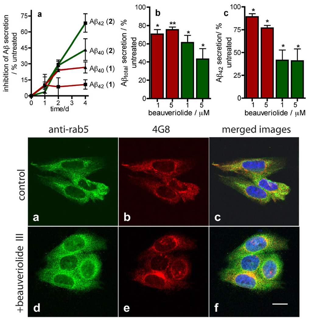
Figure 3.
Depsipeptides 1 and 2 reduce Aβ secretion from the CHO cell-line 7WD10 and do not perturb trafficking of Aβ. UPPER a Graph of time-dependent changes in Aβ40 (▲) and Aβ42 (■) secretion by 7WD10 cells when incubated with either 1 ( ) or 2 (
 ) (1 µM) as determined by ELISA analysis of Aβ40 and Aβ42 in conditioned media standardized to total protein. b Concentration-dependence of 1 (red bars) and 2 (green bars) on Aβtotal secretion from the 7WD10 cell line. c Concentration-dependence of 1 (red bars) and 2 (green bars) on Aβ42 secretion from the 7WD10 cell line. Data in a–c is expressed as the mean value ± SEM of at least triplicate separate experiments and is recorded as a percent of vehicle treated cells (0.1 % DMSO v/v). Statistical analysis was performed using a student t test and significance *P < 0.05 and **P < 0.01. LOWER Confocal microscopy of 7WD10 cells incubated with either vehicle (0.1 % DMSO, v/v) (a–c) or beauveriolide III (1 µM) (d–f) for 48 h. Cells were fixed with paraformaldehyde and immunolabeled with either a rabbit anti-Rab5 antibody with an Alexa-fluor488 (green)-labeled 20 antibody (a, d), or the murine anti-Aβ antibody 4G8, with an Alexafluor 543-labeled (red) 20 antibody (b, e). Nuclei were stained with 4',6-diamidino-2-phenylindole (DAPI). Images were recorded with a Zeiss LSM510 confocal microscope at 63 × and image analysis and merges were performed with LSM image examiner software (4.2). Note: yellow color in the merged images (c, f) corresponds to co-localized Rab5 and Aβ; Bar = 10 µm.

