Abstract
The present study had three goals: (i) to evaluate the relative quantities of aerosolized Bacillus atrophaeus spores deposited on the vertical, horizontal top, and horizontal bottom surfaces in a chamber; (ii) to assess the relative recoveries of the aerosolized spores from glass and stainless steel surfaces with a polyester swab and a macrofoam sponge wipe; and (iii) to estimate the relative recovery efficiencies of aerosolized B. atrophaeus spores and Pantoea agglomerans using a foam spatula at several different bacterial loads by aerosol distribution on glass surfaces. The majority of spores were collected from the bottom horizontal surface regardless of which swab type and extraction protocol were used. Swabbing with a macrofoam sponge wipe was more efficient in recovering spores from surfaces contaminated with high bioaerosol concentrations than swabbing with a polyester swab. B. atrophaeus spores and P. agglomerans culturable cells were detected on glass surfaces using foam spatulas when the theoretical surface bacterial loads were 2.88 × 10⁴ CFU and 8.09 × 10⁶ CFU per 100-cm2 area, respectively. The median recovery efficiency from the surfaces using foam spatulas was equal to 9.9% for B. atrophaeus spores when the recovery was calculated relative to the theoretical surface spore load. Using a foam spatula permits reliable sampling of spores on the bioaerosol-exposed surfaces in a wide measuring range. The culturable P. agglomerans cells were recovered with a median efficiency of 0.001%, but staining the swab extracts with fluorescent dyes allowed us to observe that the viable cell numbers were higher by 1.83 log units than culturable organisms. However, additional work is needed to improve the analysis of the foam extracts in order to decrease the limit of detection of Bacillus spores and Gram-negative bacteria on contaminated surfaces.
Surface sampling is performed on a frequent basis in all situations where clean environment monitoring is needed, e.g., in health care facilities and in the pharmaceutical industry and food industry. An anthrax bioterrorist event in the fall of 2001 has emphasized the importance of efficient sampling methods for detection of pathogenic microorganisms on surfaces within intentionally contaminated locations (22). Unfortunately, our knowledge on the most effective sampling methodology as well as the level of confidence we may have in the results obtained by wiping, swabbing, and other sample collection strategies is still limited (1). Moreover, in most of the studies performed so far, bacteria and/or spores were collected from test samples or coupons of various materials, inoculated with a suspension of microorganisms that had been placed and spread over the surface, and then dried (14, 15). This may not mimic the true situation of surface contamination by a pathogen that has been intentionally released. Edmonds et al. (12) recently reported lower swabbing efficiencies of different types of swab materials used for sampling glass, polycarbonate, and vinyl surfaces contaminated with dry aerosol-deposited Bacillus atrophaeus spores compared to the surfaces inoculated by spore suspensions. Solid surface contamination from exposure to aerosolized spores fits the real world better than the previous models.
Therefore, in our study we decided to generate aerosols of various concentrations of B. atrophaeus spores as well as the vegetative cells of Pantoea agglomerans inside a chamber where the bioaerosol particles were allowed to gravitationally settle on solid surfaces. The aerosolization of P. agglomerans was performed to verify the recovery of Gram-negative bacteria according to the recommendations of Budowle et al. (5). The main goal of our study was to establish the range of detection when bioaerosol-contaminated surfaces were swabbed using a commercially available foam spatula.
MATERIALS AND METHODS
Bacteria, growth conditions, and spore preparation.
Bacillus atrophaeus ATCC 9372 and Pantoea agglomerans ATCC 33243 were used in this study. The bacterial strains were kept as reference stocks at a temperature of −20°C. B. atrophaeus was grown on a solid 2×SG sporulation medium (17) for 5 days at a temperature of 35°C, followed by culture at room temperature. The extent of sporulation was monitored by phase-contrast microscopy. The cultures were stopped once 85 to 90% of the bacilli produced spores. The mixture of spores and bacteria in Roux bottles were agitated twice with glass beads in 10 ml of sterile deionized water (SDW) and then transferred to new tubes. The spores were purified from cell debris by the method of Caipo et al. (8). Briefly, lysozyme (Sigma-Aldrich, St. Louis, MO) solution (50 μg per ml) in 0.05 mol liter−1 Tris-HCl (pH 7.2) was added, and the spores were incubated overnight. The liberated spores were purified by several washes (three to five) with cold SDW. The spore slurry was stored in 50% ethyl alcohol at a temperature of 4°C. Before each experiment, the spores were washed three times in SDW, and the optical density of the spore suspension was adjusted to 1.0 when it was read at a wavelength of 600 nm with a SP6-500 UV spectrophotometer (Pye Unicam Ltd., Cambridge, England). Appropriate dilutions were made to achieve the desired spore concentrations in the suspensions for aerosol generation. The spores in the suspensions were placed on ice and subjected to five 10-s bursts of high-power ultrasound with a 30-s break between bursts; the high-power ultrasound was generated by an ultrasonic disintegrator type UD-11 Techpan (Poland) with a frequency of 22 kHz and power of 100 W. The number of viable spores was counted in a suspension after sonication by plating the spores onto Luria-Bertani (LB) agar (Sigma-Aldrich, St. Louis, MO). P. agglomerans was grown without shaking in nutrient broth (Oxoid, Ltd., Basingstoke, Hampshire, England) for 18 h at a temperature of 35°C. The liquid culture was centrifuged at 4,000 rpm for 15 min. The supernatant was removed, and the bacterial pellet was washed by centrifugation three times with a sterile saline solution. Finally, the bacterial pellet was resuspended in sterile saline solution to an optical density value of 0.6 at 600 nm, the appropriate dilutions were made, and the number of viable bacterial cells was estimated by plating.
Surface materials.
Glass plates (Glasspoint, Wadowice, Poland) and stainless steel plates (Investa, Warsaw, Poland) with the dimensions 20 by 20 cm (400 cm2) were used as surface materials and served as test samples or coupons on which B. atrophaeus spores were deposited. The stainless steel was grade 1.4301 according to European standard EN 10088, which corresponds to grade 304 according to the standard of the American Society for Testing Materials, and the steel roughness was 0.1 to 0.5 μm. The plates were divided by pen markings into four parts (quadrants) with the dimensions 10 by 10 cm (100 cm2), which were used for surface sample collection. To avoid the possibility of cross-contamination, only one quadrant on one plate was swabbed. In the experiments in which horizontal and vertical surfaces were tested, the two diagonal quadrants of the plate were swabbed. The plates (made of glass or steel) were washed with 70% ethyl alcohol, rinsed in SDW, air dried, and sterilized with an autoclave at 121°C.
Bioaerosol deposition system.
Bioaerosols were generated in a test chamber (model 830-ABB/Sp with 800-HEPA/D; Plas-Labs, Inc., Lansing, MI). The chamber was constructed of clear cast acrylic with an interior volume of 0.5 m3. The dimensions of the bottom surface were 89 cm wide by 74 cm wide. Bioaerosols were generated by a compressed-air nebulizer Monsun 2 MP2 equipped with a RF6 head (Medbryt, Warsaw, Poland). The nebulizer was placed outside the chamber and was connected with tubing with the RF6 head through a valve and HEPA filter (with a diameter of 5.5 cm). The RF6 head was placed 65 cm above the test surface. Ten-milliliter samples of a B. atrophaeus spore suspension in SDW or a P. agglomerans suspension in 0.01 M phosphate-buffered saline (PBS), pH 7.4, were aerosolized at 46.4 lb/in2 pressure, an airflow rate of 15.5 liters/min, and a liquid generation rate of 0.48 ml/min. According to the manufacturer, the nebulizer generates airborne particles with a mass median aerodynamic diameter of 1.4 μm. The aerosolized B. atrophaeus spores were allowed to gravitationally settle for 16 h onto glass and stainless steel surfaces placed in a test chamber, and P. agglomerans cells were allowed to deposit for 1 h on glass surfaces only. Additionally, wet-surface collectors made of polystyrene with the dimensions of 9 by 9 cm by 2 cm deep, containing 15 ml of SDW (for collection of spores) or PBS (for collection of vegetative cells) were placed side by side on the surfaces tested on the bottom of the test chamber. The collectors and test surfaces were placed exactly in the middle of the bottom surface of the chamber. The wet-surface collectors were used to quantify without swabbing the number of microorganisms that settle on the surface in the time span of the experiments and to avoid the possible loss of the bacteria that adhered to the swabbing material. After each trial, the chamber was decontaminated with PeraSafe (Antec International, DuPont, Sudbury, Suffolk, England) and rinsed with water, and before each experiment, a UVC lamp (Puritec LPS9; OSRAM, GmbH, Augsburg, Germany) was switched on inside the chamber for 1 h. Then, interior air was exchanged through a HEPA filtration system for 30 min. All trials were conducted at room temperature (20 to 23°C).
Surface sampling and sample processing.
A foam spatula (catalog no. 939CS01; Copan Diagnostics Inc., Corona, CA), a polyurethane macrofoam sponge wipe (LM) (Ligasano; Ligamed Medical Producte GmbH, Germany), and a polyester swab (CP) (catalog no. 159C; Copan Diagnostics Inc., Corona, CA), were used as swab materials. The original foam attached to the spatula was too wide to fit within a Falcon tube, so the foam tip was aseptically cut to a width of 2.5 cm. The LM macrofoam sponge wipe was cut to the dimensions of 3.0 by 5.0 cm and was held by forceps for sampling. All materials tested were premoistened with SDW or PBS in a sterile 50-ml Falcon screw-top tube. The surfaces (100 cm2) were swabbed in a horizontal direction and then, with the unused side of the materials, were sampled in a vertical direction. The macrofoam sponge wipe and the spatula tip were placed in a sterile stomacher bag (Seward Ltd., Great Britain) that contained 10 ml of SDW for spore extraction or PBS for extraction of P. agglomerans cells, respectively. The macrofoam sponge wipe and foam spatula were hand mixed for 1 min, and then they were squeezed with sterile forceps and removed. The polyester swabs were placed in tubes containing 5-ml volumes of SDW and sonicated in a Sonic-6 ultrasonic bath (Polsonic, Warsaw, Poland) for 12 min (at a frequency of 40 kHz and an average power of 200 W) or vortexed for 1 min for extraction of B. atrophaeus. The volume of the liquid samples was measured, and the swabs were discarded. The suspensions were serially diluted in SDW (for B. atrophaeus spores) or PBS (for P. agglomerans) prior to inoculation onto LB agar. The plates were incubated at 35°C for 24 h (B. atrophaeus) or at 35°C for 1 to 3 days (P. agglomerans). The colonies were counted, and the final results were expressed as the number of CFU per milliliter.
Bioaerosol deposition on horizontal and vertical surfaces inside a chamber.
Glass and stainless steel plates were fixed to the hand-made plastic frame (dimensions, 66 by 47 by 67 cm) in three positions: horizontal top (steel), vertical (glass), and horizontal bottom (steel) in the center of the test chamber. A B. atrophaeus spore suspension at a concentration of 1 × 10⁸ to 2 × 10⁸ CFU/ml was aerosolized as described above. After 16 h, the aerosol-deposited B. atrophaeus spores were collected with CP swab and LM macrofoam sponge wipe from six vertical, top horizontal, and bottom horizontal surfaces of 100 cm2 each. The swabs were processed as described above, extracts were diluted and plated, and the number of CFU was determined. The experiment was repeated two times, and the average number of CFU (± standard deviation [SD]) per 100 cm2 was calculated.
Surface sampling of spores and bacteria aerosolized at various concentrations.
Suspensions of B. atrophaeus spores at concentrations ranging from 2.6 × 10⁵ to 4.7 × 10⁸ CFU per ml and suspensions of P. agglomerans at concentrations ranging from 7.2 × 10⁷ to 1.4 × 10⁹ CFU per ml in a total volume of 10 ml were aerosolized inside the test chamber as described above. In each experiment, 16 h after spore aerosolization or 1 h after P. agglomerans aerosolization, the 100-cm2 surfaces of four glass plates, placed at the bottom of the test chamber, were swabbed with the foam spatulas. Four wet-surface collectors containing 15 ml of SDW (for B. atrophaeus collection) or PBS (for P. agglomerans collection) were placed adjacent to the glass plates and were opened throughout the experiments. The wet-surface collectors were tightly closed before the start of a swabbing procedure. The spores or bacteria extracted from the swab materials in the stomacher bags and the suspensions from wet-surface collectors were serially diluted and spread on LB agar plates. Five repetitions were done of each dilution series. The plates were incubated at 35°C for 24 h (B. atrophaeus) or at 35°C for 1 to 3 days (P. agglomerans). Colonies were counted, and the numbers of CFU per 100 cm2 were determined.
Direct epifluorescence filter technique.
One milliliter of the suspended P. agglomerans organisms collected 1 h after aerosolization from glass surfaces with CP swabs (eight surfaces of 100 cm2 were swabbed) and from 10 wet-surface collectors was stained by using a Live/Dead BacLight bacterial viability kit (Invitrogen, Eugene, OR) according to the manufacturer's instructions. The samples were incubated for 30 min in the dark and filtered with black polycarbonate membrane filters (25-mm diameter, 0.22-μm pore size; Invitrogen, Eugene, OR) on filter apparatus (Millipore, Bedford, MA). The filters were immobilized by BacLight mounting oil on a glass microscope slide under a coverslip and were observed with a Nikon Eclipse E200 epifluorescence microscope with the appropriate filter block for Syto-9 fluorescent nucleic acid stain (EX450-490, DM505, and BA520) and filter block for propidium iodide fluorescent nucleic acid stain (EX510-560, DM575, and BA590). Pictures were taken using a Nikon DS-Fi1-U2 camera equipped with Nikon NISF software. The total number of bacteria with intact cell membranes (fluorescent green) and bacteria with damaged cell membranes (fluorescent red) was estimated by the method of Mesa et al. (18).
Statistical analysis.
A nonparametric Kruskal-Wallis test followed by a posthoc test (Statistica v. 8.0 for Windows) was used to compare the number of spores deposited on horizontal and vertical surfaces and collected with LM macrofoam sponge wipes and CP swabs. The relative percentage (RD) of spores collected from four vertical or horizontal top surfaces compared to the horizontal bottom surface was calculated as follows: %RD = (Σ[DVT/NH]/n) × 100, where NH is the number of spores collected from the horizontal bottom surface of the test chamber, DVT is the number of spores collected from the entire vertical or horizontal top surface of the test chamber, and n is the sample size. Linear regression lines were automatically drawn using a software graphics package (SigmaPlot; Jandel, San Rafael, CA) for the number of spores and bacteria collected by the foam spatulas or deposited in wet-surface collectors versus the various concentrations of the suspensions used to produce aerosols. The percent recovery efficiencies (REs) of B. atrophaeus spores and P. agglomerans cells collected with foam spatulas were calculated according to the following equation (22): %RE = (Σ[NSW/N0]/n) × 100, where N0 is the theoretical surface organism load and is equal to the number of bioparticles that may be present on a 100-cm2 area when the total number of viable aerosolized spores or bacteria (with an acknowledgment of the potential losses on the vertical and horizontal top surfaces) would settle on the horizontal bottom surface in each experiment, NSW is the number of CFU from the foam spatula or from the wet-surface collector, and n is the sample size. The median percent recovery efficiencies of B. atrophaeus spores using the foam spatulas and in wet-surface collectors were compared using the Mann-Whitney U test (Statistica v. 8.0 for Windows).
RESULTS
Bioaerosol deposition within the test chamber.
The experiments were performed to determine the relative quantities of spores that are deposited on the vertical, horizontal top, and horizontal bottom surfaces in a chamber when the organisms were aerosolized within the test chamber. As shown in Table 1, the majority of spores were collected from the bottom horizontal surface, regardless of which swab type and extraction protocol were used. The average recovery of spores from 100-cm2 areas of vertical surfaces using LM sponge wipes and CP swabs was 7.9% of the number of CFU collected from 100 cm2 of the horizontal bottom surface (100%). The lowest number of spores (an average of 0.6%) was collected using swabs from 100-cm2 areas of the horizontal top surfaces relative to CFU recovered from 100 cm2 of the horizontal bottom surface (100%). The number of CFU collected using LM sponge wipes was significantly higher than that using CP swabs when sampling was performed on horizontal bottom and vertical surfaces. When the number of viable spores recovered from an area of 100 cm2 dropped below 3 × 10⁴ CFU, as was observed for samples from the horizontal top surface, the differences between the numbers of CFU from two different swab materials were not more significant. No difference in the recovery efficiency was observed between two agitation methods (sonication and vortexing) used for extracting spores from CP swabs. When the adjustment of the potential losses on four vertical surfaces and the horizontal top surface was taken into account, then the theoretical surface spore load, the number of spores that might be deposited on the entire horizontal bottom surface, was estimated to be 74.17% of the total number of the spores aerosolized in the test chamber used in this study.
TABLE 1.
Deposition of aerosolized B. atrophaeus spores on horizontal and vertical surfaces in the test chambera
| Surface | No. of CFU/100 cm2b |
P valuec | ||
|---|---|---|---|---|
| LM swab | CP swab |
|||
| Sonication | Vortexing | |||
| Horizontal bottom | 5.1 × 10⁶ (4.3 × 10⁶-6.2 × 10⁶) | 1.9 × 10⁶ (1.4 × 10⁶-2.2 × 10⁶) | 2.5 × 10⁶ (2.3 × 10⁶-2.7 × 10⁶) | <0.0001 |
| Vertical | 4.6 × 10⁵ (3.2 × 10⁵-7.3 × 10⁵) | 1.6 × 10⁵ (1.1 × 10⁵-2.0 × 10⁵) | 1.6 × 10⁵ (1.2 × 10⁵-2.8 × 10⁵) | 0.0006 |
| Horizontal top | 2.3 × 10⁴ (1.8 × 10⁴-3.8 × 10⁴) | 1.5 × 10⁴ (1.0 × 10⁴-2.0 × 10⁴) | 1.4 × 10⁴ (1.2 × 10⁴-2.0 ×10⁴) | 0.0752 |
B. atrophaeus spores were collected using LM macrofoam sponge wipes or CP swabs after 16 h of bioaerosol generation.
Results are expressed as medians (interquartile ranges) of CFU/100 cm2 of the surfaces tested.
P values comparing the median values in all three columns and estimated by a nonparametric Kruskal-Wallis test followed by a posthoc test are shown.
Deposition and recovery of spores from a glass surface as a function of aerosol exposure.
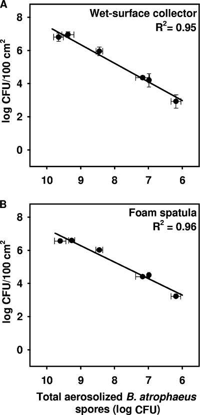
The total number of spores recovered from the wet-surface collectors and collected from glass surfaces using the foam spatulas was dependent on the concentration of the spores aerosolized. A linear regression was drawn in order to characterize the relation between aerosol exposure within the chamber and the number of spores that settled on glass surfaces and were collected using the foam spatula and in the wet-surface collector (Fig. 1). A positive correlation was noted between the number of total CFU aerosolized inside a chamber and the number of B. atrophaeus collected in wet-surface collectors (R2 = 0.96). Similarly, a positive correlation (R2 = 0.95) was also demonstrated between the number of total CFU aerosolized inside a chamber and the number of B. atrophaeus collected using foam spatulas. The theoretical surface spore loads with B. atrophaeus spores, i.e., the total number of CFU that may settle in each experiment on a 100-cm2 area, assuming that 74.17% of the total number of the spores aerosolized would be deposited on a bottom horizontal surface of the test chamber, were in the range from 2.88 × 10⁴ to 2.95 × 10⁷ CFU per 100 cm2. The limit of spore detection on a 100-cm2 area using both the foam spatula and wet-surface collector was observed when spores were aerosolized at a concentration of 105 CFU/ml in a total volume of 10 ml in our deposition system, and the theoretical surface spore load was 2.88 × 10⁴ CFU per 100 cm2. In these experiments, the number of B. atrophaeus colonies grown on the LB agar plate was below 20 when an undiluted extract from a foam spatula or liquid from wet-surface collectors was plated. In this study, neither the extract from foam spatulas (10 ml) nor the liquid taken from wet-surface collectors (15 ml) was concentrated; therefore, using a simple protocol for surface sampling, we recovered 103 B. atrophaeus spores deposited on the surface of 100 cm2 at the lowest concentration of spores aerosolized.
FIG. 1.
Linear regression analysis of the correlation between the numbers of B. atrophaeus spores collected in wet-surface collectors (A) and swabbed from glass surfaces using foam spatulas (B) and the total number of spores aerosolized within the test chamber. The number of viable spores was estimated after 16 h of settling in still air. Data points are shown as the mean log values (± SD [error bars]) per 100-cm2 area. Linear regression coefficients (R2) are shown in the upper right corner of each graph.
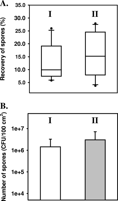
The median percent recovery efficiencies of spores from surfaces with foam spatulas and in wet-surface collectors in the experiments described above are shown in Fig. 2. As already mentioned, the majority of the spores in the test chamber were collected from bottom horizontal surface; however, the losses on four vertical and top horizontal surfaces were taken into account when the calculation of the theoretical surface spore loads were done for each experiment performed in this study. The percent recovery efficiency in this study was calculated relative to the theoretical surface spore load. The percent recovery efficiency of B. atrophaeus spores in these experiments were not stable or dependent on aerosol exposure. Thus, the median percent recovery efficiencies of spores from glass surfaces as well as in wet-surface collectors were presented in Fig. 2A as box plots, in which the median values of all measurements were marked as a straight line within the boxes. The median percent recovery efficiency of B. atrophaeus spores from surfaces using the foam spatulas was equal to 9.9% and was lower than the median percent recovery efficiency of spores in wet-surface collectors (15.2%); however, the difference was not significant (P > 0.05). As it was shown in Fig. 2B, the mean number of spores recovered with foam spatulas was two times lower than the number of spores in wet-surface collectors in all the experiments performed in this study; however, this difference was not significant (P = 0.43). The mean percent recovery efficiency of spores from the contaminated surfaces with foam spatulas was calculated also in relation to the mean number of spores that settled in the wet-surface collectors and was equal to 48.4%.
FIG. 2.
(A) Box plots showing the median, interquartile, and range values of the percent recovery efficiencies of B. atrophaeus spores from glass surfaces using foam spatulas (I) and wet-surface collectors (II). The median value is indicated by the horizontal line within a box. The 25th and 75th percentiles are indicated by the bottoms and tops of the boxes, respectively. No significant difference between the median percent recovery efficiencies of spores using foam spatulas and wet-surface collectors was determined with the nonparametric Mann-Whitney U test (P > 0.05). (B) Mean numbers of B. atrophaeus spores recovered from glass surfaces with foam spatulas (I) and in the wet-surface collectors (II) in all experiments performed in this study.
Deposition and recovery of P. agglomerans from surfaces as a function of aerosol exposure.
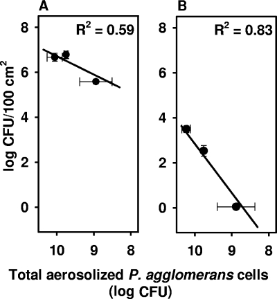
P. agglomerans was aerosolized at three different concentrations. The bacteria from wet-surface collectors and the viable cells, which were deposited for 1 h on the glass surface at the bottom of the test chamber and collected with the foam spatula, were plated on nutrient agar plates for counting. The mean numbers of culturable P. agglomerans swabbed from glass surfaces using foam spatulas were 2.4 × 10³, 2.7 × 10², and 0 per 100 cm2 when the total numbers of the aerosolized culturable bacteria were 1.4 × 10¹⁰, 5.9 × 10⁹, and 7.2 × 10⁸ CFU, respectively (Fig. 3). The theoretical surface bacterial loads, calculated similarly as for B. atrophaeus spores, were in the range from 8.09 × 10⁶ to 1.53 × 10⁸ CFU per 100 cm2. Thus, in this study we were able to recover culturable P. agglomerans cells using the foam spatulas only when the organisms were aerosolized at a concentration higher than 108 CFU/ml in a total volume of 10 ml, and the theoretical surface bacterial load was 8.09 ×10⁶ CFU per 100 cm2. At the same time, the mean numbers of culturable P. agglomerans cells were equal to 5.0 × 10⁶, 6.4 × 10⁶, and 3.9 × 10⁵ CFU per a 100-cm2 area when the bacteria were deposited in wet-surface collectors (Fig. 3). The median percent recovery efficiency of the Gram-negative rods was calculated in the same manner as for B. atrophaeus spores. The median recovery efficiency in wet-surface collectors was 4.87%, and the recovery from a glass surface using the foam spatulas was only 0.001% of the total number of P. agglomerans that might settle on the horizontal bottom surface of the test chamber.
FIG. 3.
Linear regression analysis of the correlation between the numbers of viable and culturable P. agglomerans cells collected in wet-surface collectors (A) and swabbed from glass surfaces using foam spatulas (B) and the total number of P. agglomerans aerosolized within the test chamber. The number of viable and culturable bacterial cells was estimated after 1 h of settling in still air. Data points are shown as the mean log values (± SD) per 100-cm2 area. Linear regression coefficients (R2) are shown in the upper right corner of each graph.
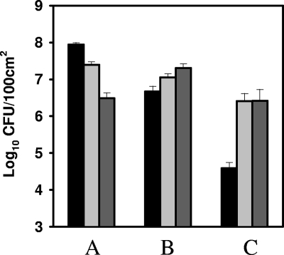
To observe how many P. agglomerans were alive or dead on the glass surfaces and in the wet-surface collectors 1 h after aerosolization, we used a Live/Dead BacLight bacterial viability kit to stain cells with intact membranes (fluorescent green) and cells with compromised membranes (fluorescent red). CP swabs were used to collect the bacteria from the surfaces, since in the extracts from the foam spatulas, even from the control samples (the sterile spatula tips extracted with SDW), many small particles (<0.5 μm) were stained fluorescent green, and thus, counting of P. agglomerans rods was unreliable. As shown in Fig. 4, after 1 h of bioaerosol deposition, the number of P. agglomerans cells with intact membranes was higher by 0.37 log unit in wet-surface collectors and by 1.83 log units in the extracts from CP swabs compared to the number of culturable P. agglomerans cells, respectively. The number of cells with compromised membranes was 0.25 log unit higher than the number of the cells with intact membranes in the wet-surface collectors. The mean recovery efficiency of P. agglomerans cells with intact membranes from glass surfaces with CP swabs was 24.22% compared to the number of P. agglomerans with intact membranes that settled within 1 h in the wet-surface collectors.
FIG. 4.
Comparison of the numbers of culturable P. agglomerans cells and bacterial cells that were stained by using a Live/Dead Bac Light bacterial viability kit that were present on glass surfaces 1 h after aerosolization. The three sets of bars show the theoretical surface load with P. agglomerans cells (A), the number of P. agglomerans cells in wet-surface collectors (B), and the number of P. agglomerans cells recovered with foam spatulas from glass surfaces (C). The numbers of culturable organisms (black bars), P. agglomerans with intact cell membranes (fluorescent green) (light gray bars), and P. agglomerans with damaged cell membranes (fluorescent red) (charcoal bars) are shown. The bars are shown as the mean log values (± SD) per 100-cm2 area.
DISCUSSION
The previous experimental studies on the collection efficiencies of different swabs used for sampling surfaces have primarily been focused on the removal of bacteria from coupons of various materials after their inoculation with liquid suspensions of Bacillus spores or vegetative P. agglomerans cells at concentrations ranging from 104 to 108 CFU per coupon (7, 12, 15, 20). However, when glass, polycarbonate, and vinyl surfaces were contaminated with Bacillus spores dispersed in aerosols, the percent recovery from the coupons was significantly lower using the same type of swab than the percent recovery from the coupons that were inoculated with spore suspensions (12). Therefore, to evaluate both the suitability of different swabs for sampling and the collection efficiency of biocontaminants from various surfaces, it seems reasonable to use the model of surface contamination with bioaerosols.
Such a model has previously been applied by others (1, 4) in aerosol chambers where the aerosolized microorganisms were permitted to settle onto a surface by gravitational force and at room temperature. A similar approach has been employed in the present study. Bioaerosols of P. agglomerans and B. atrophaeus spores were aerosolized in a test chamber and deposited for 1 and 16 h, respectively. The settling time of P. agglomerans was only 1 h, because as shown by Buttner et al. (7), after overnight settling within a chamber, very few culturable P. agglomerans could be collected from surfaces (<0.04 CFU per cm2). Most probably, the bacteria lost viability or the ability to produce colonies after aerosolization and desiccation stresses.
In this study, the recovery of B. atrophaeus spores from surfaces at three different locations within the test chamber, i.e., horizontal bottom, vertical, and horizontal top surfaces was concurrently estimated using LM, a macrofoam sponge wipe, and CP, a polyurethane swab. The most aerosolized B. atrophaeus spores were deposited on the horizontal bottom surface; however, about 7.9% of the spores (relative to the number of spores collected from a 100-cm2 area of the bottom horizontal surface) were recovered from a 100-cm2 area of vertical surface. These experiments showed that foam materials perform better in spore recovery than polyester swabs; however, with relatively low spore loads on surfaces (∼104 CFU per a 100-cm2 area), this difference is not significant. This observation confirmed the previous results (6, 20), which have demonstrated that foam swab materials recover more bacterial spores than cotton and polyester swabs do. In our preliminary experiments, we also noticed the highest recovery of aerosol-deposited B. atrophaeus spores from glass surfaces using a polyurethane macrofoam sponge wipe compared to polyester, rayon, cotton, or alginate swabs (data not shown).
Thus, in further experiments, we used a large foam spatula designed by a manufacturer for microbiological examination of environmental surfaces. According to CDC and International Organization for Standardization (ISO) recommendations, the samples were collected from 100-cm2 areas of the surfaces tested (9, 16). Wet-surface collectors were placed side by side on the surfaces tested in order to better estimate the number of viable B. atrophaeus spores and P. agglomerans cells that settled on the bottom of the test chamber and to avoid loss of bacterial cells that adhered to the swab materials during swab processing. A similar method of bioaerosol particle collection has previously been used by Sahu et al. (21). Due to overlapping bacterial colonies on agar plates and counting-based errors after direct deposition of highly concentrated bioaerosols, a wet-surface collector may be a better choice when a measurement of the number of the bioparticles deposited within the test chamber is required (10).
In a study performed by Hodges et al. (15), a good dose-response relationship has been observed between Bacillus spore recovery from stainless steel surfaces and the inoculum size; however, these experiments were performed on coupons inoculated with spore suspensions. Edmonds et al. (12) have observed, using the same method of surface contamination, that the percentage of recovery of liquid-deposited B. atrophaeus spores on glass coupons decreased from 92.7 to 42.1 as the concentration of spores deposited on surfaces dropped from 107 to 104 CFU. In the present study, glass surfaces placed at the bottom of the test chamber were contaminated with aerosolized suspensions containing from 2.6 × 10⁶ to 4.7 × 10⁹ CFU of B. atrophaeus spores and from 7.2 × 10⁸ to 1.4 × 10¹⁰ CFU of P. agglomerans. The numbers of spores and vegetative cells recovered from surfaces with foam spatulas and in wet-surface collectors were linearly dependent on the number of organisms aerosolized (Fig. 1 and 3).
The median recovery efficiency from the bioaerosol-contaminated surfaces was equal to 9.9% when the theoretical surface loads of B. atrophaeus spores were in the range from 2.88 × 10⁴ to 2.95 × 10⁷ per a 100-cm2 area. At the same time, the median efficiency of spore recovery in wet-surface collectors was ≥15.2%; however, the difference was not significant. Estill et al. (13) sampled stainless steel coupons using a polyurethane macrofoam-tipped swab and have found median recovery efficiency of aerosolized B. anthracis Sterne spores equal to 3.8% when the surface spore load was 2.7 × 10² CFU per 100-cm2 area.
The median efficiency of viable and culturable P. agglomerans cell recovery in wet-surface collectors was 4.87%. However, the foam spatulas recover only 0.001% of the total number of culturable P. agglomerans cells that may settle on a 100-cm2 area within the test chamber. Wet-surface collectors allowed detection of aerosols of Gram-negative organism with a better efficiency than sampling surfaces using foam spatulas. As the number of bacteria in wet-surface collectors did not decrease below 105 CFU per 100-cm2 area at the lowest concentration for which observations have been made, we conclude that desiccation of vegetative bacterial cells on glass surfaces probably causes this extremely low recovery of P. agglomerans from glass surfaces. A similar result has been reported by Buttner et al. (7) who demonstrated very low mean efficiency of P. agglomerans (formerly Erwinia herbicola) recovery, equal to 0.005%, when the bacteria at a concentration of 108 CFU per ml were deposited as a liquid on the glass coupons and swabbed using a macrofoam swab. Experiments performed in this study using a direct epifluorescence filter technique to count the number of viable cells (fluorescent green) in the extracts from CP swabs 1 h after aerosolization of P. agglomerans showed that the number of organisms with intact cell membranes was almost 2 log units higher than the number of culturable vegetative cells. At the same time, the number of viable cells (fluorescent green) that settled within the time frame of the experiments in the wet-surface collectors differed by only 0.37 log unit from the number of culturable P. agglomerans cells. Thus, as mentioned above, the stress of desiccation may influence the ability of P. agglomerans to grow on solid medium. However, the green-fluorescing cells with intact membranes cannot be considered with certainty as viable cells, as intermediate cellular stages with various degrees of damage to the outer membrane may also exist (2, 3). The number of dead cells with damaged membranes (fluorescent red) in the wet-surface collectors was higher than the number of viable cells, and an inverse viable/dead cell ratio was observed compared to the P. agglomerans suspension used for aerosol generation. This observation may indicate the aerosolization damage that was encountered by the bacteria in our experiments. Experiments with other fluorescent dyes are scheduled in the near future to verify whether P. agglomerans might be in a viable but nonculturable state in the extracts of foam spatulas.
One concern in this study is the lack of a method to estimate the actual total number of microorganisms that settle on a 100-cm2 area during an experiment. Thus, in the present study, the percent recovery efficiency is calculated relative to the theoretical number of organisms that might be deposited on a given surface area. In the earlier studies, Brown et al. (4) dislodged B. atrophaeus spores deposited on reference coupons by sonication, and the total number of CFU per sample was determined by plating. The average number of CFU per reference coupon was used as the total number of aerosol-deposited spores on the surface tested in the recovery efficiency formula. A similar approach has been presented by Edmonds at al. (12) who removed aerosol-deposited B. atrophaeus spores from glass control coupons by vortexing and sonication. Estill et al. (13) used Trypticase soy agar plates (60 cm2) that were placed near the coupons tested and were exposed to bioaerosol for the same settling period. Recovery efficiency was calculated as the number of CFU from the surface sample per unit of area relative to the number of CFU on the adjacent plate per unit of area. However, in our opinion, none of the methods described above is suitable to properly estimate the number of microorganisms deposited on surfaces in a wide range of concentrations in bioaerosol experiments. Spore extraction from control coupons by sonication or vortex methods do not dislodge all bacteria attached to the surface. An average recovery of liquid-deposited B. anthracis spores from reference stainless steel coupons by the sonication and vortexing methods has been shown to be only 24% when coupons were inoculated with 5.9 × 10⁶ CFU per cm2 (19). There are also many limitations of the agar plate collection technique as a method for the determination of total bacterial counts of aerosolized bacteria deposited on an agar surface (11). Thus, in our study, the percent recovery efficiency from contaminated surfaces and from wet-surface collectors was calculated relative to the total number of culturable organisms aerosolized in the test chamber (after the appropriate adjustments of the bacterial load on the horizontal bottom surface). The median percent recovery of spores from glass surfaces with a foam spatula and from the wet-surface collectors was not significantly different, though the foam was additionally processed in stomacher bags and loss of spores at this step was possible. However, the mean number of spore recovered from surfaces with foam spatulas was lower than the mean number of spores collected in wet-surface collectors in experiments performed in this study. The recovery of spores with foam spatulas expressed as a percentage of the mean number of spores collected in the wet-surface collectors was almost five times higher than the percent recovery calculated relative to the theoretical surface bacterial load. The foam spatula evaluated in this study permits reliable measurement of B. atrophaeus spore contamination on bioaerosol-exposed surfaces in a wide measuring range. However, additional work is needed to improve analysis of the foam extracts in order to decrease the limit of detection of Bacillus spores and Gram-negative bacteria on contaminated surfaces.
Footnotes
Published ahead of print on 18 December 2009.
REFERENCES
- 1.Baron, P. A., C. F. Estill, G. J. Deye, M. J. Hein, J. K. Beard, L. D. Larsen, and G. E. Dahlstrom. 2008. Development of an aerosol system for uniformly depositing anthracis spore particles on surfaces. Aerosol Sci. Technol. 42:159-172. [Google Scholar]
- 2.Berney, M., F. Hammes, F. Bosshard, H. U. Weilenmann, and T. Egli. 2007. Assessment and interpretation of bacterial viability by using the LIVE/DEAD BacLight kit in combination with flow cytometry. Appl. Environ. Microbiol. 73:3283-3290. [DOI] [PMC free article] [PubMed] [Google Scholar]
- 3.Boulos, L., M. Prevost, B. Barbeau, J. Coallier, and R. Desjardins. 1999. LIVE/DEAD BacLight: application of a new rapid staining method for direct enumeration of viable and total bacteria in drinking water. J. Microbiol. Methods 37:77-86. [DOI] [PubMed] [Google Scholar]
- 4.Brown, G. S., R. G. Betty, J. E. Brockmann, D. A. Lucero, C. A. Souza, K. S. Walsh, R. M. Boucher, M. Tezak, M. C. Wilson, and T. Rudolph. 2007. Evaluation of a wipe surface method for collection of Bacillus spores from nonporous surfaces. Appl. Environ. Microbiol. 73:706-710. [DOI] [PMC free article] [PubMed] [Google Scholar]
- 5.Budowle, B., S. E. Schutzer, J. P. Burans, D. J. Beecher, T. A. Cebula, R. Chakrabarty, W. T. Cobb, J. Fletcher, M. L. Hale, R. B. Harris, M. A. Heitkamp, F. P. Keller, C. Kuske, J. E. Leclerc, B. L. Marrone, T. S. McKenna, S. A. Morse, L. L. Rodriguez, N. B. Valentine, and J. Yadev. 2006. Quality sample collection, handling, and preservation for an effective microbial forensics program. Appl. Environ. Microbiol. 72:6431-6438. [DOI] [PMC free article] [PubMed] [Google Scholar]
- 6.Buttner, M. P., P. Cruz, L. D. Stetzenbach, A. K. Klima-Comba, V. L. Stevens, and P. A. Emanuel. 2004. Evaluation of the biological sampling kit (BiSKit) for large-area surface sampling. Appl. Environ. Microbiol. 70:7040-7045. [DOI] [PMC free article] [PubMed] [Google Scholar]
- 7.Buttner, M. P., P. Cruz, L. D. Stetzenbach, and T. Cronin. 2007. Evaluation of two surface sampling methods for detection of Erwinia herbicola on variety of materials by culture and quantitative PCR. Appl. Environ. Microbiol. 73:3505-3510. [DOI] [PMC free article] [PubMed] [Google Scholar]
- 8.Caipo, M. L., S. Duffy, L. Zhao, and D. W. Schaffner. 2002. Bacillus megaterium spore germination is influenced by inoculum size. J. Appl. Microbiol. 92:879-884. [DOI] [PubMed] [Google Scholar]
- 9.Centers for Disease Control and Prevention. 2002. Comprehensive procedures for collecting environmental samples for culturing Bacillus anthracis. Centers for Disease Control and Prevention, U.S. Department of Health and Human Services, Atlanta, GA. http://www.bt.cdc.gov/agent/anthrax/environmental-sampling-apr2002.asp.
- 10.Chang, C. W., Y. H. Hwang, S. A. Grinshpun, J. M. Macher, and K. Willeke. 1994. Evaluation of counting error due to colony masking in bioaerosol sampling. Appl. Environ. Microbiol. 60:3732-3738. [DOI] [PMC free article] [PubMed] [Google Scholar]
- 11.Chang, C. W., S. A. Grinshpun, K. Willeke, J. M. Macher, J. Donnelly, S. Clark, and A. Jouozaitis. 1995. Factors affecting microbiological colony count accuracy for bioaerosol sampling and analysis. Am. Ind. Hyg. Assoc. J. 56:979-986. [DOI] [PubMed] [Google Scholar]
- 12.Edmonds, J. M., P. J. Collet, E. R. Valdes, E. W. Skowronski, G. J. Pellar, and P. A. Emanuel. 2009. Surface sampling of spores in dry-deposition aerosols. Appl. Environ. Microbiol. 75:39-44. [DOI] [PMC free article] [PubMed] [Google Scholar]
- 13.Estill, C. F., P. A. Baron, J. K. Beard, M. J. Hein, L. D. Larsen, L. Rose, F. W. Schaefer III, J. Noble-Wang, L. Hodges, H. D. A. Lindquist, G. J. Deye, and M. J. Arduino. 2009. Recovery efficiency and limit of detection of aerosolized Bacillus anthracis Sterne from environmental surface samples. Appl. Environ. Microbiol. 75:4297-4306. [DOI] [PMC free article] [PubMed] [Google Scholar]
- 14.Frawley, D. A., M. N. Samaan, R. L. Bull, J. M. Robertson, A. J. Mateczun, and P. B. Turnbull. 2008. Recovery efficiencies of anthrax spores and ricin from nonporous or nonabsorbent and porous or absorbent surfaces by a variety of sampling methods. J. Forensic Sci. 53:1-6. [DOI] [PubMed] [Google Scholar]
- 15.Hodges, L. R., L. J. Rose, A. Peterson, J. Noble-Wang, and M. J. Arduino. 2006. Evaluation macrofoam swab protocol for the recovery of Bacillus anthracis spores from a steel surface. Appl. Environ. Microbiol. 72:4429-4430. [DOI] [PMC free article] [PubMed] [Google Scholar]
- 16.International Organization for Standardization. 2004. Microbiology of food and animal feeding stuffs - horizontal methods for sampling techniques from surfaces using contact plates and swabs. ISO 18593:2004. International Organization for Standardization, Geneva, Switzerland.
- 17.Leighton, T. J., and R. H. Doi. 1971. The stability of messenger ribonucleic acid during sporulation in Bacillus subtilis. J. Biol. Chem. 246:3189-3195. [PubMed] [Google Scholar]
- 18.Mesa, M. M., M. Macias, D. Cantero, and F. Barja. 2003. Use of the direct epifluorescent filter technique for the enumeration of viable and total acetic acid bacteria from vinegar fermentation. J. Fluoresc. 13:261-265. [Google Scholar]
- 19.Rastogi, V. K., L. Wallace, L. S. Smith, S. P. Ryan, and B. Martin. 2009. Quantitative method to determine sporicidal decontamination of building surfaces by gaseous fumigants and issues related to laboratory-scale studies. Appl. Environ. Microbiol. 75:3688-3694. [DOI] [PMC free article] [PubMed] [Google Scholar]
- 20.Rose, L., B. Jensen, A. Peterson, S. N. Banerjee, and M. J. Arduino. 2004. Swab materials and Bacillus anthracis spore recovery from nonporous surfaces. Emerg. Infect. Dis. 10:1023-1029. [DOI] [PMC free article] [PubMed] [Google Scholar]
- 21.Sahu, A., S. J. Grimberg, and T. M. Holsen. 2005. A static water surface to measure bioaerosol deposition and characterize microbial community diversity. Aerosol Sci. 36:639-650. [Google Scholar]
- 22.Sanderson, W. T., M. J. Hein, L. Taylor, B. D. Curwin, G. M. Kinnes, T. S. Seitz, T. Popovic, H. T. Holmes, M. E. Kellum, S. K. McAllister, D. N. Whaley, E. A. Tupin, T. Walker, J. A. Freed, D. S. Small, B. Klusaritz, and J. H. Bridges. 2002. Surface sampling methods for Bacillus anthracis spore contamination. Emerg. Infect. Dis. 8:1145-1151. [DOI] [PMC free article] [PubMed] [Google Scholar]


