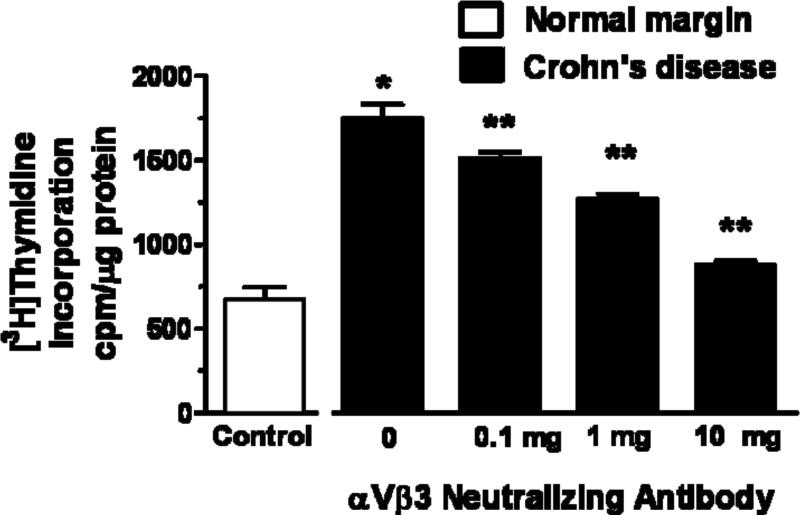
Figure 6. Smooth muscle cell proliferation is increased in stricturing Crohn's disease and dependent on endogenous IGF-I and αVβ3 integrin ligands.

Panel A: Basal [3H]thymidine incorporation is increased in muscle cells isolated from stricturing Crohn's disease compared to normal margins. Proliferation is inhibited by the αVβ3 integrin antagonist, echistatin (100 nM), or by the IGF-I receptor tyrosine kinase inhibitor, AG1024 (10 μM). Panel B: [3H]thymidine incorporation in muscle cells isolated from stricturing Crohn's disease is inhibited in a concentration-dependent fashion by an αVβ3 integrin neutralizing antibody. Results are expressed in cpm/μg total cell protein. Values represent the mean ± SEM of 5 separate experiments. * denotes P < 0.05 vs control muscle cells from normal margins, ** denotes P < 0.05 vs control muscle cells from stricturing Crohn's disease, *** denotes P < 0.05 vs control muscle cells from normal margins.

