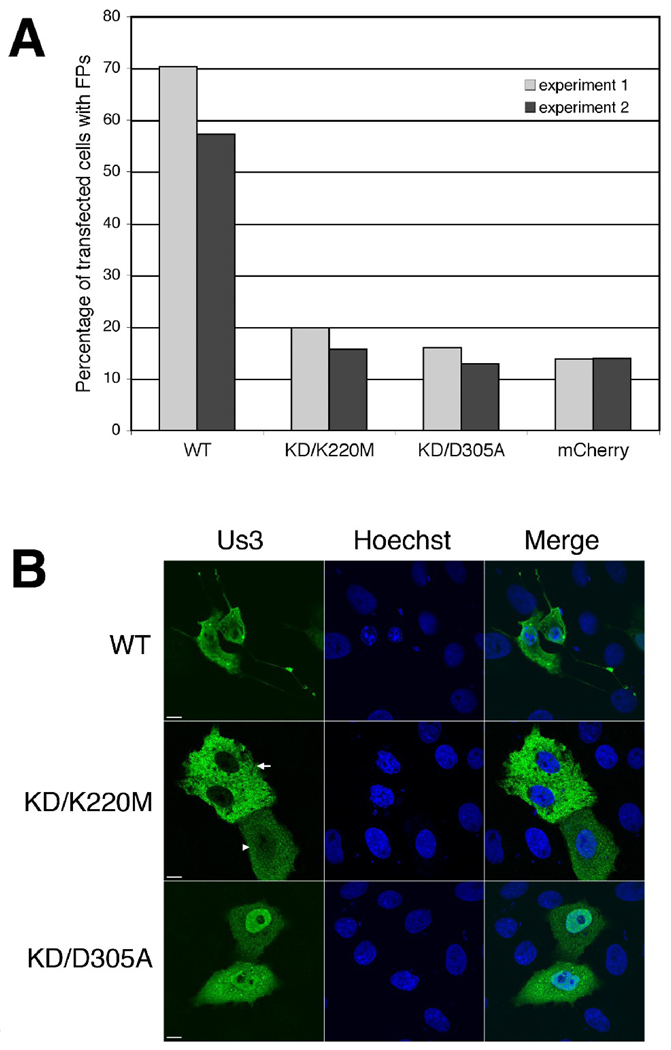
Figure 5.
Us3 kinase activity is required for HSV-2 Us3-induced FP formation. A. Percentage of transfected cells with FPs. Vero cells were transfected with plasmids encoding the indicated proteins. At 24 hours post transfection, cells were fixed and stained with rat polyclonal antiserum specific for HSV-2 Us3 followed by staining with an Alexa 488 conjugated donkey anti-rat secondary antibody. Nuclei were stained with Hoechst 33342. Stained cells were examined by confocal microscopy and 50 independent fields of cells containing at least one transfected cell were scored for the presence or absence of FPs. Results from two independent experiments are shown. Numbers of cells scored in experiment 1 and experiment 2 were as follows: for WT – 111 and 124; for KD/K220M – 151 and 179; for KD/D305A – 138 and 163; for mCherry – 123 and 187. B. Representative images of transfected cells used in this analysis. Arrow and arrowhead indicate the two different subcellular localization patterns of Us3 observed in cells transfected with a plasmid encoding KD/K220M Us3. Scale bar is 10 µm.

