
Figure 3.
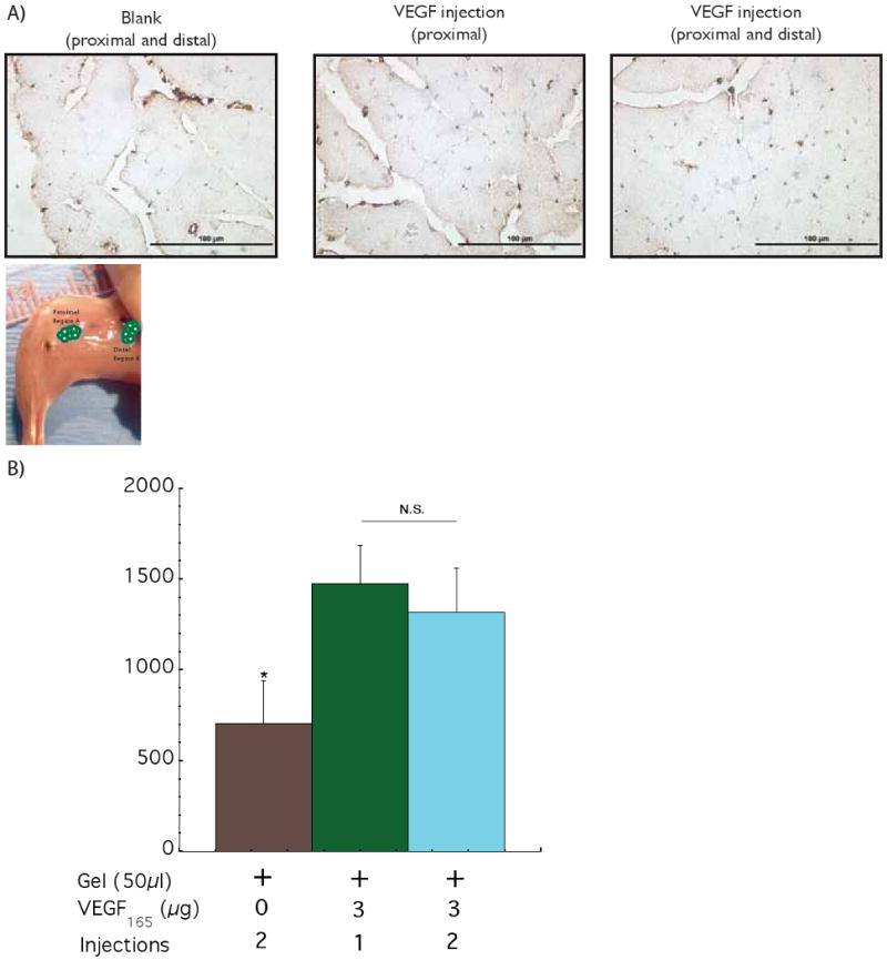
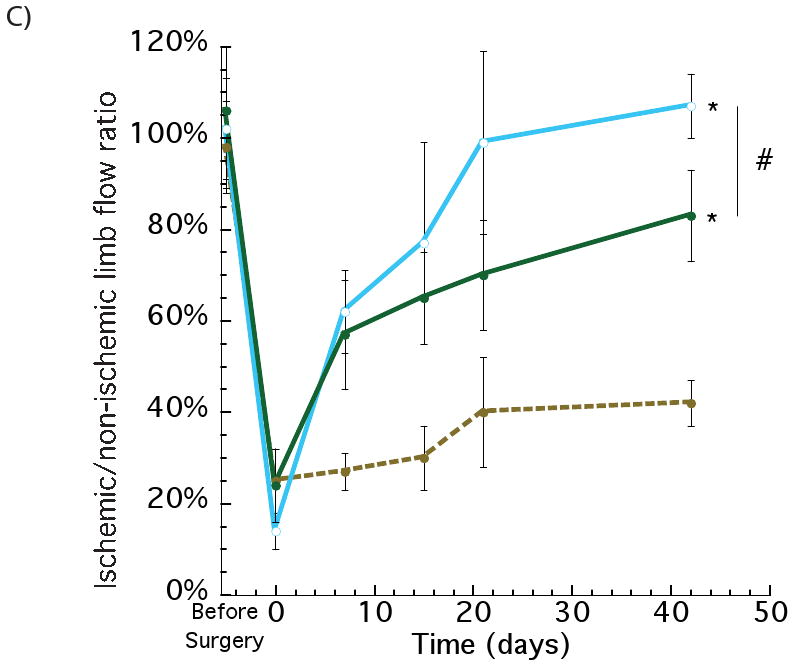
Assessing the effect of spatial distribution of VEGF delivery on the ischemic ApoE-/- hindlimbs. Photomicrographs of representative tissue sections from hindlimbs of ApoE-/- mice at postoperative 42 days, immunostained for the endothelial marker CD31 (A). Quantification of blood vessels densities after 6 weeks with 2 injections (proximal and distal) of control blank alginate hydrogel (+ 0 2); 1 injection of alginate loaded with VEGF (proximal) (+ 3 1) and 2 injections of alginate loaded with VEGF (proximal and distal) (+ 3 2) (B). Similar values of blood vessel densities were obtained for the 1 and 2 injections of VEGF. Perfusion profiles of hindlimbs at various experimental time points with 2 injections of control blank gel (●); VEGF delivered from alginate gels (1 injection - proximal) (●) and VEGF delivered from alginate gels (2 injections – proximal and distal) (○) (C). The two injections of VEGF elicited an increase in the regional perfusion, as compared with a single injection (VEGF loaded), even though the total VEGF dose was the same between the two conditions. Mean values are presented with standard deviations, * indicates statistically significant differences (p<0.05), as compared to control gels, and # represents statistically significant differences (p<0.05) between conditions.