Abstract
Style is a familiar category for the analysis of art. It is less so in the history of anatomical illustration. The great Renaissance and Baroque picture books of anatomy illustrated with stylish woodcuts and engravings, such as those by Charles Estienne, Andreas Vesalius and Govard Bidloo, showed figures in dramatic action in keeping with philosophical and theological ideas about human nature. Parallels can be found in paintings of the period, such as those by Titian, Michelangelo and Hans Baldung Grien. The anatomists also claimed to portray the body in an objective manner, and showed themselves as heroes of the discovery of human knowledge. Rembrandt’s painting of Dr Nicholas Tulp is the best-known image of the anatomist as hero. The British empirical tradition in the 18th century saw William Cheselden and William Hunter working with techniques of representation that were intended to guarantee detailed realism. The ambition to portray forms life-size led to massive volumes, such as those by Antonio Mascagni. John Bell, the Scottish anatomist, criticized the size and pretensions of the earlier books and argued for a plain style adapted to the needs of teaching and surgery. Henry Gray’s famous Anatomy of 1858, illustrated by Henry Vandyke Carter, aspired to a simple descriptive mode of functional representation that avoided stylishness, resulting in a style of its own. Successive editions of Gray progressively saw the replacement of Gray’s method and of all his illustrations. The 150th anniversary edition, edited by Susan Standring, radically re-thinks the role of Gray’s book within the teaching of medicine.
Keywords: anatomical teaching, Andreas Vesalius, Gray’s Anatomy
Introduction
We are used to the idea that ‘style’ is a key element in the study of art, even if it seems less central than it used to be. Considered in its broadest sense, style speaks not only of the maker’s mode of presentation but also about aspects of production, patronage, intended reception, visualization and visual language – in short, about every aspect of the process of communication between the originators of the image and the viewers. Style is not normally taken as one of the prime criteria when we analyze a scientific activity. In the specific field of scientific illustration, it has been seen as having some limited degree of relevance, if only in relation to the era of the illuminated manuscript and earlier phases of the printed text, when the provision of lavish picture-books for noble patrons was one of the standard types of production. Considered from the standpoint of the prevailing orthodoxies in much 19th-century and subsequent science, style is at best regarded as a rather irrelevant adornment to the business of communicating information and at worst as a positive liability. However, every made image of what is seen – every representation of nature or attempt to model some aspect of the world in visual terms – unavoidably has its own style, in as much as it has a visual ‘air’ or ‘aura’ through which its origins are recognizable (Kemp, 1991). For discussions of style as a critical term, see Shapiro (1953), Compagnon (1998), Ginzburg (2002), and Elsner (2003).
The tendency to regard evident stylishness as an irrelevance or encumbrance in scientific illustration has arisen as the result of a concerted ambition in science and technology from about 1850 to achieve ‘style-less’ images in which there has been nothing more to the presentation than the direct communication of objective information in the most functional manner. This aspiration apparently contrasts markedly with the overt espousing of style in Renaissance and Baroque illustration, in which the production of a fine display through the visual equivalent of rhetoric was either an explicit or implicit goal. I will also be arguing that the ‘style-less’ manner is as much a style as any mode of presentation and exhibits its own kind of contrived rhetoric.
I will be covering a range of material from the Renaissance to Gray’s Anatomy (with a brief notice of what has subsequently happened to this famous and enduring brand name), but I will in no sense be undertaking a historical survey. A convenient illustrated survey is provided by Roberts & Tomlinson (1992). Nor will I be addressing the rather belated role of photography in anatomical illustration, which I have discussed elsewhere (Kemp, 1997). My entry to the debates will be via the Edinburgh anatomist and surgeon Dr John Bell, whose Engravings of the Anatomy of the Bones, Muscles and Joints, published in 1794, is prefaced by one of the most perceptive discussions of the parameters for anatomical illustration in any printed text. Bell, brother of the more famous Charles, worked as an independent surgeon and teacher after falling foul of James Gregory in the Edinburgh Royal Infirmary. He was one of the earliest to devote sustained attention to how anatomy could best be communicated visually to aspiring surgeons (Kaufman, 2005). Let us look at the issues as raised by his preface, in his own order.
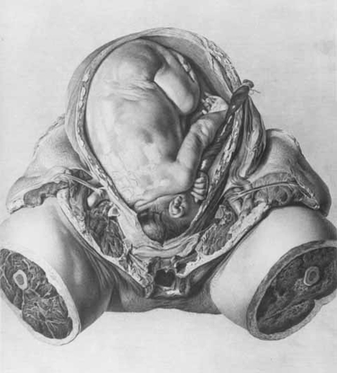
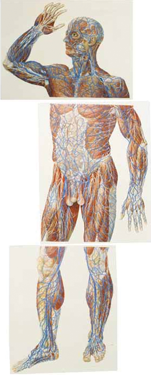
He begins with an outline of Cheselden’s famous account (1733) of the perceptual problems of an adult who had been newly enabled to see (Bell, 1794; p. ii). Amongst the obstacles encountered by the previously blind subject was his difficulty in believing that an image of something the size of a head could be contained within a small locket. Cheselden’s Lockeian evidence about our need to learn how to see is used by Bell to justify his provision of relatively small illustrations in a modestly sized book. The apparently obvious point that an effective representation need not be the same size as the object itself did indeed need to be made at the time, in the face of the fashion for real-size anatomical illustrations. The illustrations of midwifery by Smellie (1752) and the book on the gravid uterus by Hunter (1774; see Fig. 1) were obvious cases in point, but remarks later in Bell’s preface indicate that he had the scheme of the Italian anatomist, Paolo Mascagni, more immediately in mind: ‘I am sensible, that those, who cannot understand these plates [in Bell’s own book], will hardly profit even by that stately anatomical figure of full six feet high, which, being cut in copper, with googes, and chisels and mallets, and all kinds of instruments, must establish a reputation for its author; which, if not high, will not fail to be at least of a lasting kind’. The reference is to Mascagni’s only partially realized project for his massive Anatomia universa (see Fig. 2), which hardly bids fair to be a convenient and affordable handbook for students. It was known to Bell in various published guises before its final, if less than complete, publication on a large scale.
Fig. 1.
Plate VI, drawn by Jan van Rymsdyk, from Anatomia uteri humani gravidi [The Anatomy of the Human Gravid Uterus], William Hunter (1774).
Fig. 2.
Demonstration of the Vessels and Muscles from the Front (three untrimmed plates arranged to form complete figure), drawn by Antonio Seratoni, from Anatomia universa Paolo Mascagni (1823–31).
Bell is even more critical of anatomical books without plates. During the 18th century, such important texts as Alexander Monro’s The Anatomy of the Humane Bones or Haller’s eight-volume Partium corporis humani continued to be issued without illustrations (Monro, 1726; von Haller, 1778). In Bell’s view, an anatomy without plates is ‘no better than a book of geography without maps’ or a Euclid without diagrams (Bell, 1794; p. iii). His own aim was to bring the written word and the drawn image into such harmony that they are ‘wrought into one perfect whole’ such that we have ‘one idea presented in a double form’. Above all, the anatomist should resist ‘making an abstract subject of one belonging to the senses chiefly’. This injunction is in keeping with his constrained view of the role of theory in anatomy, which at best can be used to ‘connect the whole’ and at worst can seduce the student from the ‘close demonstration of the parts’ (Bell, 1794; p. vi). This is not to say that illustrations should be included for illustration’s sake, and he is highly critical of the practice by which the author of an anatomical text might annex admired plates from previous publications, as ‘directed by his bookseller’, availing himself of whatever ‘will make the handsomest figure’ (Bell, 1794, p. v). He cites the serial and progressively debased use of plates from Vesalius as exemplifying this tendency.
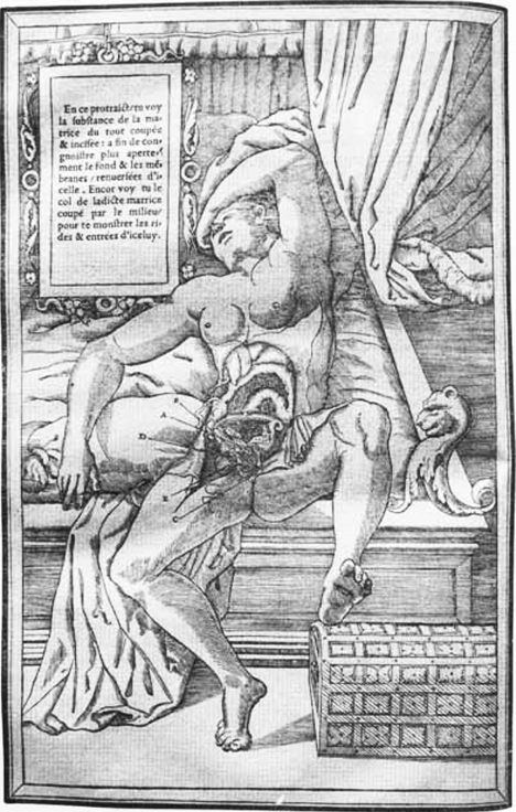
Bell was also fully alert to the problem that the achieving of a ‘close demonstration of the parts’ is not a simple matter of veridical depiction: ‘even in the first invention of our best anatomical plates, we see a continual struggle between the anatomist and the painter, the one striving for elegance of form, the other insisting on accuracy of representation’. As the result of this struggle, we see incongruous demonstrations of anatomy ‘monstrously compounded betwixt the image of the painter and the sober remonstrances of the anatomist’. It is clear that he is thinking particularly of some of the early texts, like Estienne (Fig. 3) or Valverde – and perhaps even Vesalius! (see below) – in which the anatomized figures act out an implicit drama. He sarcastically describes
Fig. 3.
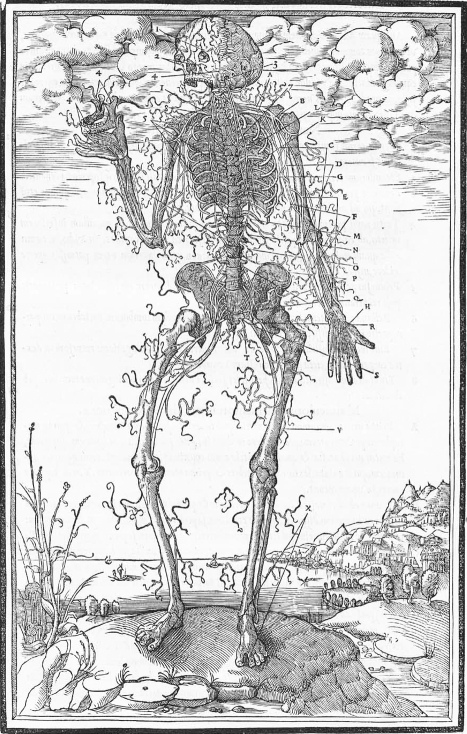
Demonstration of the Abdomen of a Woman to Show the Womb, from La Dissection des parties du corps humain, Charles Estienne (1546).
‘…sturdy and active figures, with an absurd contrast of furious countenances and active limbs, combined with ragged muscles, and naked bones and dissected bowels, which they are busily employed in supporting, forsooth, or even demonstrating with their hands.’
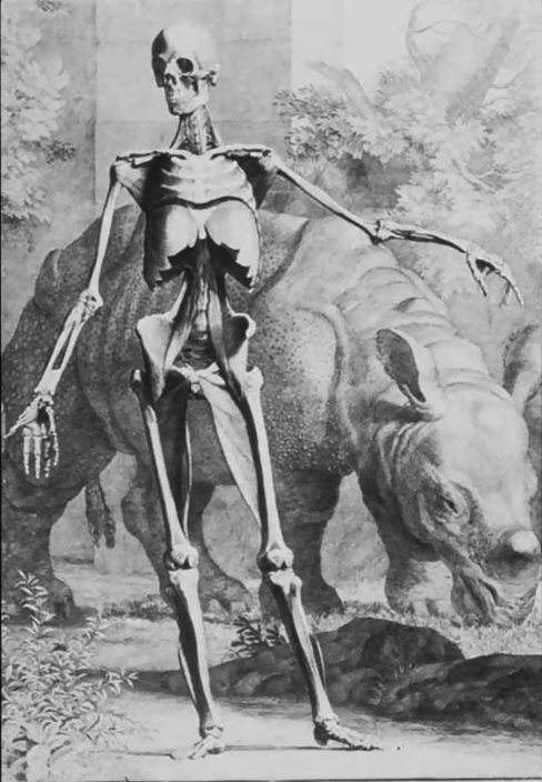
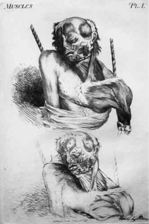
Like William Hunter, Bell is additionally concerned with the tension between the representation of the typical and the particular. Should the anatomist, in Hunter’s words, concentrate on ‘a simple portrait in which the object is represented exactly as it is seen’, or strive to achieve ‘the representation of the object under such circumstances as were not actually seen, but conceived in the imagination’ in such a way that the illustrator can ‘exhibit in one view, what could only be seen in several objects’? (Hunter, 1774; preface; for Hunter’s ‘style’, see Kemp, 1993). At the latter extreme for Bell stood Albinus’s splendid tables of the human figure (Fig. 4), in which the textural particularities of flesh and bone, of tissue and ligament, and the qualities of an individual specimen have been so suppressed that everything is ‘rounded down to the smoothness of ivory’, and the figure looks ‘like a statue anatomised’ rather than a real body (Bell, 1794; referring to Albinus, 1747).
Fig. 4.
Muscle-Man with Rhinoceros, drawn by Jan Wandelaar, from Tabulae skeleti et musculorum corporis humani, Bernard Siegfried Albinus (1747).
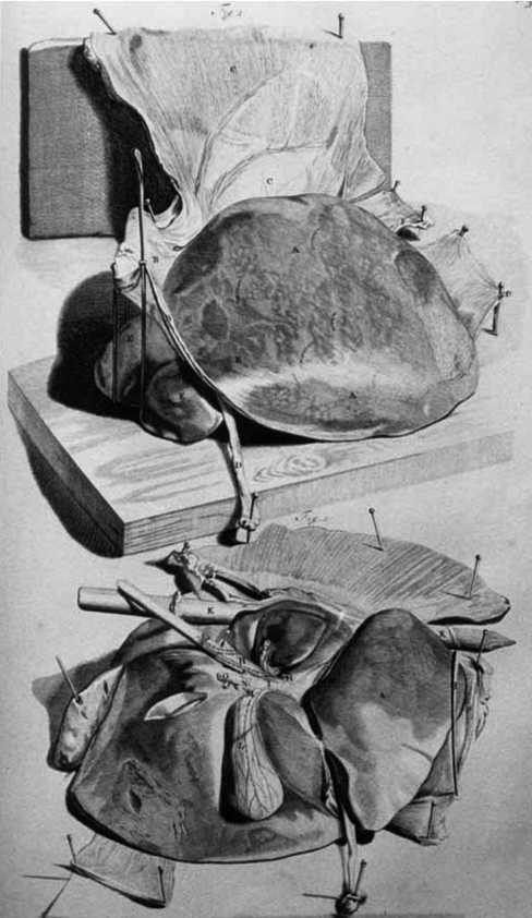
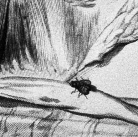
At the other extreme stood Bidloo’s striking plates (Figs 5 and 6), in which the ‘master-hand of the painter prevails almost alone’ in an artistic confrontation with the dissected and mounted parts of the body (Bell, 1794, p. ix, referring to Bidloo, 1685). We have ‘the very subject before us! The tables, the knives, the apparatus, down even to the flies that haunt the places of dissection’, and ‘thus we have prefect confidence in the drawing’. But judged as an exposition of the form and features of the human body, the cumulative effect of the bits and pieces is ‘disorder and confusion’, requiring the knowledge of both anatomist and painter to interpret in a coherent manner what is displayed (Bell, 1794, x).
Fig. 5.
Demonstration of the Liver, drawn by Gérard de Lairesse, from Anatomia humani corporis, Govard (Gottfried) Bidloo (1685).
Fig. 6.
Detail of Fly on the Dissection of the Abdomen drawn by Gérard de Lairesse, from Anatomia humani corporis, Govard (Gottfried) Bidloo (1685).
Bell is not, however, wholly hostile to striking and even stylish presentation. He commends ‘statuaries or painters’ for ‘studying the anatomy of the human body with a perseverance and success which may well put us to shame’. Michelangelo’s ‘bold and terrible pictures of action and strength’ and his emphatic anatomical characterizations are particularly to be admired ‘as correct and true’, though less well adapted for ‘female forms’ (Bell, 1794, p. xv).
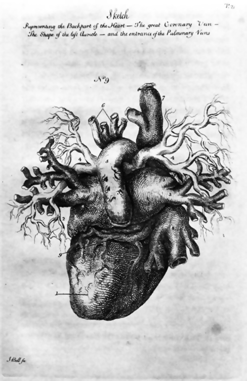
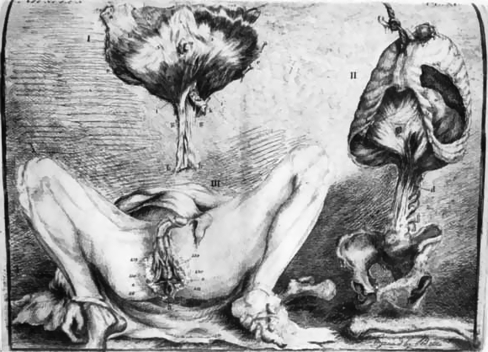
Bell’s proclaimed ambitions for his own illustrations are modest aesthetically yet efficient functionally: ‘The author surely will not be accused of such want of taste and relish for elegant drawing or engraving, as to hold these plates out as excelling in what is beautiful; yet, may he not hope, that they are not wanting in what is useful’ (see Figs 20–22; Bell, 1794, p. xviii). He explains: ‘I have drawn the plates with my own hand. I have engraved some of the plates and etched almost all of them, which I mention only to show, that they have their chance of being correct in the anatomy, and that whatever, by my interference, they may have lost in elegance, they have gained, I hope, in truth and accuracy’ (Bell, 1794, p. xx).
Fig. 20.
Sketch Representing the Backpart of the Heart, from The Anatomy of the Human Body, John Bell (1794–1804).
Fig. 22.
Dissection of the Genital Region with the Abdomen, Thorax and Diaphragm, from Engravings Explaining the Anatomy of the Human Body, John Bell (1797).
His emphasis upon illustrations that are ‘simple, intelligible and plain’, such as to permit the student ‘to enter the dissecting room with confidence’, is all of a piece with his drive to simplify the language of anatomical description. He is scathing about anatomists’ tastes for ‘peculiar and affected language, with needless terms of art’, which constitute the ‘barbarous jargon’ which is found in the ‘trashy language of school books’ (Bell, 1794, p. xxi). This emphasis upon plainness in word and image was shared with his brother, Charles Bell, who consciously renounced Latin, recognizing that the ‘changed frame of society’ demanded a different language from that which had previously given ‘a liberal tone to the men of influence in the several states, and a disposition to polite literature and science’ (Bell, 1806; pp. 7 and 10). What the Bells are assaying, in theory at least, is a plainness which eschews overt stylishness and indeed tends strongly towards non-style. Their books, accordingly, are smallish in scale, bound in non-luxurious materials, and printed in a businesslike manner on paper of ordinary quality. As soon as we pick up a book like Charles Bell’s The Hand, issued as a volume in Bohn’s Scientific Library, we cannot doubt that we are in a different world from that which gave birth to the great regal picture books such as William Hunter’s Anatomia uteri humani gravidi (Bell, 1833).
Bell’s preface provides a telling introduction to the enduring issue of ‘style’ and ‘non-style’. In the following sections, I will pay particular attention to the examples he cogently identified, before looking at Gray as the natural heir to his enterprise.
The heroic style of anatomical humanism
Of all the books in which ‘active limbs’ are ‘combined with ragged muscles, and naked bones and dissected bowels’, Estienne’s De dissectione of 1545 is the most extreme in its apparent contrast between what we may regard as ‘anatomical content’– sometimes limited to a smallish insert in the woodcuts – and elaborate figures posturing in fancy settings (Fig. 3). However, if we read Estienne’s preface, we find him making claims which are fundamentally the same as Bell’s about the role of visual images in bringing the real forms before our eyes, the true sensory organs for visible things, more powerfully than is possible with words, which best suit ‘the spirit and the memory’ (Estienne, 1545; pp. 6–7; see Kellett, 1964 for the artistic sources of the illustrations). He also explains that his representations will serve as practical aids to the surgeon, and that he is eschewing the literary stylishness of Cicero in favour of a directness which appeals to a different part of our understanding. How can we make sense of such apparently Bell-like claims in the face of the actual presentation of the human figure in his plates?
One clue is provided by his argument that depictions serve to make the absent seem to be present. This is precisely the argument used in Alberti’s humanist book On Painting, which itself builds upon a precedent in Cicero’s On Friendship (Alberti, 1435; especially 60). Another clue is provided by his compilation in 1553 of a humanist dictionary of famous people from the classical past and of more recent eras, including Galen, who is described as a ‘medicus excellentissimus’(Estienne, 1606; entry for ‘Galenus’) and who is cited throughout Estienne’s anatomical text. What Estienne is doing is to provide a grand ‘history’ of the human body, in the Renaissance sense of ‘historia’, the term used by Alberti to denote a story which signifies great things. The figures – whether men acting out heroic dramas in grand settings such as all’antica landscapes, or women anatomized in bedrooms – correspond in tone to the author’s definition of the nobility and grandeur of the human estate in relation to God’s creation, a definition that he derives from the ancient Stoic authors whom he cites in his preface (for the philosophical nature of Renaissance anatomy, see Carlino, 1999a,b; see Sawday, 1997 for an alternative interpretation). When he depicts the nerves (Fig. 7), twitching with febrile energy, he explains that they were created to resonate with the ‘concordance and harmony’ of universal design, like the strings of a musical instrument (Estienne, 1545, p. 50).
Fig. 7.
Demonstration of the Nerves, from La Dissection des parties du corps humain, Charles Estienne (1546).
The vision is one of the whole organism as the dynamic expression of the world as a whole. Throughout the greater part of his three books Estienne studiously avoids showing separate bits of the body, though he acknowledges that smaller features are less visible than they might be (Estienne, 1545, p. 262). What appeared to be a grotesque comedy to Bell was for Estienne a manifestation of necessary decorum in terms of a heroic kind of visual rhetoric.
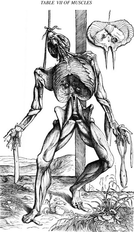
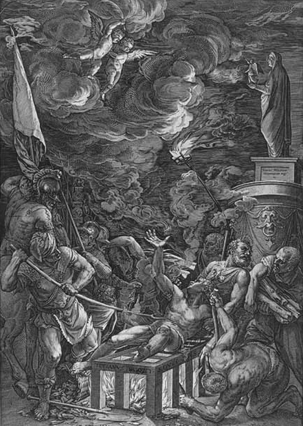
Although Andreas Vesalius’s great De Humani corporis fabrica in 1543 is generally seen as providing a stark contrast to Estienne’s volume, the philosophical rhetoric is shared. The famed sequence of muscle-men (Fig. 8) demonstrates heroically their god-given bodies in the spirit of the much-cited Sibylline tag, nosce te ipsum– know thyself, which was inscribed on the Temple of Apollo at Delphi. The panoramic sequence of bodies undertaking their myological striptease in a landscape rich in ruined ancient architecture is as much a part of a historia as Etienne’s animated figures (Kemp, 1996). The Titianesque male heroes of this drama were drawn by Jan Steven van Kalkar, who spent a period in the great Venetian painter’s workshop (Simons & Kornell, 2008), demonstrating that the reference to Titian himself as author of the illustrations is mistaken. A martyr such as St Lawrence, portrayed memorably by Titian for the Jesuit church in Venice (Fig. 9), is a clear prototype for the visual rendering of a redeeming death, modelled ultimately on Christ, providing the religious underpinning for the violent portrayal.
Fig. 8.
7th Plate of the Muscles, drawn by Jan Steven von Kalkar, from De humani corporis fabrica, Andreas Vesalius (1543).
Fig. 9.
Cornelius Cort, engraving after Titian (1567), Chiesa de’ Gesuiti, Venice.
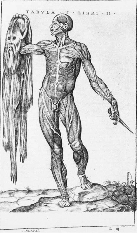
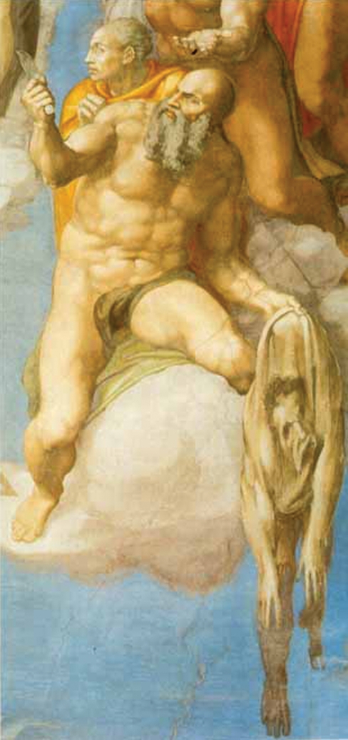
Bell’s admired Michelangelo played a comparable role for the Spanish anatomist Jan Valverde de Hamusco, who adopted the motif of a figure holding its own flayed skin from St Bartholomew in the Last Judgment in the Sistine Chapel (Valverde de Hamusco, 1556). The drooping pelt of the saint (Fig. 10), martyred by flaying, bears the distorted portrait of the artist himself. Valverde’s self-flayer (Fig. 11), brandishing a dagger, knowingly adopts a pose similar to that of the renowned Apollo Belvedere in the Vatican, which had itself provided a major source of inspiration for Michelangelo’s David. At one level, these interlocking artistic motifs are simply manifestations of the fashionable Renaissance style all’antica, but at other levels the style keys philosophically into the deepest questions of the nature of the human condition, into the issues of self-knowledge and life and death, that provided the fraught spiritual and moral background to early dissection.
Fig. 10.
Flayed Man holding his own Skin, drawn by Gaspar Beccara (?), from Historia de la composición del cuerpo humano, Jan Valverde de Hamusco (1556).
Fig. 11.
St Bartholomew from the Last Judgement, Michelangelo (1537-41), Sistine Chapel, Vatican.
The climax of this humanist tradition is Bernhard Seigfried Albinus’s Tabulae sceleti et musculorum corporis humani of 1747, which bids fair to be the most self-consciously poised anatomical book ever produced (Fig. 4) (Albinus, 1747). Albinus himself outlines at some length and with no little pride the incredible effort that went into the production of the plates by himself and his artist, Jan Wandelaar. The aim, as I have described elsewhere, was a seamless combination of precision, refinement and anatomical synthesis, which resulted in images of great polish far removed from the flesh-and-blood reality of dissection (Kemp, 1993; for highly suggestive discussions of the issues of seeing, knowing and representing in 18th-century anatomy and art, see Stafford, 1993; and for the 19th century, see Daston & Galison, 2007). That this ‘ivory’ style was not automatically the product of Wandelaar’s pen can be confirmed by looking at one of the artist’s illustrations for Fredrik Ruysch, which emphasizes the elaborate detail and minute ramifications which Albinus’s Dutch predecessor so prized (Ruysch, 1720, p. 46). Albinus claimed that, far from being ‘needless and unnecessary’, the supreme effort to synthesize the forms of the body in all their inner beauty was entirely appropriate to reveal the magnificence of God’s handiwork – the same rationale as Bidloo and Estienne had adduced in support of their very different methods. The lordly magnificence of ‘man’ in the context of brute creation is underscored by the so-called ‘Dutch Rhinoceros’ grazing in the background of an elegant muscle-man. Again the pose is reminiscent of the Apollo Belvedere or like one of the majestic full-length portraits that Pompeo Batoni provided in Rome for young aristocrats on their Grand Tour (Bowron & Kerber, 2007).
What is particularly interesting in our present context is that this apparently supreme example of the stylish style actually involved a highly elaborate attempt to overcome the problem of the intervention of the artist’s own eye and hand. He devised a method based on the artists’ traditional drawing-frame containing a grid (or ‘veil’ as Alberti had called it) to achieve systematic control over the rendering from a precisely established viewpoint (Kemp, 1993). As with Vesalius, stylishness and accuracy were not seen as conflicting aspirations.
The rhetoric of the ‘real thing’
Alongside the stylish heroism of the Vesalian ‘stories’ ran a recurrent insistence on direct representation of the ‘real thing’. Whatever the bookish sources for traditional features in the illustrations, as is readily apparent in the works of all Renaissance anatomists, they repeatedly emphasized that their pictures were based on direct observation. There was a specifically northern European dimension to the visual tropes of realism that were adopted by Renaissance illustrators. It is significant that both Vesalius and Kalkar were northerners working in Italy.
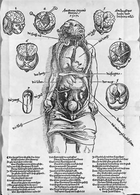
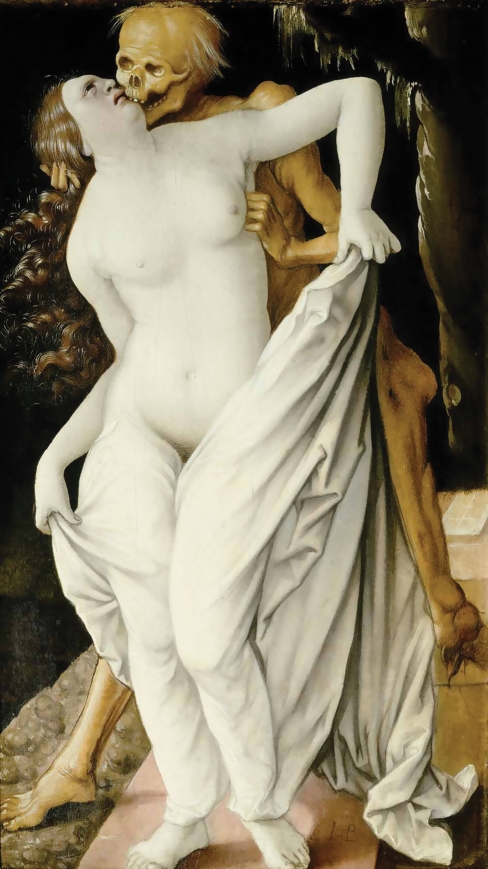
The tone was set by Hans Wächtlin’s unprecedented woodcut of a man with a dissected thorax, abdomen and cranium in 1517 (Fig. 12). The anatomy was, as an inscription on the earliest state of the print tells us, performed in Strasburg by Wendelin Hock von Brackenau. It was published in Hans von Gersdorff’s book of wound surgery in the same year, and a year later in Lorenz Fries’s medical textbook (von Gersdorff, 1517, Fries, 1518). The customary attribution of the original drawing to Hans Baldung, who was active in Strasburg, seems unlikely given the weak drawing of the arms, but it certainly belongs in his orbit. He specialized in small paintings of sexualized and macabre subjects for humanist patrons (Fig. 13). The deliberately gruesome directness of portrayal in Wächtlin’s woodcut (Fig. 12) is not only in tune with the double punishment of the criminal (both executed and dissected) but also serves to convince us that we are virtual witnesses to the visceral act. The sequential studies of the brain that surround the main image are particularly potent in this latter respect.
Fig. 12.
Dissection of the Thorax, Abdomen and Cranium, woodcut, Hans Wächtlin (1517).
Fig. 13.
Death and the Maiden, Hans Baldung Grien (1518–20), Öffentliche Kunstsammlung, Basel.
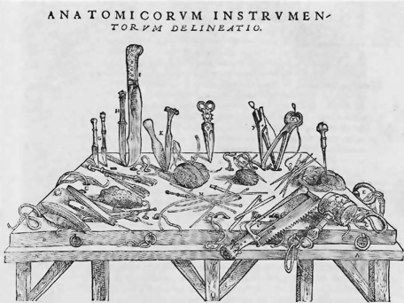
Vesalius and his illustrator may be seen to be fusing the Italianate and northern traditions. Alongside the humanist grandeur of the figures, redolent of ancient Roman sculpture, he is at pains in both text and woodcuts to stress that his is the direct dissector of the forms that he is disclosing to us in all their graphic conviction. The woodcut of his uncompromising array of tools placed on and stabbed into a Galenic vivisection board for pigs (Fig. 14) plays an important role in setting this tone of direct portrayal. His plates are made with a full awareness of the disturbing nature of his heroic quest to ‘know thyself’.
Fig. 14.
Tools and Vivisection Board, drawn by Jan Steven von Kalkar from De humani corporis fabrica, Andreas Vesalius (1543).
More than a century later, the rhetoric involved in Bidloo’s Anatomia humani corporis is different in tone, though devoted to the expression of many of the same ideals that motivated Vesalius and Estienne (Bidloo, 1685). Bell emphasized that Bidloo’s plates spoke of a direct confrontation between the artist’s eye and the dissected organs, and this was indeed just the effect that was intended (Fig. 5). The overt display of the blocks, tools, pins (including a bent pin on the right of the upper study of the liver), carefully differentiated textures rendered skillfully by the engraver, cast shadows, and even in one instance the intrusion of the hand of a demonstrator holding back the brain to show forms beneath, are all intended to speak of an uncompromising directness of portrayal – to give us confidence that we are seeing the real thing.
The visual devices were themselves rhetorical in effect (using what I term ‘the rhetoric of the real’). They also knowingly alluded to a tradition of trompe l’oeil realism which was sanctioned by antique precedent. The fly (Fig. 6), singled out for scorn by Bell, is just the kind of trick that Pliny records in ancient art, and there can be little doubt that Bidloo’s distinguished illustrator, Gherard de Lairesse, was alert to such an allusion. Lairesse was a theorist of art in his own right and had done much to earn his nickname of the ‘Dutch Poussin’ (Roy, 1992).
Such devices of the ‘rhetoric of the real’ continued to be regular standbys of the anatomical illustrator’s art. As we will see, William Hunter was a master of the mode.
The Anatomist as self-fashioning hero
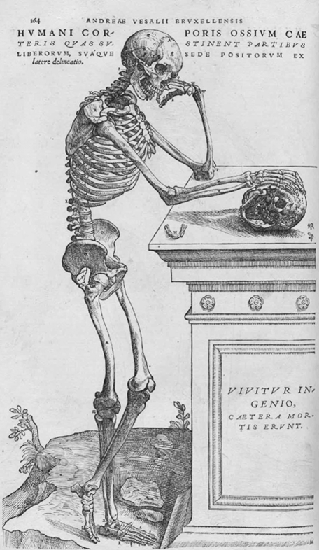
Vesalius and the other conspicuous producers of magnificent humanist picture-books of anatomy were not prone to modesty. The quest for fame, a motif inspired by antiquity, was a notable characteristic of the Italian Renaissance cult of the male individual who achieved status through the exercise of his ingegno (innate talent or ‘genius’) and virtù (worth and merit) (for self-fashioning, see Greenblatt, 2005). In his frontispiece, Vesalius has himself depicted disclosing the secret of life in a woman’s womb in the dramatic context of a great temple-theatre, where he holds the centre stage, surrounded by awed spectators. When he depicted a skeleton contemplating a skull (Fig. 15), he qualified the obvious memento mori with a tag from an eclogue by Virgil: ‘Vivitur ingenio, caetera mortis erunt’ (Genius lives on, all the rest will perish: Virgil, Elegiae in Maecenatem 1.38). That Vesalius’s ingenium would cheat death, enabling him to live in the eyes of others across successive generations, has of course proved to be the case – to a degree that even he could not have envisaged.
Fig. 15.
Skeleton, drawn by Jan Steven von Kalkar, from De humani corporis fabrica, Andreas Vesalius (1543).
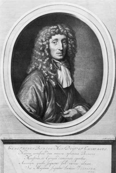
Bidloo was no more inclined to be reticent than Vesalius. In his engraved portrait at the start of his splendid and costly volume (Fig. 16), the bewigged anatomist, wearing a fine cravat, stares self-confidently at the reader, holding his incisive razor. The inscription denotes that he is the true successor to Vesalius. His text opens with ringing praise of the bodily microcosm and concludes in fine rhetorical style with a valedictory flourish extolling the supremely great Creator of the edifice of the human body. The great anatomists were men who saw themselves as being at the heart of the age-old quest to understand the most glorious creation of the supreme artifex.
Fig. 16.
Portrait of Govard Bidloo drawn by Gérard de Lairesse, from Anatomia humani corporis, Bidloo (1685).
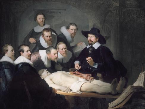
Nowhere is this clearer than in Rembrandt’s Anatomy of Dr Tulp (Fig. 17), the most dramatic of the series of Dutch surgeons undertaken for their guild premises. There has been much discussion on ‘errors’ in Rembrandt’s depiction, but, allowing for the fact that it is a boldly painted image and not a diagrammatic illustration, it serves as an effective representation of the dissected arm. (For the other paintings in the series, see Kemp & Wallace, 2000; for Rembrandt’s Tulp, see Schupbach, 1982). The air, like that of Vesalius’s frontispiece, is one of awe. A cluster of Tulp’s fellow surgeons react with contrived amazement at his revelation of the mechanism of the hand. This does not reflect the standard order of dissection, which began with the soft internal organs. Tulp has consciously chosen the hand as ‘the instrument of instruments’, to quote Aristotle (Kemp, 2000). Specifically he is dealing with the mechanism of the fingers and thumb. The extraordinary device of the interpenetration of the flexor tendons in the hand – extolled by Galen – is conspicuously evident. As a pioneer of the study of primates, Tulp would have understood that the precision grip facilitated by the opposition of thumb and forefinger was a crucial feature that distinguished humans from the homo silvestris (the ‘wild man of the woods’ or orangutan) (Tulp, 1641; see Corbey, 2005, pp. 38–40; and Kemp, 2007, pp. 137–8) (it is likely that Tulp’s homo silvestris was a chimpanzee). For Tulp, as for his predecessors, anatomy was a philosophical science, not merely a body of technical knowledge for the surgeon.
Fig. 17.
Anatomy of Dr Tulp, Rembrandt (1632), The Hague, Maristhuis. For a larger version of this image, see Ingham, this issue (2010).
The unvarnished truth, as large as life
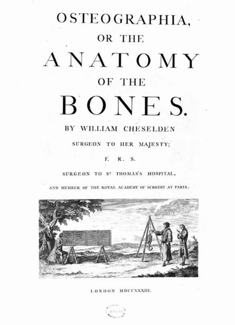
In the 18th century, the powerful empiricism that dominated British scientific thought found specific expression in anatomy in what can be regarded a ‘proto-photographic’ method. It involved particular processes and methods of depiction designed to guarantee objectivity and clarity. Its first manifestation was William Cheselden’s use of a camera obscura for his Osteographia in 1733 (Fig. 18) (Cheselden, 1733; see Kemp, 1993; for Chesleden and artists, see Bignamini & Postle, 1991).
Fig. 18.
Title Page of Osteographia, William Cheselden (1733).
The fact that Cheselden paraded the device in operation on the title page of his book emphasizes that he wants the reader to know that he has introduced an unprecedented level of objectivity. The artist’s hand is to be subordinated to the non-subjective eye of the camera. Cheselden was well aware, however, that the translation of the image on the ground glass plate of the camera into a drawing and then into engraved form involved special graphic skills. He was alert to the way that his illustrators’ somewhat different graphic habits were best adapted to particular features. Shinevoet was ‘neat and expressive and well suited to such things as he was mostly employed in’, but he was inferior in ‘stile’ to Gerard van der Gucht. Cheselden’s open discussion of such issues of representation indicates that he is aspiring to inculcate a special form of visual trust between the anatomist and the viewer of his plates.
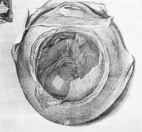
This form of contract between anatomist and virtual witness reached its peak in what I have called the ‘warts and all’ style favoured by John and William Hunter. It was expressed most tellingly by their master illustrator, Jan van Rymsdyk (Thornton, 1982; Kemp, 1993). William Hunter – as Professor at the Royal Academy of Arts and as teacher of medicine – was an uncompromising advocate of the truth to be obtained by minutely scrupulous looking and rendering. Presenting his life-size demonstration of the Anatomy of the Gravid Uterus in 1774 (Fig. 1), Hunter emphasizes throughout that he is presenting the particular specimen rather than synthesizing observations from a number of dissections, even when the method works against what he might ideally wish to show. He insists that his illustrator shows the eight-paned window of his dissecting room in the moistly shiny membrane over the head of the fetus (Fig. 19) – because it was really there. This is an extreme example of the ‘rhetoric of reality’.
Fig. 19.
Fetus and Membranes, drawn by Jan van Rymsdyk, from Anatomia uteri humani gravidi [The Anatomy of the Human Gravid Uterus], William Hunter (1774).
We find related if distinctly national varieties of the minutely detailed style in both Germany and Italy, though without Hunter’s obsessive insistence upon the depiction of the vagaries of an individual specimen as seen in individual circumstances. Hopffer’s illustrations in 1775 for the Observationes anatomicae by J. G. Walter, the Berlin anatomist, and Paolo Mascagni’s treatise on the lymphatics published in Siena in 1787, are both visually splendid examples of the international style, with intensely detailed plates of the tube-work of the body in fully rendered glory, accompanied by outline diagrams with keys, and demanding improbable levels of patience from all concerned – not least the reader (Walter, 1775; Mascagni, 1787).
The point is not necessarily that every minute observation is of immediate utility, but rather that a scrupulously empirical investigator is under an ethical imperative to leave nothing out. Accordingly, an illustrative technique must be devised which would effect a graphic translation of the anatomist’s habits of remorselessly detailed scrutiny. The incredible techniques devised by the late 18th-century engravers stand alongside increasing refinements in the preparations of specimens and making of models, particularly the wax constructions which could achieve such astonishing levels of illusion (see Riva et al. 2010; Ballestriero, 2010; Mazzotti et al. 2010).
A natural corollary to the extreme demand for veridical representation was the requirement that anatomies should be shown real size. The result, in projects such as Hunter’s great obstetrical atlas or Mascagni’s Vasorum lymphaticum, were books of a very specific kind, which were too large and expensive to be considered handbooks, or even regular teaching aids in most normal contexts.
The height of magnificent impracticality was reached in Mascagni’s incompletely realized project for an Anatomia universa (Fig. 2), which ultimately tends to ally itself more with Albinus’s synthesis than Hunterian empiricism (Mascagni, 1823–31; also in reduced and more practical versions, Mascagni, 1816, 1833). Clearly intending to create the anatomy to end all anatomies and to secure world-wide fame, Mascagni commissioned Antonio Serantoni –‘draftsman, engraver and modeller in wax’– to produce grandiose sets of plates in huge folios, three for each complete figure, which were to be elaborately coloured and assembled into a series of demonstrations, each over 6 feet high (as Bell testified). Each of the three ‘strata’ of dissection was shown from the front and back, together with views of the skeleton and separate studies of various organs. As Serantoni testified, the aim was to provide the viewer with ‘a deep understanding of the component parts of the human machine’ (Mascagni, 1833, preface).
Unlike Hunter, Mascagni was, in the manner of Continental Neoclassicism, aiming to show a perfectly proportioned archetype rather than a flawed individual. The means to realize such an overtly heroic ambition could only arise within a certain kind of context of patronage and market. Mascagni’s publications, like Hunter’s Anatomy of the Human Gravid Uterus, openly brandish the author’s patronage by the highest in the land. The large-scale works by the British and Continental anatomists were prepared ‘with great care and expense’, as Smellie testified, and were generally sold by subscription in Britain (Smellie, 1752, advertisement for the plates at end of text). Smellie advertised 26 plates by Rymsdyk, ‘drawn… as large as the human subjects themselves’, which were to be marketed in ‘Ireland, France and Holland’ and were to be available with English or Latin texts at a subscription price of two guineas, one on order and the other on receipt. Such a price was by no means small, and hardly brought the work within the budget of aspiring midwives, although subsequent re-publication of his plates undoubtedly brought them before a wider audience. Mascagni, for his great enterprise, was at one stage promised enthusiastic support from Napoleon, for whom Mascagni’s ‘universal’ ambition and heroic air of scientific finality had obvious appeal.
Bell, Gray and the surgical anatomies
Such was the grandiose world of anatomical illustration which John Bell surveyed from his skeptical standpoint in Edinburgh around 1800. Teaching outside the official medical establishment, Bell’s espousal of a certain kind of democratic plainness makes a good deal of sense. When we look at his own illustrations, we see him demonstrating his own particular way in which directness of representation was signalled for the viewer. There is of course no attempt to make the figures act as if alive, and the style of the drawing manifests a certain kind of naturalism – not unlike the manner in which trees were drawn by his Edinburgh contemporary, Alexander Nasmyth – without ever pretending to be anything other than a hand-made rendering. (For Nasmyth’s graphic style, see Kemp, 1970.) It is characteristic that he openly labels his etching ‘Representing the Backpart of the Heart’ as a ‘Sketch’ (Fig. 20).
The language in which he describes the depicted dissections aspires to a comparable plainness, if not always with total success. In demonstrating the muscles of the face (Fig. 21), upon which his brother was to write the supreme treatise, he acknowledges that we see
Fig. 21.
Dissection of the Muscles of the Face, from Engravings Explaining the Anatomy of the Human Body, John Bell (1797).
‘…the mouth open, the lips loose and shrivelled; the angles of the mouth drooping down, the cheek sunk; and the eye also closed, and sunk down within its orbit. – And the countenance is deformed, and the traits of individual character and beauty quite gone – but still enough remains to explain what those muscles are, upon which chiefly the interesting variety of expression and form depends’
(Bell 1794, 83).
The conscious renouncing of stylishness does itself involve the adoption of a recognizable style. One male anatomy is set in a space that contains enough subtle hints to identify it as an Edinburgh interior of the period, making a visual point as polemical as the texts Bell issued in his dispute with Alexander Monro secundus, the official teacher at the University. Another (Fig.22) exploits dramatic directness to underline the air of reality. He is aiming to provide illustrations that are both striking and will serve a specific purpose for the aspiring surgeon who is learning his trade in the dissecting room.
Although it was not immediately apparent, the future was to lie more with Bell’s studied plainness than with the grand naturalism of Hunter or the more classicizing grandiosity of Mascagni. This is not to say that suites of illustrations on a large and demonstrative scale ceased to be produced. The new medium of lithography permitted an ease of production and a particular kind of pliable naturalism which was eagerly and rapidly taken up by ambitious anatomists in France, Germany and Britain (for French and German examples, see Roberts & Tomlinson, 1992, pp. 534–58). The most spectacular of the large-scale British enterprises involved the Irish surgeons Richard Quain and Joseph Maclise, who produced a large two-volume Anatomy of the Arteries in 1841–44 (Quain, 1841–44). Joseph was the younger brother of Daniel Maclise, the painter whose prestigious career included commissions for the new Houses of Parliament in London (Ormond, 1972). Joseph’s own illustrations for Quain demonstrate an aptitude for figure draftsmanship which would not disgrace a professional artist. Quain was keen that Maclise’s drawings should lack nothing ‘in spirit and effect’. When Joseph published his own free-standing Surgical Anatomy in 1851, he proclaimed that ‘a picture is a form of proposition which solves itself. It is as an axiom encompassed in a framework of self-evident truth’ (Maclise, 1851). Daniel Maclise’s painting openly aspired to the same kind of axiomatic truth to nature, in the manner that was advocated in an extreme form by the Pre-Raphaelite painters at this time (Weston, 2001). The scale and visual magnificence of the illustrations might seem to align Quain’s atlas with Hunter’s in terms of patronage and audience, but it seems likely that Quain could now look to a far more substantial market in the professional schools of medicine in Universities. The University of St Andrews, for example, still possesses multiple copies of Quain’s Anatomy, with both bound and unbound plates, and their worn condition testifies to their regular use over the years.
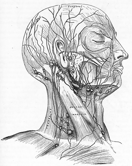
It is the context of the increasing institutionalization of medical teaching in Universities within established courses of regimented learning – including the mind-numbing ‘naming of the parts’– that helps to explain the ultimate success of the style-less style. It is this context that made it possible for an institutionalized classic, like Gray’s Anatomy, to be edited and re-edited over the years by successive professors, each of whom pass the torch jealously from generation to generation. Henry Gray’s Anatomy. Descriptive and Surgical, with 363 ‘drawings by H. V. [Vandyke] Carter, late Demonstrator of Anatomy at St George’s Hospital’ first appeared in a fat but unpretentious quarto volume in 1858, bound in matter-of-fact brown cloth (Gray, 1858). As Gray and successive editors emphasized, the aim was restrictedly practical, avoiding any taint of philosophic anatomy in favour of introducing such verbal and visual descriptions as would be useful for the aspiring surgeon and clinician. The tone of the text is sober to the point of anonymous flatness, while the woodcut diagrams use business-like lines of no more than three or four thicknesses to achieve a steady register of unseductive description, which varies little in its graphic conventions throughout the whole book (Fig. 23).
Fig. 23.
The Superficial Lymphatics and Glands of the Head, Face and Neck drawn by Henry Vandyke Carter, from Anatomy: Descriptive and Surgical, Henry Gray (1858).
The fact that the drawings of the lymphatics are acknowledged to have been based upon Mascagni only serves to highlight Gray’s distance in style from his predecessors. The avoidance of any appealing views of the whole body – even the depiction of a complete skeleton – and his functional setting of the illustrations of the detailed parts of the body within the pages of printed text consistently negate any tendency to think that we are dealing with the ‘arty’ production of a picture book. There is little modelling in light and shade, no attempt to place figures in graceful poses, and no evocative backgrounds.
The most laconic of prefaces is followed by the table of contents and list of illustrations, before the text opens with an 11-line definition of the branches of Anatomy. Then the reader is plunged in medias res with a detailed account of osteology. After an unrelenting progress through the various systems, his text ends abruptly with an account of the rectovesical fascia. The only conclusion is provided by a prodigious 30-page index. The whole book presents a heroically disciplined exercise in intellectual and visual restraint. Its overt functionalism unequivocally exudes the air of institutionalized science instruction in the mid-19th century. As a consequence, it has a special style all of its own.
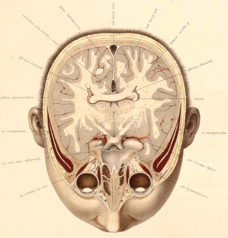
In most respects, Carter’s woodcuts for Gray’s Anatomy were deeply and consciously unadventurous and deliberately constrained in their visual means, predominantly depicting gross anatomy as a guide to the undertaking and viewing of dissections as preparatory to surgery. It is characteristic that Gray and Carter made only limited and rudimentary use of sections, perhaps the most striking of the 19th-century developments in anatomical illustration. Sectional anatomy had been attempted as early as the Renaissance, but it was only in the early 19th century that de Riemer developed a method for sectioning frozen bodies in such a way that the organs remained in their true locations (Fig. 24). (For an outline of the history of cross-sectional anatomy, see Eycleshymer & Schoemaker, 1923; pp. ix–xiii.) Particularly interesting from our present standpoint was Braune’s technique of depicting the sections directly on a transparent paper placed over a thin layer of ice on the surface of the section. As he explained, ‘when the drawings concern such an elaborate mechanism as the human body, every line must be true to nature and copied with the greatest care’ (Braune, quoted by Eycleshymer & Schoemaker, 1923, p. xi). Published in 1867 and 1872, followed by a number of subsequent editions and translations, his sections combined a high level of visual sophistication with an air of high scientific objectivity which gave them an obvious appeal – even if the reconstruction of the sections into a complete picture of the organs required a considerable act of visualization.
Fig. 24.
Horizontal Cross-Section of the Head, drawn by C. Schmiedel, from Topographisch-anatomischer Atlas nach durchschnitten an gefrornen Cadavern, Wilhelm Braune (1872). Courtesy of the US National Library of Medicine.
By the time Gray’s Anatomy appeared for the 22nd time in 1923, under the long-term editorship of R. Howden of Durham, it had grown to 1378 pages, with 1265 illustrations, of which 587 involved some degree of colour (Gray, 1923; for a full account of the making and editions of Gray’s book, see Richardson, 2008). A ‘signed’ photograph and brief biography of Gray certified the pedigree of the enterprise through its founder. By this time, we have to wait until page 162 for the osteological descriptions with which Gray had begun, having been first taken through histology and embryology. Over the years an accretion of revised texts, with reworked and supplementary drawings, meant that the original Gray was difficult to discern, and by the 27th edition in 1938, for which Johnston and Whillis were responsible, the volume had become a monument to changing conventions and techniques of illustration in a way that totally obscured the illustrative coherence which had been assiduously maintained by Gray himself. X-radiographs were used for the first time, which Johnston explains as ‘an innovation which some may regard as overdue, but it must be remembered that in the past an X-ray photograph often proved more confusing than informative to the student in the preclinical period’ (Gray, 1938, preface). Other new illustrations were based on the ‘beautiful drawings’ by A. K. Maxwell. Typical of what has happened is the provision of a reticently coloured illustration by Maxwell of the thigh, based on the 11th edition of Quain’s Anatomy, followed three pages later by an illustration of the right saphenous opening, one of Gray’s few surviving originals, which had originally performed a quite different function in one of the concluding sections of the 1858 text and which has now been enhanced with red arteries and blue veins. The two illustrations rely upon a quite different relationship between illustrative means and the seen object, and it is difficult to make coherent sense of the conflicting details. Gray’s famous book had become a hodge-podge of styles and little short of a visual disaster.
Successive editors have availed themselves of all the latest technologies of examination and techniques of representation. The function of Gray’s book had been reconsidered in the context of modern teaching and reference material. The 150th anniversary edition in 2008, edited by Susan Standring (Gray, 2008) and subtitled The Anatomical Basis of Clinical Practice, is a huge volume of 1551 pages, directed predominantly at a postgraduate audience. Nothing of Gray remains, other than the reassuring brand name that denotes enduring authority, and the book’s systematic attuning to its pedagogic function. Gray himself featured no histology, no embryology and no neuroanatomy, which occupy almost the whole of the first 400 pages of the new edition. Nor did he include any accounts of physiology, function or pathology, which now loom large. There is now little that is purely ‘descriptive’ in Gray’s understanding of the term, and there is none of his asides directed at the surgeon. Many of the new, high-tech illustrations of forms are predicated on and even illustrative of mechanical function and physiology rather than restricted to observed morphology. The radical changes, not least the shift from systematic to regional anatomy, mirror the massive transformation in what is considered to be the best presentation of appropriate knowledge for students of medicine at a particular level in the system of teaching. Along with the new content comes the new style that is needed for the new age – a style needs to speak visually of the very latest technologies.
Conclusion
I think it is true to say that every one of the anatomists we have encountered up to the time of Gray was aiming to produce what portraitists call a ‘speaking likeness’. Key questions in judging the nature of this likeness, as we have seen, repeatedly concern who is speaking in what language through what medium to whom and in what context. I would strenuously resist simple generalizations about the motives (explicit and implicit) of the protagonists in achieving such communication as they succeeded in establishing. The various permutations of intellectual, visual, economic, institutional, and political factors which bore in on the perceptual and representational processes involved in the making and reading of the illustrations varied greatly for different anatomists and illustrators working in different places at different times and on different projects. However, there were clearly styles of enterprise which were common to particular phases of anatomical illustration, whether these phases are characterized in large collective terms, such as humanist and modern, or more precisely by localized identifications of place and date. One of the virtues of entering the business of characterizing anatomical illustrations through style is that its very elasticity as a concept does not force the historian into rigid preconceptions about ‘the central issue’ in scientific representation – whether defined by social attitude, religious or political conviction, patronage, institutional structures, view of science, philosophical stance, or whatever. Rather, style allows a richness of categorization on the largest and smallest of scales, ranging from gross generalizations about the modern ‘non-style’ down to minute observations about the humanist fly in Bidloo’s dissecting room.
References
- Alberti LB. In: On Painting. Kemp M, editor. London: Penguin; 1435. translator Grayson C. [Google Scholar]
- Albinus BS. Tabulae sceleti et musculorum coporis humani. Basel: J. and H. Verbeek; 1747. [Google Scholar]
- Ballestriero R. Anatomical models and wax Venuses: art masterpieces or scientific craft works? J Anat. 2010;216:223–234. doi: 10.1111/j.1469-7580.2009.01169.x. [DOI] [PMC free article] [PubMed] [Google Scholar]
- Bell J. Engravings Explaining the Anatomy of the Human Body. Edinburgh: J. Patterson; 1794. [Google Scholar]
- Bell J. The Anatomy of the Human Body (with Charles Bell) Edinburgh: Cadell and Mudie; 1794. 4 vols. –1804. [Google Scholar]
- Bell J. Engravings Expressing the Anatomy of the Bones, Muscles and Joints. Edinburgh: Cadell and Mudie; 1797. [Google Scholar]
- Bell C. Essays on the Anatomy of the Expression in Painting. London: Longman Hurst Rees and Orme; 1806. [Google Scholar]
- Bell C. The Hand; its Mechanism and Vital Endowments as Evincing Design. London: Bell and Daldy; 1833. [Google Scholar]
- Bidloo G. Anatomia humani corporis. Amsterdam: Someren, Dyk, Hendrick & Boom; 1685. [Google Scholar]
- Bignamini I, Postle M. The Artist’s Model Exhibition Catalogue. Nottingham University: Kenwood House London; 1991. [Google Scholar]
- Bowron P, Kerber P. Pompeo Batoni exhibition catalogue Museum of Fine Arts Houston. New Haven: Yale University Press; 2007. [Google Scholar]
- Braune W. Topographisch-anatomischer Atlas nach Durchschnittern an gefrornen Cadavern. Leipzig: E. A Finke; 1872. [Google Scholar]
- Carlino A. Books of the Body. Chicago: Chicago University Press; 1999a. [Google Scholar]
- Carlino A. Paper Bodies: a Catalogue of Anatomical Fugitive Sheets 1538-1687. London: Wellcome Institute; 1999b. [PMC free article] [PubMed] [Google Scholar]
- Cheselden W. Osteographia or the Anatomy of the Bones. London: W Cheselden; 1733. [Google Scholar]
- Compagnon A. Paris: Seuil; “le Stile”; pp. 195–230. [Google Scholar]
- Corbey R. The Metaphysics of Apes: Negotiating the Animal-Human Boundary. Cambridge: Cambridge University Press; 2005. [Google Scholar]
- Daston L, Galison P. Objectivity. New York: Zone Books; 2007. [Google Scholar]
- Elsner J. Style. In: Nelson R, Shiff R, editors. Critical Terms for the History of Art. Chicago: Chicago University Press; 2003. [Google Scholar]
- Estienne C. De Dissectione partium corporis humani. [French translation La Dissection des Parties du Corps Humain] Paris: Simon de Colines; 1545. Carolus Stephanus. [Google Scholar]
- Estienne C. Dictionarum historicum geographicum poeticum. Geneva: Iacob Stoer; 1606. [Google Scholar]
- Eycleshymer A, Schoemaker D. A Cross-Section Anatomy. New York: Appleton-Century-Crofts; 1923. [Google Scholar]
- Fries L. Spiegel der Artzny. Strasburg: Schott; 1518. [Google Scholar]
- von Gersdorff H. Feldbüch der Wundartzney. Strasburg: Schott; 1517. [Google Scholar]
- Ginzburg C. Style: inclusion and exclusion. In: Ginzburg C, editor. Wooden Eyes. London: Verso; 2002. pp. 27–54. [Google Scholar]
- Gray H. Anatomy. Descriptive and Surgical. London: Parker and Son; 1858. [Google Scholar]
- Gray H. In: Gray’s Anatomy. 22nd edition. Howden R, editor. London: Longman; 1923. [Google Scholar]
- Gray H. In: Anatomy. Descriptive and Surgical. 27th edition. Johnston TB, Whillis J, editors. London: Longman; 1938. [Google Scholar]
- Gray H. In: Gray’s Anatomy: The Anatomical Basis of Clinical Practice. 40th edition. Standring S, editor. London: Elsevier; 2008. [Google Scholar]
- Greenblatt S. Renaissance Self-Fashioning. From Moore to Shakespeare. Chicago: University of Chicago Press; 2005. [Google Scholar]
- von Haller A. De partium corporis humani. Bern: Societas typographicae; 1778. [Google Scholar]
- Hunter W. Anatomia uteri humani gravidi (The Anatomy of the Human Gravid Uterus) Birmingham: J. Baskerville; 1774. [Google Scholar]
- Ingham K. Art and the theatre of mind and body: how contemporary arts practice is re-framing the anatomo-clinical theatre. J Anat. 2010;216:251–263. doi: 10.1111/j.1469-7580.2009.01166.x. [DOI] [PMC free article] [PubMed] [Google Scholar]
- Kaufman M. John Bell (1763–1820), the ‘father’ of surgical anatomy. J Med Biogr. 2005;13:73–81. doi: 10.1177/096777200501300204. [DOI] [PubMed] [Google Scholar]
- Kellett C. Perino del Vaga et les illustrations pour l’Anatomie d’Estienne’. Aesculapius. 1964;27:74–79. [Google Scholar]
- Kemp M. Alexander Nasmyth and the style of graphic eloquence. Connoisseur. 1970;173:93–100. [Google Scholar]
- Kemp M. ‘Intellectual ornaments’ style function and society in some instruments of art. In: Pittock J, Wear A, editors. Interpretation and Cultural History. London: Macmillan; 1991. pp. 135–152. [Google Scholar]
- Kemp M. ‘The Mark of Truth’: looking and learning in some anatomical illustrations from the Renaissance and eighteenth century. In: Bynum W, Porter R, editors. Medicine and the Five Senses. Cambridge: Cambridge University Press; 1993. pp. 85–121. [Google Scholar]
- Kemp M. Temples of the body and temples of the cosmos: vision and visualisation in the Vesalian and Copernican revolutions. In: Baigrie B, editor. Picturing Knowledge. Historical and Philosophical Problems Concerning the Use of Art in Science. Toronto: Toronto University Press; 1996. pp. 40–85. [Google Scholar]
- Kemp M. ‘A perfect and faithful record’. Mind and body in medical photography before 1900. In: Thomas A, editor. Beauty of Another Order. New Haven: Yale University Press; 1997. pp. 120–149. Exhibition catalogue National Gallery of Canada, Otttawa. [Google Scholar]
- Kemp M. The handyworke of the incomprehensible creator. In: Sherman C, Lukehart P, editors. Writing on Hands. Memory and Knowledge in Early Modern Europe. Washington, DC: Dickinson College and Folger Shakespeare Library; 2000. pp. 22–27. Exhibition catalogue. [Google Scholar]
- Kemp M. The Human Animal in Western Art and Science. Chicago: Chicago University Press; 2007. [Google Scholar]
- Kemp M, Wallace M. Spectacular Bodies. The Art and Science of the Human Body from the Renaissance to Now. Los Angeles: University of California Press; 2000. Exhibition catalogue, Hayward Gallery London. [Google Scholar]
- Maclise J. Surgical Anatomy. London: John Churchill; 1851. [Google Scholar]
- Mascagni P. Vasorum lymphaticorum corporis humani. Siena: Pazzini Carli; 1787. [Google Scholar]
- Mascagni P. Anatomia per uso degli studiosi di scultura et pittura. Florence: Giovanni Marenigh; 1816. [Google Scholar]
- Mascagni P. Anatomia universa. Pisa: Nicolo Capurro; 1823. 9 vols, –32. [Google Scholar]
- Mascagni P. Anatomia universale del professore Paulo Mascagni. Florence: Batelli e figli; 1833. [Google Scholar]
- Mazzotti G, Falconi M, Teti G, et al. The diagnosis of the death of Venerina. J Anat. 2010;216:271–274. doi: 10.1111/j.1469-7580.2009.01151.x. [DOI] [PMC free article] [PubMed] [Google Scholar]
- Monro A. Anatomy of the Humane Bones. Edinburgh: W Munro; 1726. [Google Scholar]
- Ormond R. Daniel Maclise 1806–1870. London, Dublin: National Portrait Gallery, National Gallery of Ireland; 1972. exhibition catalogue. [Google Scholar]
- Quain R. The Anatomy of the Arteries of the Human Body. London: Taylor and Walton; 1841. 2 vols, –44. [Google Scholar]
- Richardson R. The Making of Mr. Gray’s Anatomy. Oxford: Oxford University Press; 2008. [Google Scholar]
- Riva A, Conti G, Solinas P, et al. The evolution of anatomical illustration and wax modelling in Italy from the 16th to early 19th centuries. J Anat. 2010;216:209–222. doi: 10.1111/j.1469-7580.2009.01157.x. [DOI] [PMC free article] [PubMed] [Google Scholar]
- Roberts K, Tomlinson J. The Fabric of the Body. European Traditions of Anatomical Illustration. Oxford: Clarendon Press; 1992. [Google Scholar]
- Roy A. Gérard de Lairesse (1640–1711) Paris: Arthena; 1992. [Google Scholar]
- Ruysch F. Adversariorum anatomico-medico-chirurgicorum. Amsterdam: Janson-Waesberg; 1720. [Google Scholar]
- Sawday J. The Body Emblazoned. Dissection and the Human Body in Renaissance Culture. London: Routledge; 1997. [Google Scholar]
- Schupbach W. The Paradox of Rembrandt’s ‘Anatomy of Dr. Tulp’. London: Wellcome Institute; 1982. [PMC free article] [PubMed] [Google Scholar]
- Shapiro M. Style. In: Kroeberger J, editor. Anthropology Today. Chicago: University of Chicago Press; 1953. [Google Scholar]
- Simons P, Kornell M. Annibal Caro’s after-dinner speech (1536) and the question of Titian as Vesalius’s illustrator. Renaissance Quarterly. 2008;61:1069–1097. doi: 10.1353/ren.0.0297. [DOI] [PubMed] [Google Scholar]
- Smellie W. A Treatise on the Theory and Practice of Midwifery. London: D. Wilson; 1752. [Google Scholar]
- Stafford B. Body Criticism. Imaging the Unseen in Enlightenment Art and Medicine. Cambridge, MA: MIT Press; 1993. [Google Scholar]
- Thornton J. Jacob van Rymsdyk. Cambridge: Cambridge University Press; 1982. [Google Scholar]
- Tulp N. Observationum medicae. Amsterdam: Elsevier; 1641. [Google Scholar]
- Valverde de Hamusco J. Historia de la Composición del Cuerpo Humano. Rome: A de Salamanca and A. Lafreri; 1556. [Also published in Italian in 1556 and Latin in 1589] [Google Scholar]
- Vesalius A. De Humani Corporis Fabrica. Venice: Senensem and Germanum; 1543. [Google Scholar]
- Walter J. Observationes anatomicae. Berlin: G.A. Lange; 1775. [Google Scholar]
- Weston N. Daniel Maclise: an Irish Artist in Victorian London. Dublin: Four Courts Press; 2001. [Google Scholar]


