Abstract
Although the contribution to anatomical illustration by Vesalius and his followers has received much attention, less credit has been given to Veslingius and particularly Fabricius. By 1600, Fabricius had amassed more than 300 paintings that together made the Tabulae Pictae, a great atlas of anatomy that was highly admired by his contemporaries. Many of his new observations were incorporated into subsequent books, including those by Casserius, Spighelius, Harvey and Veslingius. Also of importance were the Tabulae by Eustachius (1552), which, although only published in 1714, greatly influenced anatomical wax modelling. In 1742, Pope Benedict XIV established a Museum of Anatomy in Bologna, entrusting to Ercole Lelli the creation of several anatomical preparations in wax. Felice Fontana realised that the production of a large number of models by the casting method would make cadaveric specimens superfluous for anatomical teaching and in 1771 he asked the Grand Duke to fund a wax-modelling workshop in Florence as part of the Natural History Museum, later known as La Specola. Fontana engaged Giuseppe Ferrini as his first modeller and then the 19-year-old Clemente Susini who, by his death in 1814, had superintended the production of, or personally made, more than 2000 models. In 1780, the Austrian Emperor Joseph II visited La Specola and ordered a great number of models for his Josephinum museum; these were made by Fontana with the help of Clemente Susini and supervised by the anatomist Paolo Mascagni. It is, however, in Cagliari that some of Susini’s greatest waxes are to be found. These were made when he was free of Fontana’s influence and were based on dissections made by Francesco Antonio Boi (University of Cagliari). Their distinctive anatomical features include the emphasis given to nerves and the absence of lymphatics in the brain, a mistake made on earlier waxes. The refined technical perfection of the anatomical details demonstrates the closeness of the cooperation between Susini and Boi, whereas the expressiveness of the faces and the harmony of colours make the models of Cagliari masterpieces of figurative art.
Keywords: anatomical illustrations, Boi, Cagliari, Fontana, Josephinum, La Specola, Susini, wax modelling
Introduction
Anatomical wax modelling started late in the 17th century in Italy on the basis of a long tradition of Italian anatomical illustration and aimed to convey, in the best possible way, the discoveries that had made anatomy the most advanced of the biological sciences. From the time of Vesalius’s ‘revolution’ (1543), which was to make the anatomist step down from his raised chair and perform what is now called autopsy (what I am seeing with my own eyes), to the beginnings of wax modelling, the manner of illustrating human anatomical specimens underwent a considerable evolution (Kemp, 2010, this issue). However, whereas the contribution to anatomical illustration by Vesalius (1514–1564) and his followers, such as Juan de Valverde (1520?–1580), Pieter Pauw (1564–1617), Volcher Coiter (1534–1576), Felix Platter (1536–1614), Caspar Bauhin (1560–1624) and Giulio Casseri (Casserius, 1552–1616), has been greatly emphasized, less credit has been given to Gerolamo Fabrici (Fabricius, 1533–1619) and Johann Vesling (Veslingius, 1598–1649), who taught anatomy in Padua, or to Eustachius (1505–1574), professor of anatomy in Rome (Roberts & Tomlinson, 1992). (The reason why Eustachius, who lived in the 16th century, is last in this list is because his main work surfaced only in 1714.) Before considering the history and importance of anatomical waxes, this article first considers the main features of anatomical illustration during this period.
Hieronymus Fabricius ab Aquapendente and his Tabulae Pictae
Gerolamo Fabrici d’Acquapendente (Fabricius, 1533–1619), anatomist and surgeon, pupil and successor of Gabriele Falloppia, held the chair of Anatomy in Padua for 50 years. His major contributions were on the human and comparative anatomy of the organs of sense, and on embryology. He was the first to describe the disappearance of the ductus arteriosus and of the umbilical vessels; he also discovered the organ in the fowl that now bears his name (bursa of Fabricius) and gave the name ‘ovarium’ to the organ in the hen that produces the eggs. In the De Venarum Ostiolis he described, and illustrated with beautiful copper-plate engravings, the valves of the veins, although he retained the old concept of Galen that venous blood flowed away from the heart. His Aristotelean research programme greatly influenced his students, among whom were Julius Casserius (1552–1616), Adrianus Spigelius (Spieghel, 1578–1625), William Harvey (1578–1657) and many others from all over Europe. Although Casserius became Fabricius’s fierce academic rival and Harvey reached conclusions on the valves of the vein opposite to those of his erstwhile teacher, all of their published works were based on Aristotle’s philosophy (Cunningham, 1997).
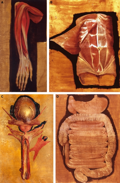
As he reported in the dedication of the De Visione Voce Auditu in 1600, Fabricius was preparing an atlas of both human and animal anatomy of which he had prepared more than 300 tables in carta regia, the largest paper format then in use. He states that, in contrast to those published by Vesalius, the body parts in his Tabulae were represented in their natural size and, more importantly, in their natural colours. Although greatly admired by Fabricius’s contemporaries, who, like his students, had free access to them, the Tabulae disappeared after his death and were only rediscovered in 1909 when Giuseppe Sterzi traced them to the Marciana, the State Library of Venice, through a document that stated that Fabricius had willed the Tabulae to the Republic of Venice (Riva et al. 2000; Ongaro, 2004; Rippa Bonati, 2004). The Marciana Tabulae include 169 oil-painted illustrations collected in eight files, with the other 43 being in three volumes that also contain five of Fabricius’s published works. Most of these pictures are still impressive for their realism and artistic value (Fig. 1A–D). The Tabulae are unlabelled and are the work of several unknown artists (Premuda, 1993; Kemp, 2004) but their quality (Riva, 2004) confirms both the great admiration that Fabricius’s contemporaries had for him and Sterzi’s statement that these pictures represent the most important anatomical work of the 16th to 17th centuries (Sterzi, 1909).
Fig. 1.
Fabricius: Tabulae Pictae. (A) Rari 116.16 (De anatomia muscolorum totius corporis): muscles of the right forearm; (B) Rari 117 1-2 (De anatomia): abdominal muscles and fasciae; (C) Rari 117.23 (De anatomia): male genital apparatus; (D) Rari 117.11–12 (De anatomia): small and large intestines, with demonstration of the rugae. Courtesy of the Marciana Library, Venice.
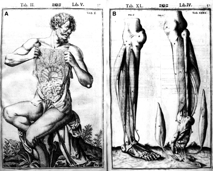
Analyses of the paintings by our group have demonstrated that several anatomical observations, apparently first reported many decades after Fabricius’s death, were actually first observed much earlier as they are reproduced in Fabricius’s Tables. These include the foramen of Monro, Sylvian fissure, arachnoid layer, bulbourethral glands and certain muscles (Riva, 2004; Riva et al. 2006; Collice et al. 2008). It should also be noted that many anatomical details, first seen in the Tabulae, are also present in the copper-engraved figures prepared by Casserius and published by Bucretius in 1627 as illustrations to Spigelius’s textbook and in Vesling’s Syntagma (Riva et al. 2001; Riva, 2004; Murakami et al. 2007). Comparison of Fabricius’s Tabulae (Fig. 1A–D) with the engravings (Fig. 2A,B) of his former pupil Casserius highlights the differences: Casserius, following Vesalius, dramatized his anatomical figures so that they were standing in ornate landscapes as if alive, whereas Fabricius’s paintings are simple representations of anatomical preparations but still clearly works of art. The great skill of the unknown artists in drawing and colouring the specimens seems to anticipate the wax models that would be made by Clemente Susini almost two centuries later (Kemp, 2004).
Fig. 2.
Casserius: Tabulae Anatomicae (1627). (A) Tab. II Lib. V: the figure, holding his own omentum, is standing in a beautiful garden; (B) Tab. XL Lib. IV: the dissected legs are standing on a lawn. Courtesy of Biblioteca Comunale Bonetta, Pavia.
Johannes Vesling and his Syntagma Anatomicum
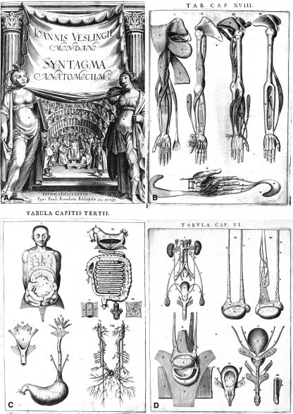
Although Johannes Vesling was born into a German, Catholic family in Mindel, Westphalia in 1598, he studied medicine in Leyden and Bologna. He then spent several years in Egypt as personal physician to Alvise Cornaro, the Venetian consul in Cairo, before returning to Italy where he taught human anatomy, first in Venice and then in Padua, where he held the chairs of both Anatomy and Botany. He died in 1649 and was buried in the cloister of the Basilica del Santo in a sumptuous baroque sarcophagus. His book Syntagma Anatomicum was issued in octavo in 1641 with no figures and in quarto with 24 full-page copper-plates in 1647 (both editions were published by Paolo Frambotti of Padua). The frontispiece of the Syntagma represents a public dissection held by Vesling in the old anatomical theatre built by Fabricius (Fig. 3A).
Fig. 3.
Johannes Vesling: Syntagma Anatomicum (1647). (A) Frontispiece: Vesling is depicted during a dissection held in Fabricius’s anatomical theatre in Padua; (B) Tab. Cap. XVIII: muscles of right upper arm, and of the left hand; (C) Tab. Cap. III: organs of the digestive tract and diagram of visceral nerves; (D) Tab. Cap. VI: male urogenital system. (A) Courtesy of Biblioteca Pinali Antica, Padua. (B–D) Courtesy of Thomas Fisher Rare Book Library, University of Toronto.
Although not his pupil, Vesling praises Fabricius in the preface, mentioning his close relationship with the illustrious Paduan School. Vesling describes, as clearly as possible and for the benefit of medical students, the parts of the body as they are seen at dissection. He also included a convincing description of the physiology of the heart and blood circulation based on Harvey’s Exercitatio Anatomica de Motu Cordis et Sanguinis in Animalibus. His description of the lymphatic circulatory system and his assertion that four pulmonary veins normally empty into the left atrium of the heart are observations ‘of particular scientific significance’ (Eimas, 1990). He also described the cerebral circle (predating Willis) and pancreatic duct (Roberts & Tomlinson, 1992). Vesling’s treatise was the most used anatomy textbook in Europe during the period 1650–1750 (Castiglioni, 1941). It was republished a number of times and translated into several languages including an English version by Nicholas Culpeter in 1653. In 1741 a Dutch version of Syntagma was the first illustrated Western anatomical tract to reach Japan (Ogawa, 1964; Murakami et al. 2007).
According to Choulant (1852), Vesling’s figures often served as models for illustrating anatomy textbooks later published in Northern Europe. The success of the book owes much to the simplicity and diagrammatic nature of the figures (Fig. 3B–D): ‘superficialities have been rejected, there are no landscapes’ (Roberts & Tomlinson, 1992). Although they did not have access to Fabricius’s Tabulae Pictae, the same authors commented that Veslingius’ pictures were the first not to be based on those in Vesalius’s De Corporis Humani Fabrica published more than a century earlier. Even if Vesalius was the author of the anatomical revolution, it was Fabricius and Vesling who freed anatomical figures from theatrical attitudes and ornate landscapes, making them more realistic and appropriate for medicine and surgery.
Eustachius and his Tabulae
Singer (1957) has pointed out that, ‘had these plates by Eustachius appeared in 1552 when they were made, his name would have stood with Vesalius as one of the founders of modern anatomy’. He goes on to say that they are more accurate than those of Vesalius and contain such a wealth of discoveries that, for originality, Eustachius has only Leonardo and Vesalius as superiors among the early anatomists. Bartolomaeus Eustachius (Bartolomeo Eustachi) was born at San Severino Marche and took his MD at La Sapienza University in Rome but had to return home on the death of his father in 1532. In 1539, he briefly held the post of second physician of the town of San Severino before his fame resulted in a call to the court of Urbino, then a flourishing centre of the humanities and science. In 1547, he became personal physician to the Duke’s brother, cardinal Giulio Della Rovere (a boy of 16, known as Cardinal d’Urbino) whom, 2 years later, he followed to Rome. Around 1550, he joined La Sapienza and held the chair of practical medicine there (1555–1566), with Pier Matteo Pini as first dissector. Despite his poor health, allegedly the cause of his bad temper, Eustachius had a successful medical practice becoming not only papal archiater (chief physician) but also physician to several important people, including Carlo Borromeo and Filippo Neri, both of whom were later sanctified. He died in 1574 in Fossato di Vico, while travelling to join Cardinal d’Urbino who had requested his medical services.
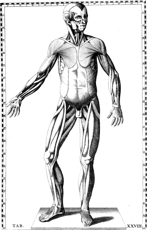
During his life, Eustachius published only the Opuscula Anatomica (Venetiis, 1563/4) containing six libelli (booklets), among which those on the kidney, teeth, auditory organ and venous system are of seminal importance. In 1552, under the direction of Eustachius and Pier Matteo Pini, 47 tables were drawn by Giulio de’Musi and then engraved in copper. Although treasured by Eustachius, they were unpublished and lost for many years. Unlike Vesalius, who used woodcut illustrations, Eustachi was one of the first to use copper engraving, which could show finer detail. Eustachius’s elegant illustrations (Fig. 4) have less dramatic poses than those of Vesalius and lack landscape backgrounds. To avoid superimposed lettering, the plates have graduated borders that allow grid references to be made, a method used by cartographers. Like those of Vesalius, however, the standing anatomical figures were represented as alive but with realistic rather than dramatic poses. The plates were eventually found and acquired by Pope Clemente XI through the efforts of Giovanni Maria Lancisi (1654–1720) who published them in 1714 with new captions to replace the lost originals. They stimulated such interest and praise among scientists that further editions, with new legends, were printed from the original blocks or from re-engravings. Particularly appreciated was the version edited in 1744 by the Dutch anatomist Bernard Sigfrid Weiss (1697–1770) whose name was latinized to Albinus. Three years later, Albinus published his famous atlas, based on Eustachius’s work; this greatly influenced Ercole Lelli, the founder of the Bologna school of ceroplastics, as well as Paolo Mascagni and Felice Fontana of La Specola Museum.
Fig. 4.
Eustachius: Tabulae Anatomicae (1714): Tab. XVIII, muscular figure. Courtesy of Biblioteca Centrale Biomedica, Cagliari.
Anatomical wax modelling: Zumbo and the school of Bologna
The use of wax modelling as a teaching aid for human anatomy is generally ascribed to the Sicilian abbot Gaetano Giulio Zummo (1656–1701, better known as Zumbo) (Lanza et al. 1979; Lemire, 1990; Chen et al. 1999; Ballestriero, 2007, 2010). In 1695, Zumbo moved to Genova where he was asked to reproduce in wax the dissections made by Guillaume Desnoues (1650–1735), chief surgeon and anatomist in the main hospital of the republic. Desnoues opened museums in Paris, and then in London, to exhibit wax anatomical models reproducing his dissections to the paying public. He maintained that wax preparations could allow people to learn anatomy while avoiding the horror of dissection. Following Desnoues’ example, several museums exhibiting human wax models to the public were then opened in central Europe, France and Britain, mostly for profit (Lemire, 1990; Musajo Somma, 2007; Bates, 2008). Of the anatomical models produced by Zumbo, only two magnificent heads survive today: one is at La Specola (see Ballestriero, 2010, this issue) and the other is in the Natural History Museum of Paris.
In the early 18th century, an academy for student instruction was founded by Luigi Ferdinando Marsili (1658–1630) in the Institutum Scientiarum et Artium of Bologna; it consisted of an anatomical chamber containing dried human preparations and was not part of the University. As the specimens deteriorated with use, Ercole Lelli (1702–1766), a talented artist who had a great interest in anatomy, was commissioned to create a series of anatomical models made of durable materials. Between 1732 and 1734 Lelli, on his own initiative and without asking for money, carved in umbrella pine the famous ‘scorticati’; these two magnificent wooden ecorchés (figures showing the muscles after the skin had been removed) were placed on either side of the chair in the Anatomical theatre of the Archiginnasio, the University palace. He also made wax models of a normal and a horseshoe kidney that were greatly praised by cardinal Prospero Lambertini (1675–1758) who later became Pope Benedict XIV (Ruggeri & Pontoni, 2005).
In 1742, Lambertini ordered a Museum of Anatomy to be established in the Institutum and entrusted to Lelli the creation of a considerable number of anatomical preparations in wax, including eight life-size statues in the style of those seen in Eustachius’ and Albinus’ tables (Maraldi et al. 2000). Over his 9-year programme of work, Lelli was helped by both a surgeon and, for 3 years, Giovanni Manzolini (1700–1755) who became very skilled in the art of wax modelling. In 1745, Manzolini left Lelli and continued producing anatomical preparations on his own, with the help of his wife Anna Morandi (1716–1774). After his death in 1755, Anna Morandi continued making wax models, achieving fame not only in Italy but also in European scientific circles (Focaccia, 2008). Unlike Lelli, who mostly devoted himself to osteology and myology, the Manzolinis preferred to reproduce the organs of sense and of the urogenital and cardiovascular systems. The Bolognese waxworks were initially made over human skeletons but later models were entirely artificial; they were, however, single works modelled by the artist on the basis of an anatomical preparation (Dacome, 2006). The works of Lelli, Manzolini and Morandi can now be seen in the Palazzo Poggi, the original site of the old Institutum that operated from 1711 to 1799 (Simoni, 2005).
Anatomical wax modelling: the school of Florence
In 1761, the publication in Venice of the De Sedibus et Causis Morborum by Giovanni Battista Morgagni (1682–1771) put an end to the holistic conception of Hippocrates and Galen by stressing the importance of organ pathology. This gave a new importance to surgery, which until then had been regarded as a minor discipline practiced by uneducated people who could not read Latin, then the standard language for medical textbooks. As they usually had a scanty knowledge of anatomy, particularly of internal organs (Belloni, 1980), there was an urgent need to instruct trainee surgeons in anatomy. This was a time when there were no freezing facilities nor chemicals able to preserve dissected bodies and it was Abbot Felice Fontana (1730–1805) who, having seen the Bolognese waxes when he was a student there, thought of producing a large number of anatomical models by the casting method for teaching purposes. Fontana was a brilliant scientist who was first a professor of logic and then of physics in the state University of Pisa. From 1766, he was in charge of the court cabinet of physics, the nucleus of the Natural History Museum that the Grand Duke Peter Leopold (1747–1792, youngest son of the Empress Marie Therese of Hapsburg, 1717–1780) planned to establish in Florence (Martelli, 1977).
The Bolognese technique of wax modelling had already been introduced to Florence in 1770 (Castaldi, 1947; Contardi, 2002) by Giuseppe Galletti (?–1819), a surgeon from the hospital of Santa Maria Novella in Florence. After seeing the obstetrical models made of wax and terra cotta manufactured in Bologna by Giovanni Manzolini and Giovanni Battista Sandi following a request of the surgeon Giovanni Antonio Galli (1708–1782), Galletti hired the sculptor Giuseppe Ferrini and produced some obstetrical and anatomical models that were noticed by Fontana. In 1771, Fontana asked the Grand Duke to fund a workshop as part of the museum. Peter Leopold was at first quite unreceptive to the idea as he was repelled by dissection but Fontana succeeded in persuading him by suggesting that a complete collection of anatomical models would make cadaveric specimens superfluous (Martelli, 1977; Lanza et al. 1979). Fontana engaged Ferrini in his workshop as his first modeller (contrary to Galletti’s wishes) and then the 19-year-old Clemente Susini (1754–1814) as second modeller, together with the dissector Antonio Matteucci and the painter Claudio Valvani, who, initially at least, made the drawings and explanatory tables (Castaldi, 1947; Martelli, 1977; Contardi, 2002; Musajo Somma, 2007). Numerous other dissectors and modellers joined the group, and individuals became specialized in tasks such as placing blood vessels, lymphatics, nerves or viscera (Azzaroli, 1977).
Unlike the Bolognese waxes, which usually contained the skeleton, the Florentine models were entirely made of wax, of various kinds and mixtures. The procedure for making the models was long and complex and only a brief summary is given here (for further details, see Hilloowala et al. 1995; Poggesi, 1999; Musajo Somma, 2007). The wax model was a close copy of a dissection made by the anatomist on the cadaver; Fontana himself did the initial dissections and was later joined by Antonio Matteucci, Tommaso Bonicoli (1746–1802), Filippo Uccelli (1770–1832) and the great Paolo Mascagni (1755–1815). Most dissections were patterned after the illustrations of famous European anatomists such as Albinus (1697–1770), Georg Thomas Asch (1729–1827), Petrus Camper (1722–1789), William Cheselden (1688–1752), David Cornelis De Courcelles (1810–?), Franz Joseph Gall (1758–1828), Albrecht von Haller (1708–1777), William Hunter (1718–1783), Johan Friedrich Meckel Jr (1781–1833), Alexander Monro Secundus (1733–1817), Johann Ernst Neubauer (1742–1777), Johann Georg Roederer (1726–1763), William Smellie (1697–1763), Samuel Thomas Sommering (1755–1830), Jan Swammerdan (1637–1680), Felix Vicq d’Azyr (1748–1794), Johann Gottfried Zinn (1727–1759), Domenico Cotugno (1736–1822), Antonio Scarpa (1747–1832), Felice Fontana and Paolo Mascagni themselves (Lanza et al. 1979; Lemire, 1990; Mazzolini, 2004; Musajo Somma, 2007; Massey, 2008).
Scarpa noted in 1786 that each model was made from a cadaver and this was confirmed in contemporary documents in the filze dei conti, the La Specola archives (see Scarpa, 1938; Martelli, 1977; Märker, 2006; Zanobio, 2007). Adult bodies were supplied by the hospital of Santa Maria Novella and child corpses by the Degli Innocenti orphanage (Märker, 2006). Specimens were made from either plaster moulds of the dissected material or a copy of the specimen executed in clay or rough wax. In the former case, a rope was inserted between the specimen and plaster so that the halves of the cast could be removed before the plaster had completely hardened. The inner surface of this negative mould was covered with several layers of translucent wax to which pigments had been added. A heavy filling material, sometimes with iron bars as supports, was often placed into the mould to ensure stability and the mould could also be filled completely with wax. The two pieces were then reassembled and the edges heated so that they would fuse. The plaster cast was then removed leaving the wax model, which was then refined by the modellers and had vessels and nerves added (the plaster casts could be used again – many are still stored in the museum). Most of the completed models were attached to tables of precious wood (mostly rosewood) covered by a silk blanket (Fig. 5) and given explanatory drawings with the main anatomical structures being indicated by numbers.
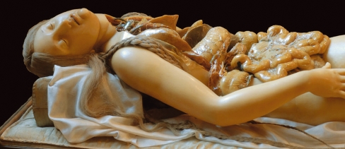
Fig. 5.
Detail of the statue representing ‘deep lymphatic vessels in a female subject’, ordered from Fontana by Scarpa and executed by Susini in 1794. Note the sleeping, serene expression of the face, typical of Fontana’s manner. Museum of the History of the University, Pavia. Photography by Alberto Calligaro.
The quality of the resulting figure depended, of course, on the skill of the modeller, who made the appropriate mixture of pigments and waxes for each preparation so as to imitate the original texture of the specimen and thus achieve a realistic result. The young Susini was soon seen to be particularly skilled and, when Ferrini left for the court of Naples in 1782, he was named first modeller, an assignment that he kept until his death in 1814, by which time he had personally made or superintended the production of more than 2000 models (Martelli, 1977; Lemire, 1990). He was succeeded by Francesco Calenzuoli (1769–1847) who had been his assistant since 1784.
The Imperial and Royal Museum of Physics and Natural History in Florence, the first of its kind to be open to the public, was inaugurated on 21st February 1775. Because of the observatory installed in its roof in 1780, the museum came to be known as La Specola (Azzaroli, 1977). At its opening, the anatomical section consisted of six large rooms exhibiting 137 showcases containing 486 models. There were also 200 drawings in colour with numbered lines and 157 explanatory hand-written sheets (Martelli, 1977; Musajo Somma, 2007). Wax models of various subjects continued to be created in the studio on the site of the museum until 1893, when, following the death of the last chief modeller Egisto Tortori (1829–1893), the workshop ended its activity. La Specola currently holds more than 1400 models grouped in 513 showcases representing human anatomical preparations that include 18 full-size entire human figures, in 65 showcases housing models of comparative anatomy and in five showcases containing waxworks by Gaetano Zumbo (Poggesi, 1999).
After Susini’s death, models continued to be made in the studio for other Florentine institutions such as the Technical Institute and the Museums of Botany and of Pathological Anatomy, and also for other Italian and foreign Universities. These included Cagliari, Bologna, Pavia, Pisa, Genoa, Perugia, Turin, Budapest, Leyden, Paris, Montpellier, London, Uppsala, Stockholm, New Orleans (whose models disappeared soon after 1900, Hilloowala et al. 1995) and possibly (but we were unable to obtain firm evidence of this) Charleston, Cairo and Lausanne (Castaldi, 1947).
The Josephinum collection
In 1780, the Austrian Emperor Joseph II (1741–1790), the elder brother of Grand Duke Peter Leopold, visited Florence and La Specola in the company of Giovanni Alessandro Brambilla (1728–1800), his personal surgeon and adviser. The emperor ordered such a large number of models that Peter Leopold vetoed the request on the grounds that it would have interfered with the Museum’s activity. The order was then taken on by Fontana himself who organized a second workshop at his home. Fontana hired many workers and was aided by both Clemente Susini and Paolo Mascagni, who acted as supervisor for the project. Over about 5 years, 1192 models were made and transported to Vienna. This required several journeys (the last in the autumn of 1788) that were long and laborious as the waxes had to be transported through Italy and over the Alps (Schmidt, 1996).
According to Lemire (1990), 150 models, among them several obstetrical preparations, were made from original casts, whereas the rest were made using those already stored at La Specola. The same author states that, although the collection as a whole looks even more magnificent than that of Florence, the pressure under which the wax modellers had to proceed in order to cope with the Emperor’s commission resulted in some models being of lower quality than the originals. Moreover, due to the harsh climate of Vienna, the models deteriorated and many had to be repaired. Today, there are 365 showcases containing 867 models of which 16 are entire human figures (Lemire, 1990; Lukić et al. 2003). The waxes were placed in the Caesareo-Regia medico-chirurgica Academia Josephina, which was inaugurated on November 7, 1785 with an opening address entitled ‘The pre-eminence and use of surgery’ read by the first director, Giovanni Alessandro Brambilla.
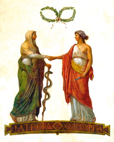
Brambilla used his influence on Joseph II to ensure that Latin was taught to Austrian surgeons so that they could study scientific texts and so be on an equal footing with physicians who attended universities where Latin was compulsory. First they were taught Latin, then anatomy and the other basic and clinical disciplines (Belloni, 1980). It was also thanks to Brambilla, who was born in the village of San Zenone al Po (province of Pavia, then part of the Austrian Empire), that the University of Pavia could secure the most brilliant Italian scientists such as Alessandro Volta (1745–1827), Lazzaro Spallanzani (1729–1799) and Antonio Scarpa (1752–1832), a friend of Fontana. It is not surprising that in the ceiling of the Anatomical Theatre, which was later named after Scarpa, there is a fresco (Fig. 6) commissioned by Brambilla depicting two ladies, each clad in the long gown of people learned in Latin, shaking hands and celebrating the parity between surgery and medicine (Zanobio, 1980).
Fig. 6.
Fresco commissioned by Giovanni Alessandro Brambilla in the ceiling of Pavia anatomical theatre (now Aula Scarpa) to celebrate the newly achieved parity between Surgery and Medicine. Photography by Alberto Calligaro.
The Cagliari collection
History
The Cagliari collection (Fig. 7) is very small in terms of the number of pieces (23 showcases for a total of 64 preparations) compared with the great collections of Florence and Vienna. It is, however, no less important as the models reflect the mature work of Susini (Ballestriero, 2010, this issue) and the ‘great artist’s last approach to his artistic vision’ (Cattaneo, 1970, 2007). According to the tags attached to each of the 23 showcases that form the collection, the models were made between 1803 and 1805, a time when Tuscany, despite the high-sounding name of ‘Kingdom of Etruria’, was reduced to a puppet state ruled by the French. By then, Fontana, in his early 70s, though still director of La Specola, had lost interest in wax modelling and was deeply engaged in the private workshop donated to him by Napoleon, who had been fascinated by his project of producing wooden anatomical models that could be touched, dismounted and reassembled (Castaldi, 1947; Martelli, 1977; Mazzolini, 2004; Märker, 2006). In 1804 Fontana was officially relieved of the task of supervising the wax modelling and was succeeded by Filippo Uccelli, who was elected to the position of anatomy teacher in the museum of La Specola (Märker, 2006).
Fig. 7.
Exhibition Room of the Museum of Cagliari. Photography by Gabriele Conti.
It was during the last years of the 18th century that the status of the modellers seems to have gradually changed from that of artisans to that of artists; they, and Susini in particular, started to receive recognition as the true authors of the La Specola waxes that in previous years had usually been reported as the works of Fontana (Märker, 2006). Thus, over the 3 years that he worked on the Cagliari commission with Francesco Antonio Boi, Susini was no longer under Fontana’s tutorship (Fig. 5) and was at last free to fully express himself. This view is supported by the fact that all 23 of the Cagliari showcases bear the date and Susini’s signature; this was unusual as, to our knowledge, only a handful of models in the collections of Florence and Bologna are signed by Susini, and just one in the Josephinum (Schmidt, 1996).
The models were ordered from the Florentine workshop by Carlo Felice of Savoy, Viceroy of Sardinia, through the Sardinian anatomist Francesco Antonio Boi who spent a period of leave at the department of surgical anatomy of the Santa Maria Novella hospital (Castaldi, 1947). Boi was born in 1767 to a family of farmers in the village of Olzai in the Barbagia di Ollolai in the district of Nuoro, central Sardinia. Because of his proficiency in elementary school, he was, according to the custom of the time, directed toward ecclesiastical studies. At the age of 18, however, he left the Seminary of Oristano and went to Cagliari to study medicine. Despite the fact that, in order to earn his living, he had to work as preceptor in the house of the Chief Customs Officer, he obtained his medical degree in 1795. He soon acquired a good reputation and in 1799 was appointed to the chair of Human Anatomy which, since its institution in 1764, had been given to professors of other subjects (Castaldi, 1947; Sorgia, 1986; Dodero, 1999). As no students enrolled for the anatomy lessons in 1801, Boi obtained financial support from the Viceroy Carlo Felice to take sabbatical leave on the Italian peninsula to improve his knowledge of anatomy. He went first to Pavia, where the chair of Anatomy was then held by Antonio Scarpa, the most illustrious Italian anatomist of the time. He then moved to Pisa and on to Florence where, although there was no university, anatomical studies were flourishing at the Arcispedale di Santa Maria Nova under the direction of Paolo Mascagni, who had recently moved there after periods at the Universities of Pisa and Siena.
It is to Boi’s sojourn in Florence that we owe the collection of Cagliari; Boi commissioned the waxes from Clemente Susini by personal order of Carlo Felice and, although no documentary evidence has been found, it seems likely that this was why Boi continued to be funded by the Sardinian government until 1805, when the models were completed. Meloni Satta (1877) states that it was Boi himself who performed the dissections reproduced by Susini. The waxes cost 14 800 lire, funded from Carlo Felice’s personal budget (Cara, 1872). This was a very substantial sum; Susini, as chief modeller of La Specola Museum, earned just 1440 lire per year (Castaldi, 1947) and the high price may be related to the cost of the wax. Unlike most of the models produced by the Florentine studio, those of Cagliari are mainly made of solid wax; the exceptions are model III, two livers and one lung that are internally void. Moreover, although we do not know its exact composition, the wax mixture used for the models of Cagliari must have been especially prepared in order to survive the high summer temperatures of southern Sardinia. Unlike those in other collections, the Cagliari models have survived more than 200 years without the need for restoration (Manzoli & Mazzotti, 1987; Lemire, 1990; Riva, 2007).
The wax anatomical models arrived in Cagliari in 1806 and that same year Boi resumed his teaching in the Medical School (Castaldi, 1947). Susini’s wax models were placed in the Museum of Antiquities and Natural History, founded by Carlo Felice, later called ‘Museo d’antichità della Regia Università degli studj di Cagliari’, which was on the ground floor of Palazzo Belgrano (Sorgia, 1986; Bullitta, 2005). In 1857, the main body of the museum was moved to another building but the wax models were granted to the University and placed under the charge of the Professor of Anatomy to be used for teaching purposes. In 1991 they were transferred to the present location in Cagliari Citadel of Museums (Riva, 2007).
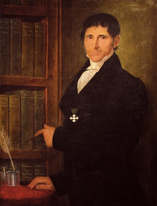
Boi had a very successful academic and professional career. In 1818 he was named ‘Archiater of the Kingdom of Sardinia’, an office comparable to that of a modern Minister of Health; in 1824 he was ennobled and in 1841 the cross of the Ordine Mauriziano, one of the most prestigious honours of the Kingdom, was bestowed on him, as can be seen in his official portrait, now exhibited in the museum. In this portrait (Fig. 8) Boi indicates, with his left index finger, a shelf containing six textbooks, four in Latin and two in French, each bearing the name of the author and title engraved in gold. These are: Marie Francois Xavier Bichat (1771–1802) Anatomie generale; Felix Vicq d’Azyr (1748–1794) Tabulae Anatomicae; Peter Johann Frank (1745–1821) Epitome de Curandis Morbis; Paolo Mascagni (1755–1815) Vasorum Absorbentium Historia; Anthelme Balthasar Richerand (1779–1840) Physiologie; and Samuel Thomas Soemmerring (1755–1830) De Corporis Humani Fabrica. According to Castaldi (1947), these books represent the medical works most liked by Boi. As a whole, they are a testimony to Boi’s broad European cultural background; only one, not surprisingly the work on lymphatics by his erstwhile teacher Mascagni, is authored by an Italian. Boi died in Cagliari in 1855.
Fig. 8.
Portrait of Francesco Antonio Boi. Unknown painter of the second half of the 19th century. Photography by Alessandro Cadau.
The collection
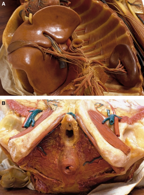
All of the models (Table 1) are unique and differ from those produced in the La Specola wax workshop both earlier and later. They are all from human dissections, with the single exception of the boiled bovine tongue of showcase XVI, prepared according to Malpighi in order to show the lamination of its external layers (Zanobio, 2007). No whole human figures are represented. The most complete preparations are those contained in cases III and XII, which demonstrate the head and trunk of a female and a male cadaver, respectively. A distinctive feature of the collection is the importance given to both visceral and somatic nerves, which are accurately shown in more than one-third of the models. The representation of nerves in table XII, particularly those of the cardiac, celiac (Fig. 9A) and pelvic plexuses, compete with – or even surpass in precision – the most celebrated textbooks of the first half of the 19th century. A model that demonstrates the exceptional skill of Boi as a dissector is the preparation of the female perineum of case III (Fig. 9B); this shows the structure of the female external genital organs with details not matched until the recent study by O’Connel et al. (1988).
Table 1.
The 23 showcases of the Cagliari collection (Riva et al. 1997).
| I | Preparations of general and microscopic anatomy (21 pieces) |
| II | Deep dorsal muscles from sacrum to occiput |
| III | Head and body of a young girl showing in particular muscles, vessels (mostly arteries), left pectoral lymph nodes, right mammary gland and perineum |
| IV | Diaphragm muscle |
| V | Muscles of the hip as seen from the front |
| VI | Muscles of the hip as seen from the back |
| VII | (1) Plantar aponeurosis of the foot; (2) interosseous muscles of the foot as seen from the sole |
| VIII | (1) Deep layer of the plantar muscles of the foot; (2) middle layer of the plantar muscles of the foot |
| IX | (1) Muscles of the pharynx as seen from the back; (2) palate and nasopharynx as seen from the bottom |
| X | (1) Open pharynx as seen from the back; (2) larynx as seen from the front; (3) hyoid bone (broken) as seen from the top |
| XI | (1) Pharynx cavity; (2) laryngeal and pharyngeal branches of the vagus nerve, and ansa hypoglossi; (3) laryngeal nerves |
| XII | Male figure showing the head (with demonstration of the encephalic trunk), neck, thorax, abdomen pelvis and relevant viscera, genital organs and left upper limb with dissected shoulder joint. Vessels and nerves, with those of the viscera in particular, have been reproduced in considerable detail |
| XIII | Male head and neck showing the surface of the encephalon with the convolutions and superficial vessels of the brain, divisions of the trigeminal nerve, and hypoglossal nerve |
| XIV | Organ of touch (four pieces). Two models of left hand sectioned at the wrist and seen from their palmar aspect: one is covered by skin and the other is dissected to show its muscles and nerves. The other two models are enlargements of the two distal phalanges of a longitudinally sectioned finger and of the last phalanx of the thumb with the nail removed and its epidermis raised to show the dermal ridges |
| XV | Organ of smell (two pieces). Parasagittal sections of the nasal cavity, one showing the nasal septum and the other showing the nasal conchae, together with preparations of their relevant nerves |
| XVI | Organ of taste (three pieces). (1) Dissected head and neck, showing the encephalic trunk, arteries and nerves of orbit, face, tongue and neck; (2) boiled veal tongue, with its covering removed on the right side to demonstrate its lamination; (3) human tongue with its vascular supply; the epithelium is raised so as to show the chorion of the mucosa with its papillae |
| XVII | Organ of hearing (12 pieces). Twelve enlarged parts of the external, middle and internal ear with muscles and nerves |
| XVIII | Organ of vision (12 pieces). (1) Head with the lateral wall removed and the skull opened to show the encephalic trunk and nerves of the orbit; (2) orbit with the ocular globe muscles and vessels; (3) lacrimal apparatus; (4) enlarged fragment of the superior eyelid seen from its conjunctival surface; (5–12) eight variously dissected eyeballs |
| XIX | Liver; stomach, duodenum, pancreas and spleen, with their vessels |
| XX | Male urogenital system with arteries and veins |
| XXI | Female urogenital system with arteries and veins; as in the following models, the thighs are frontally sectioned close to the groin in order to show the muscles with their fasciae and nerves and vessels |
| XXII | Female urogenital system with relevant vessels and opened uterus during pregnancy |
| XXIII | Opened female abdomen with vessels and the uterus at the end of pregnancy |
Fig. 9.
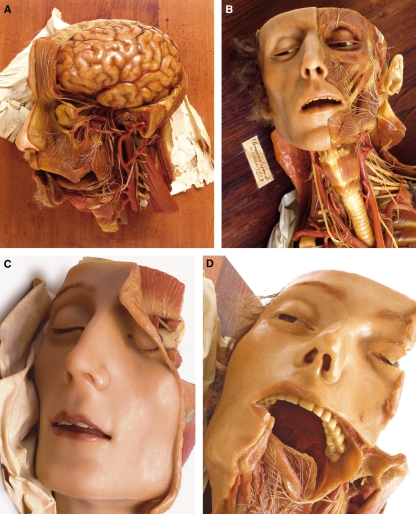
Examples from the Cagliari anatomical wax collection. (A) Detail of case XII model showing the coeliac plexus; note that the hepatic artery is double. (B) Detail of case III model: dissection of the female perineum. Note the relationship of the bulbs of the vestibule (officially renamed corpora spongiosa clitoridis by the Federative International Committee on Anatomical Terminology, 2007) with the urethra and clitoris. Photography by Dessì & Monari.
A finding that distinguishes the waxes of Cagliari from those of Florence and Vienna, and even from those of Bologna made in 1810, is the absence of lymphatics in the brain (Fig. 10A). Lymphatics are represented in brain preparations of these other collections and are said to be based on a mistake of Paolo Mascagni, who erroneously depicted them in his textbooks (Lukić et al. 2003).
Fig. 10.
Cagliari collection. (A) Detail of case XIII model: the brain is devoid of lymphatics; note also the detailed representation of nerves and the correct configuration of cerebral convolutions. (B) Detail of case XII model; note the tag with Susini’s signature and date. (C) Detail of case XVIII model. (D) Detail of case XVI model. Photography by Dessì and Monari.
Considering the preparations as a whole, one cannot but be impressed by the organic unity of the collection, which reflects an intelligent selection of topics with respect to their scientific and didactic usefulness. All of the models fit perfectly with the requirements that Antonio Scarpa laid down for the use of the waxes in anatomy and are in his letters (Zanobio, 1979, 2007). His view was that the models can be conveniently used for demonstrating: (i) all parts of the human body that cannot be preserved for long periods; (ii) all parts that cannot be entirely demonstrated from a single point of view; and (iii) all parts that are hardly visible and especially those that require the use of a microscope. A further statement made by Scarpa in a letter (1786) addressed to Gregorio Fontana (1735–1803), brother of Felice Fontana, attests that no model was produced in La Specola in the absence of the cadaver (Scarpa, 1938). This statement finds support in the presence of such anatomical variations as the double hepatic artery seen in case XII (Fig. 9A). The fact that variants are represented in these models is of great relevance for the teaching of clinical anatomy because, as a rule, they are not illustrated in modern anatomy textbooks.
Conclusion
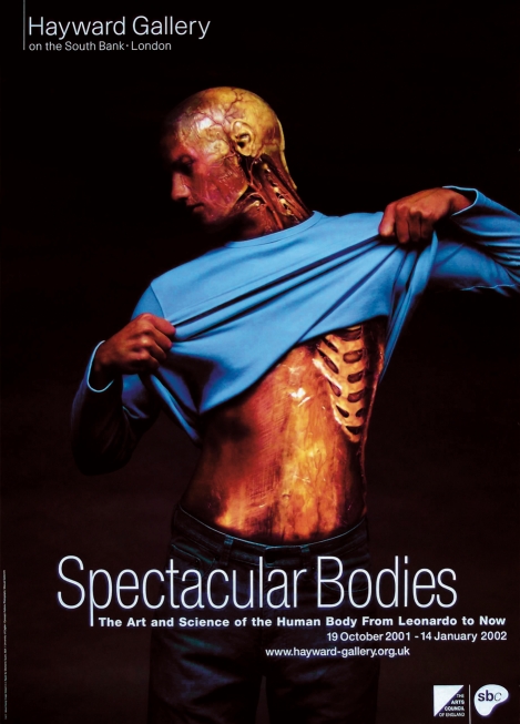
The 17th–18th century origins of anatomical waxes lay firstly in the need to provide more visual information than was possible in the two-dimensional illustrations then available and secondly in the lack of effective preservation techniques for cadavers, which made dissection of deteriorating bodies highly unpleasant. This is shown by the fact that many of the waxes display autolysis, the early works of Zumbo showing even more advanced stages of putrefaction. It is interesting to note that what started out as a practical craft soon started to become an art (Ballestriero, 2010, this issue). The quality of the reproductions became better and better, the waxes were treated with respect and often expensively mounted in high-quality cases, and it soon became clear that there was no better way of teaching human anatomy to students. The waxes at La Specola and Cagliari are still used for this purpose as they are far more lifelike than embalmed cadavers. They are also a lot more beautiful! Even today, we can admire the artistry of people who devoted their lives to wax modelling, and to the very skilled surgeons who made the dissections. This is particularly well demonstrated in the Cagliari waxes; here, the refined technical perfection with which anatomical details are reproduced confirms the close cooperation between Susini and Boi. Moreover, the realistic expressiveness of the faces, which really do seem to be portraits, together with the harmony of colours (Fig. 10A–D) make the models of Cagliari true masterpieces of figurative art. The exceptional quality of the Cagliari collection has been greatly appreciated in important exhibitions on art and anatomy (Fig. 11) and repeatedly acknowledged (Lanza et al. 1979; Lemire, 1990; Mazzolini, 2004; Musajo Somma, 2007).
Fig. 11.
Advertising poster based on model III of the Cagliari Collection. Spectacular Bodies (curators: M. Kemp and M. Wallace), Hayward Gallery, London, 19 October 2001–14 January 2002. Concept by Publicis. Photography by Masuad Golsorkhi.
Acknowledgments
We are greatly indebted to Gillian Morris Kay and Jonathan Bard for carefully editing the text. To Jonathan Bard we owe the section headed Conclusion.
References
- Azzaroli ML. La ceroplastica nella scienza e nell’arte. Atti del 1 Congresso Internazionale. Firenze: Leo S Olschki Editore; 1977. La Specola. The zoological museum of Florence University; pp. 1–22. [Google Scholar]
- Ballestriero R. The art of ceroplastics. Clemente Susini and the collection of the anatomical wax models of the University of Cagliari. In: Riva A, editor. Flesh and Wax. The Clemente Susini’s Anatomical Models in the University of Cagliari. Nuoro: Ilisso; 2007. pp. 35–45. [Google Scholar]
- Ballestriero R. Anatomical models and wax venuses: art masterpieces or scientific craft works? J Anat. 2010;216:223–234. doi: 10.1111/j.1469-7580.2009.01169.x. [DOI] [PMC free article] [PubMed] [Google Scholar]
- Bates AW. ‘Indecent and demoralising representations’: public anatomy museums in mid-Victorian England. Med Hist. 2008;52:1–22. doi: 10.1017/s0025727300002039. [DOI] [PMC free article] [PubMed] [Google Scholar]
- Belloni L. Per la storia della medicina. Bologna: Arnaldo Forni Editore; 1980. [Google Scholar]
- Bullitta P. L’Università degli Studi di Cagliari dalle origini alle soglie del terzo millennio. Cagliari: Telema Mythos Edizioni; 2005. [Google Scholar]
- Cara G. Notizie sul museo di antichità della Regia Università di Cagliari. Cagliari: Tipografia Simon; 1872. [Google Scholar]
- Castaldi L. Francesco Boi (1767–1860), primo cattedratico di Anatomia Umana a Cagliari e le Cere Anatomiche fiorentine di Clemente Susini. Firenze: Leo S. Olschki; 1947. [Google Scholar]
- Castiglioni A. A History of Medicine. New York: Alfred A Knopf; 1941. [Google Scholar]
- Cattaneo L. Le Cere Anatomiche di Clemente Susini dell’Università di Cagliari. San Casciano Val di Pesa: Università di Cagliari; 1970. [Google Scholar]
- Cattaneo L. Presentation of the collection of Cagliari. In: Riva A, editor. Flesh and Wax. The Clemente Susini’s Anatomical Models in the University of Cagliari. Nuoro: Ilisso; 2007. pp. 35–45. [Google Scholar]
- Chen J, Amar A, Levy M, et al. The development of anatomical art and sciences. The Ceroplastica anatomic models of La Specola. Neurosurgery. 1999;46:883–891. doi: 10.1097/00006123-199910000-00031. [DOI] [PubMed] [Google Scholar]
- Choulant L. History and Bibliography of Anatomic Illustration in its Relation to Anatomic Sciences and the Graphic Arts. Chicago, IL: The University of Chicago Press; 1852. [Google Scholar]
- Collice M, Collice R, Riva A. Who discovered the Sylvian fissure? Neurosurgery. 2008;63:623–628. doi: 10.1227/01.NEU.0000327693.86093.3F. [DOI] [PubMed] [Google Scholar]
- Contardi S. La casa di Salomone a Firenze: l’imperiale e reale Museo di fisica e storia naturale. Firenze: Leo S Olschki; 2002. pp. 1775–1801. [Google Scholar]
- Cunningham A. The Anatomical Renaissance. The Resurrection of the Anatomical Projects of the Ancients. Aldershot: Scholar Press; 1997. [Google Scholar]
- Dacome L. Waxworks and the performance of anatomy in mid-18th-century Italy. Endeavour. 2006;30:29–35. doi: 10.1016/j.endeavour.2006.01.004. [DOI] [PubMed] [Google Scholar]
- Dodero G. Storia della medicina e della sanità pubblica in Sardegna. Cagliari: Aipsa; 1999. [Google Scholar]
- Eimas R. Heirs of Hippocrates. Iowa City: University of Iowa Press; 1990. [Google Scholar]
- Focaccia M. Anna Morandi Manzolini, una donna tra arte e scienza. Firenze: Leo S Olschki; 2008. [Google Scholar]
- Hilloowala R, Lanza B, Azzaroli Puccetti ML, et al. The Anatomical Waxes of La Specola. Firenze: Arnaud; 1995. [Google Scholar]
- Kemp M. Il mio bell’ingegno. L’Anatomia visiva. In: Rippa Bonati M, Pardo-Tomàs J, editors. Il teatro dei corpi, le pitture colorate di anatomia di Fabrici d’Acquapendente. Milano: Mediamed Edizioni Scientifiche; 2004. pp. 83–107. [Google Scholar]
- Kemp M. Style and non-style in anatomical illustration: from renaissance humanism to Henry Gray. J Anat. 2010;216:192–208. doi: 10.1111/j.1469-7580.2009.01181.x. [DOI] [PMC free article] [PubMed] [Google Scholar]
- Lanza B, Azzaroli Puccetti ML, Poggesi M, et al. Le Cere Anatomiche della Specola. Firenze: Arnaud; 1979. [Google Scholar]
- Lemire AM. Artistes et mortels. Paris: Chabaud; 1990. [Google Scholar]
- Lukić IK, Glunčić V, Ivkić G, et al. Virtual dissection: a lesson from the 18th century. Lancet. 2003;362:2110–2113. doi: 10.1016/S0140-6736(03)15114-8. [DOI] [PubMed] [Google Scholar]
- Manzoli FA, Mazzotti G. Il museo di anatomia umana. In: Tega W, editor. Storia illustrata di Bologna, i musei dell’Università. Bologna: AIEP Editore; 1987. pp. 201–220. [Google Scholar]
- Maraldi NM, Mazzotti G, Cocco L, et al. Anatomical waxwork modelling: the history of the Bologna anatomy museum. Anat Rec. 2000;261:5–10. doi: 10.1002/(SICI)1097-0185(20000215)261:1<5::AID-AR3>3.0.CO;2-U. [DOI] [PubMed] [Google Scholar]
- Märker A. The anatomical models of La Specola: production, uses, and reception. Nuncius. 2006;21:295–321. [PubMed] [Google Scholar]
- Martelli A. La ceroplastica nella scienza e nell’arte. Atti del 1 Congresso Internazionale. Firenze: Leo S Olschki; 1977. La nascita del reale gabinetto di fisica e storia naturale di Firenze e l’anatomia in cera e legno di Felice Fontana; pp. 103–134. [Google Scholar]
- Massey L. On waxes and wombs, eighteenth-century representation of the gravid uterus. In: Panzanelli R, editor. Ephemeral Bodies, Wax Sculpture and Human Figure. Los Angeles: Getty Research Institute; 2008. pp. 83–106. [Google Scholar]
- Mazzolini R. Plastic anatomies and artificial dissections. In: Chaderavian S, Hopwood N, editors. Models the Third Dimension of Science. Stanford: Stanford University Press; 2004. pp. 43–70. [Google Scholar]
- Meloni Satta P. Ricordi storici: effemeride sarda. Sassari: G Dessì; 1877. [Google Scholar]
- Murakami M, Rippa Bonati M, Riva A. Fabricius’s De venarum ostiolis, 1st translation into Japanese. Invited lecture with a critical introduction. J Phys Soc Japan. 2007;69:54–70. in Japanese. [Google Scholar]
- Musajo Somma L. In cera. Anatomia e medicina nel XVIII secolo. Bari: Progedit; 2007. [Google Scholar]
- O’Connel HE, Hutson JM, Anderson CR, et al. Anatomical relationship between urethra and clitoris. J Urol. 1988;159:1892–1897. doi: 10.1016/S0022-5347(01)63188-4. [DOI] [PubMed] [Google Scholar]
- Ogawa T. History of Medicine. The First Anatomical Record in Japan. Zoh-shi (in Japanese) Tokyo: Chuko Shinsho; 1964. [Google Scholar]
- Ongaro G. Fabrici: dai manoscritti alla stampa. In: Rippa Bonati M, Pardo-Tomàs J), editors. Il teatro dei corpi, le pitture colorate di anatomia di Fabrici d’Acquapendente. Milano: Mediamed Edizioni Scientifiche; 2004. pp. 156–169. [Google Scholar]
- Poggesi M. La collezione ceroplastica del museo ‘La Specola’. In: Lamers-Schütze P, Havertz Y, editors. Encyclopedia anatomica. Köln: Taschen; 1999. pp. 6–46. [Google Scholar]
- Premuda L. Storia dell’iconografia anatomica. Oreggio: Ciba-Geigy; 1993. [Google Scholar]
- Rippa Bonati M. Anatomia in mostra. In: Rippa Bonati M, Pardo-Tomàs J, editors. Il teatro dei corpi, le pitture colorate di anatomia di Fabrici d’Acquapendente. Milano: Mediamed Edizioni Scientifiche; 2004. pp. 17–30. [Google Scholar]
- Riva A. Priorità anatomiche nelle Tabulae Pictae. In: Rippa Bonati M, Pardo-Tomàs J, editors. Il teatro dei corpi, le pitture colorate di anatomia di Fabrici d’Acquapendente. Milano: Mediamed Edizioni Scientifiche; 2004. pp. 147–155. [Google Scholar]
- Riva A. The collection of wax anatomical models by Clemente Susini at the University of Cagliari. In: Riva A, editor. Flesh and Wax. The Clemente Susini’s Anatomical Models in the University of Cagliari. Nuoro: Ilisso; 2007. pp. 9–14. [Google Scholar]
- Riva A, Segawa A, Lai I, et al. The Clemente Susini collection of wax models of the University of Cagliari. Ital J Anat Embryol. 1997;102:77–84. [Google Scholar]
- Riva A, Orrù B, Testa Riva F. Giuseppe Sterzi (1876–1919) of the University of Cagliari: a brilliant neuroanatomist and medical historian. Anat Rec (New Anat) 2000;261:105–110. doi: 10.1002/1097-0185(20000615)261:3<105::AID-AR5>3.0.CO;2-H. [DOI] [PubMed] [Google Scholar]
- Riva A, Orru’ B, Pirino A, et al. Iulius Casserius (1552–1616): The self-made anatomist of Padua’s golden age. Anat Rec (New Anat) 2001;265:168–175. doi: 10.1002/ar.1151. [DOI] [PubMed] [Google Scholar]
- Riva A, Riva L, Conti G. The first coloured atlas of anatomy: discoveries in Fabricius’s tabulae pictae. Morphol Newsl (Moscow) 2006;3–4:132–134. [Google Scholar]
- Roberts KB, Tomlinson JDW. The Fabric of the Body. European Tradition of Anatomical Illustrations. Oxford: Clarendon; 1992. [Google Scholar]
- Ruggeri A, Pontoni L. L’insegnamento dell’anatomia nelle sedi universitarie. In: Campanili G, Guarino M, Lippi G, editors. Le arti della salute. Milano: Skira; 2005. pp. 457–469. [Google Scholar]
- Scarpa A. In: Epistolario (1772–1832) Sala G, editor. Pavia: Società Medico Chirurgica; 1938. [Google Scholar]
- Schmidt G. Sul contributo di Paolo Mascagni alla collezione viennese di cere anatomiche nel Josephinum. In: Vannozzi F, editor. La scienza illuminata, Paolo Mascagni nel suo tempo. Siena: Nuova Immagine Editrice; 1996. pp. 101–110. 1755–1815) [Google Scholar]
- Simoni F. Le cere anatomiche del museo di palazzo Poggi a Bologna. In: Campanili G, Guarino M, Lippi G, editors. Le arti della salute. Milano: Skira; 2005. pp. 469–470. [Google Scholar]
- Singer C. A Short History of Anatomy and Physiology from the Greeks to Harvey. New York: Dover Publications; 1957. [Google Scholar]
- Sorgia G. Lo studio generale cagliaritano. Storia di una università. Cagliari: Università degli Studi; 1986. [Google Scholar]
- Sterzi G. Le ‘Tabulae Anatomicae’ ed i loro codici marciani con note autografe di Hieronymus Fabricius ab Aquapendente. Anat Anzeiger. 1909;35:338–348. [Google Scholar]
- Zanobio B. Le Cere Anatomiche di Clemente Susini dell’Università di Cagliari, testimonianza di una stagione della scienza italiana. Rass Med Sarda. 1979;82:1–11. [Google Scholar]
- Zanobio B. Giovanni Alessandro Brambilla nella cultura medica del Settecento Europeo. Pavia: Istituto Editoriale Cisalpino-La Goliardica; 1980. [Google Scholar]
- Zanobio B. The anatomical wax models of the university of Cagliari as witnesses of a season of Italian science. In: Riva A, editor. Flesh and Wax. The Clemente Susini’s Anatomical Models in the University of Cagliari. Nuoro: Ilisso; 2007. pp. 47–50. [Google Scholar]


