Abstract
The correspondences and disparities between how artists and anatomists view the body have historically been a source of creative collaboration, but how is this imaginative interdisciplinarity sustained and expressed in a contemporary context? In this review I suggest that contemporary artists engaging with the body, and the corresponding biomedical and architectural spaces where the body is investigated, are engendering innovative and challenging artworks that stimulate new relationships between art and anatomy. Citing a number of examples from key artists and referencing some of my own practice-based research, I posit that creative cross-fertilization provokes a discourse between mediated public perceptions of disease, death and the disposal of morbid remains, and the contemporary reality of biomedical practice. This is a dialogue that is complex, rich and diverse, and ultimately rewarding for both art and anatomy.
Keywords: anatomical theatre, art and bioscience, art and neuroscience, contemporary art, dissecting room, history of art, interdisciplinarity, site-specific.
Introduction
As an artist-theorist the body and its spaces have fascinated me for the best part of a decade. This ongoing engagement has produced a number of collaborative partnerships, examples of which I will discuss in this review, to share what might broadly be referred to as my practice-led research. I will also make reference to a number of contemporary artists working in this area to illustrate some of the processes involved in embarking on collaborative practice across art and anatomy. Michel Foucault postulated (2003: p. 179) that: ‘Western medicine dates precisely from the moment clinical experience became the anatomo-clinical gaze’. Foucault’s phrase is reflective of the epistemological body’s transformation into a clinical and ordered system of pathological investigation. He was describing the period in the 18th century when the rational and methodological language and ordering of the diseased and dissected body becomes the medicalized body as we might recognize it today: the body of nosology.
I have borrowed Foucault’s phrase and have broadened its meaning to reflect the discourse between and across anatomical and clinical domains and have extended it to encompass the actual anatomical space itself; from the anatomy theatre proper to contemporary teaching and operating theatres, and to the medical research laboratory. I do not strictly adhere to the term anatomy in this review but also refer to the biosciences in their broader context. This is a reflection of the fact that many aspects of contemporary arts practice in this area extend beyond traditional anatomy to include collaborations with medical research laboratories, neurology departments, hospitals, and many other specialist facilities engaging in the biological sciences. These collaborations encompass both practice and theory and often involve public engagement. It may be expedient if I use my own artistic process to illustrate this.
As an artist I work across theory and practice, moving between historical and written research and the production of visual artworks. More often than not I work site-specifically, that is, in direct response to a particular site or space, for example the dissecting room or anatomy museum. My work is exhibited wherever possible back in the site of origin rather than through the more usual route of showing in the gallery space. The artwork produced is open to the public and is usually accompanied by a series of talks and associated public engagement events. My practice is that of process. My studio bears many resemblances to the laboratory; it is a space for research and experimentation. I engage in both artistic and scientific speculation, and I take a creative yet rigorous approach to the subjects I study. I am a serial collaborator, keen to engage my scientific partners in the creative and speculative process. This type of approach demonstrates what Graeme Sullivan (2005: p. 4) suggests is evidence of an emerging generation of artist-theorists:
The contemporary artist these days is part theorist, performer, producer, installer, writer, entertainer, and shaman, who creates in material, matter, media, text and time, all of which take shape in real, simulated and virtual worlds.
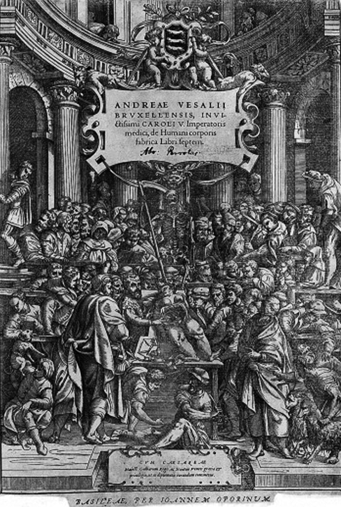
Sullivan goes on to trace this historical legacy back to the Enlightenment, a period when some of the most creative and enduring collaborative partnerships were developed. However, I would argue that it was during the Renaissance that the artist-theorist or, if we take the case of Leonardo, the artist-scientist, first gained ground and the imaginative, creative, and philosophical relationship between art and anatomy began to flourish. In terms of my own research I am particularly interested in art where allegory plays a part: the telling of one story through another, or, the deconstruction of meaning through the surface illusion of other meanings. It is therefore fitting that I begin my enquiry with one of the finest allegorical anatomical illustrations, the Epitome or title page to the great anatomist Andreas Vesalius’De Humani Corporis Fabrica.
Theatres of the body
The Epitome (Fig. 1) to the 1543 Vesalian image De Humani Corporis Fabrica by artist Jan Stephan van Calcar contains a micro-history of the politics, culture and philosophy of its time. Jonathan Sawday suggests that the Vesalian theatricum anatomicorum was ‘…an extravagant idealization of anatomy’ (1995: p. 66) in which, by cutting open and exposing the womb of the female cadaver Vesalius is making a comment on ‘…the conjunction of the womb and the tomb within the ‘magnificent temple’– Copernicus’ own phrase for the universe itself.’ (1995: p. 71).
Fig. 1.
The Epitome or title page to Andreas Vesalius’ 1543 De Humani Corporis Fabrica by artist Jan Stephan van Calcar, courtesy of The Wellcome Library, London.
Allegory was built into the very fabric of the anatomy theatre, which was originally a space designed as a locus not only of epistemological exposition but also of metaphorical unfolding, as was so abundantly evident in theatres like Leiden and Padua. Foucault (2003; Original edition 1963) and Sawday (1995) have postulated the architecture and spatial dynamics of the early anatomical theatre influenced not only the physical design of our contemporary medical theatres, but also the epistemology of how the body was perceived, ordered, and depicted. The historical theatre of anatomy came to symbolize the emergence of a new knowledge about the body, representing a paradigm shift in scientific investigation, a paradigm that was not only intensely theatrical in design but also in the performative aspects of the anatomical space.
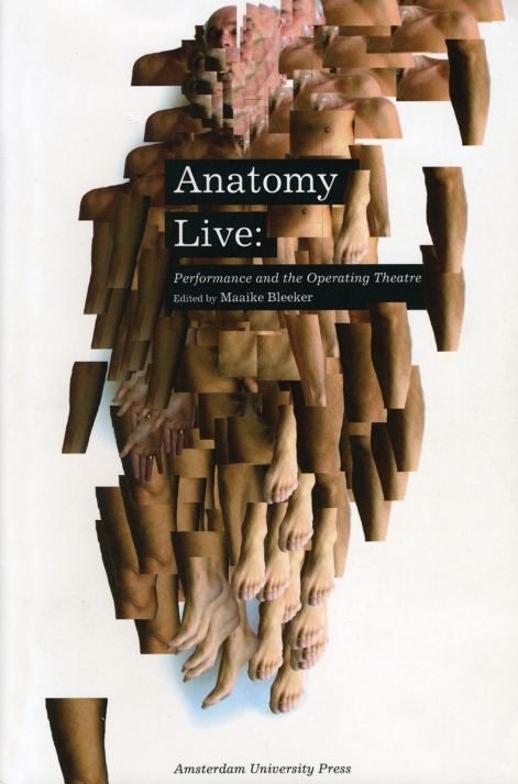
The significance of the theatre of anatomy, and of the dissection of the body as a performative act, has been brought to wider public attention with exhibitions like The Quick and The Dead (Petherbridge, 1997), Spectacular Bodies (Kemp & Wallace, 2000), Anatomy Acts: How We Come to Know Ourselves (Patrizio, 2006) and Anatomy Live: Performance and The Operating Theatre (Bleeker, 2008; Fig. 2). This last example is particularly germane with regard to the rich diversity and interdisciplinarity that has come to fruition in drama and theatre studies where the anatomical theatre is considered a theatre proper and the anatomical body and its entourage ‘players’. To this list I would tentatively add my own Anatomy Lessons (2004), discussed further on, as an example of contemporary arts practice where interdisciplinary collaboration with anatomists and the anatomical theatre forms the basis of a visual investigation into contemporary anatomical spaces and practices. If we look more broadly at the body, medicine and death, then other examples of artistic and biomedical collaborations include notable examples such as Confronting Mortality with Art & Science: Scientific and Artistic Impressions on What the Certainty of Death Says about Life (Pollier-Green et al., 2007), which is refreshing in that it illuminates the less well publicized practice of contemporary medical and anatomical illustration. There are of course a number of well known artists who have made the subject of death and the body the focus of their enquiry, Damien Hirst among them, but Hirst, like many other artists of his generation, is making work ‘in response to’ his subjects rather than ‘in collaboration with’ and in this review I am more concerned with the cross-fertilization of art and anatomy.
Fig. 2.
Book cover, Anatomy Live: Performance and the Operating Theatre, Ed. Maaike Bleeker, University of Amsterdam Press, 2008.
Artists working in anatomical and biomedical domains have, until relatively recently, been few and far between. The reasons for this are complex and are bound up with art historical movements and moments that led inexorably from the figurative to the modern. One of the consequences of this was a diminishing interest in life-drawing with its component studies in drawing from cadavers and/or dissected body parts as an aide to understanding the form and structure of the body. Put simply, many artists moved away from the dissecting room of their own accord, moving from realism to abstraction. I suggest that it was the late British artist Helen Chadwick who instigated a sea change in art and biomedical collaboration. The result of her collaboration with the fertility lab at Kings College London was ‘Unnatural Selection’ (1995), a series of exquisite photographic works based on embryos rejected as ‘faulty’ in the IVF selection process. The work was made even more poignant following the artist’s premature death and a generation of artists, myself among them, were influenced by Chadwick’s biomedical artworks. Mary Horlock (2004: p. 44) suggests that Chadwick, ‘used science to extend the parameters of her art, not necessarily expecting to find solutions or answers, but seeking the most pertinent framing for her questions.’ I concur with her argument that Chadwick’s theoretical approaches and transdisciplinary methodologies, ‘foreshadowed a broader shift in the 1990s which saw a burgeoning interest in science and a growing number of collaborative ventures between scientists and artists’ (Horlock, 2004: p. 44). It is my contention that as art has become more interdisciplinary, and acceptance grows of the role of the artist as practice-based ‘researcher’ with a seriousness of purpose, a revitalized affinity has evolved between the arts and biosciences, and indeed, between art and science generally. The past decade has witnessed an ever-growing number of collaborations and a greater sense of trust has been established between the arts and biosciences. Thus, the anatomical space in all its many guises is once again the focus of artistic attention.
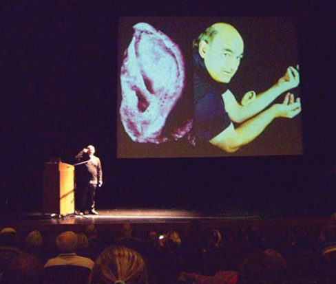
The examples I’ve mentioned thus far have what might be described as an art-historical focus, but artists working with organizations such as SymbioticA in Western Australia have a much more hands-on approach to all things bodily. An interdisciplinary research group working in the School of Anatomy and Human Biology at the University of Western Australia, SymbioticA have pioneered what they refer to as ‘tissue culture art’. As the name suggests, this is art made by engineering cell cultures and there is great interest amongst artists for working with tissue art, or, as it is often referred to, bio-art. SymbioticA is rare in enabling artists to experiment with wet biology practice in an actual working lab and their annual residency programme ensures that there is no shortage of applicants. An artist who has benefited from collaboration with SymbioticA is Stelarc, whose ‘ear on arm’ (Fig. 3) is an example of his approach to manipulating and re-fashioning what he believes to be essentially a ‘machine body’. Although we do not have an organization like SymbioticA in Britain, we do have various agencies that have demonstrated sustained and comprehensive support for collaborations across the arts and biosciences, the Wellcome Trust being the most visible and by far the most financially supportive of artists working in this area.
Fig. 3.
The performance artist Stelarc explains his artwork Extra Ear: Ear on Arm Project at the CIANT/Leonardo conference Mutamorphosis in Prague, 2007.
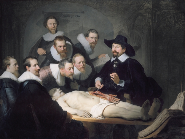
One of the collaborations supported by the Wellcome Trust was Mapping Perception (2003), made by the artist Andrew Kotting and the neurophysiologist Mark Lythgoe, which explored questions of perception, selfhood, and normality through an exploration of Kotting’s relationship with his daughter Eden who suffers from the rare physio-neurological condition Joubert syndrome. Kotting and Lythgoe re-staged Rembrandt’s famous 1632 The Anatomy Lesson of Dr Nicolaes Tulp (Fig. 4) with Eden as the subject of anatomical scrutiny and investigation to raise questions about the nature of medical knowledge and understanding and definitions of what is defined as ‘normal’. The choice of the Tulp painting was a good one, for this image, perhaps more than any other within the great suite of Dutch anatomy lesson paintings, is a densely coded and richly evocative allegory of medical knowledge and philosophical understanding. It is also a key image in terms of my own practice-based research, as I will now demonstrate.
Fig. 4.
Rembrandt, 1606–1669, The Anatomy Lesson of Dr Nicolaes Tulp, 1632, courtesy of Mauritshuis, The Hague.
The lesson
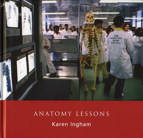
Anatomy Lessons (touring exhibition; 2003–2005, Fig. 5) followed on from previous interdisciplinary projects Just Another Day (2000) and Death’s Witness (2001) in which I collaborated with surgeons and technicians at Morriston Hospital in Swansea. It was while observing reconstructive hand surgery in the Burns and Plastics Unit that I found myself musing on the theatrical ‘grammar’ of the modern day surgical operating theatre. By grammar I refer to the nuanced systems which apply in the space of the operating theatre: the dramatic lighting, the passivity of the draped patient, and the hierarchy of the medical practitioners, the ‘players’, all of which are reminiscent of Rembrandt’s Tulp, right down to the significance of the hand itself. In art historical terms the hand is a well-established motif for philosophical progress, as noted by Martin Kemp and Marina Wallace (2000: p. 28) who suggest that, ‘For artists the hand was a communicative device second only in eloquence to the face.’ I became interested in how the performative nature of the operating theatre and its precursor, the theatre of anatomy, influence the acts that occur within it – the locus of the spectacle performed. The result of these musings was Anatomy Lessons, which was funded by the Wellcome Trust and the Arts and Humanities Research Council and was an ambitious and multi-layered project involving collaboration with anatomical theatres, museums, and dissecting rooms in Britain and Europe.
Fig. 5.
Book cover, Anatomy Lessons, Karen Ingham, 2004.
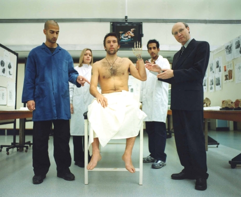
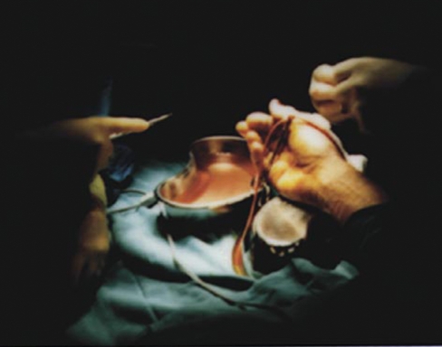
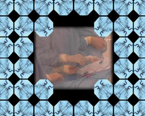
In researching the project I was concerned with the history of the anatomical theatre and with the re-staging, in the form of photographic tableaux vivants, of the Dutch suite of Anatomy Lesson paintings within a contemporary context. This aspect of the project involved negotiation and interaction with anatomists and their staff in Edinburgh, Cardiff, Dublin and London, and with the cross-fertilization of the site-specific artworks in medical museums and arts domains. In The Anatomy Lesson of Professor Moxham (Fig. 6) the professor and his team are paying homage to Rembrandt’s Tulp through an image which re-appropriates the visual grammar of the original painting, only in Moxham’s anatomy lesson the instruments of dissection are digital, not surgical. If, however, you look closely at the nearby teaching monitor a real image of reconstructive hand surgery can be seen (Fig. 7).
Fig. 6.
The Anatomy Lesson of Professor Moxham, from Anatomy Lessons, Karen Ingham, 2004.
Fig. 7.
Image of reconstructive hand surgery from Just Another Day, Karen Ingham and Ffotogallery, 2000.
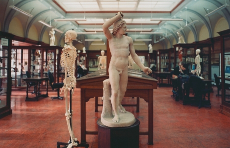
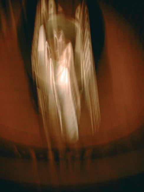
As I have written elsewhere (Ingham, 2004:18), it would seem that to successfully re-construct the body we must accept the anatomist’s maxim ‘know thyself’ by first de-constructing the human form. No exploration of the spaces of anatomy would be complete without the ubiquitous skeleton, and in Anatomy Lessons, the skeleton suite of images is a reminder of the iconic role the skeletal figure has played in the historical, and indeed contemporary, representation of the anatomical arts. In the eclectic Edinburgh Anatomy Museum (Fig. 8) we see an example of art and anatomy standing side by side in perfect partnership in the form of a skeleton and a classical sculpture of a male nude. Also in the Edinburgh museum is the skeleton of the grave robber William Burke, of the infamous Burke and Hare. As Ruth Richardson noted in Death, Dissection and the Destitute (2001: p. 143) Burke received a particularly fitting punishment for his crimes: following public hanging his body was dissected and his skeleton put on public display. I was fortunate to be able to extend the Anatomy Lessons project to collaboration with the exquisite Paduan Theatre of Anatomy, now a museum, at the University of Padova in Italy. This much cited anatomical theatre is a stunning example of the Renaissance anatomy theatre and nothing quite prepares the spectator for the architectural and allegorical resonance of the space. In 2000, the British artist John Isaacs produced the video artwork ‘A Cyclical Development of Stasis’, which was shot in the Paduan theatre and inter-cut with a high-tech dissecting room in Essen. His title plays on the oxymoronic notion of developmental stasis, progression and movement developing from something that is still and dead. As Martin Kemp and Marina Wallace posit (2000: p. 158) the piece is important in that it is an enquiry into ‘…both positions of objectivity and subjectivity, the dissector and the dissected.’ My own response to this influential space was to create a digital video performance, Orpheus Rising (Fig. 9), which played on the themes of death and resurrection in the Greek myth of Orpheus and Eurydice. In a short video ‘loop’ a male figure appears resignedly to wait for death in the deep dissecting pit. This aspect is shot from his point of view and appears in almost documentary style, the footage hand-held and ‘grainy’. This is followed by the figure’s ascension up through the tiered balconies, which are flooded with symbolic light: an allegory of death, dissection and resurrection.
Fig. 8.
Edinburgh Anatomy Museum, from Anatomy Lessons.
Fig. 9.
Orpheus Rising, from Anatomy Lessons.
The reading of any image is heavily dependent on interpretation and contextualization, and it was the contextualization of the artworks in Anatomy Lessons that was a key objective of the project: namely, the exhibition of the works back in their site of origin, and the cross-fertilization of art and medicine through the simultaneous siting of the artworks in both the medical and art domains. Cultural critic Hugh Adams speaks of the complex role contemporary artists are expected to negotiate, and argues that for artists making interventions, they must do so ‘...at a multiplicity of points, social as well as artistic...’ (Adams, 2003: p. 10) For Adams, Anatomy Lessons is one of a body of works that illustrates how an artist may function at the interface of psychological, geographical and architectural space. He describes the typologies as a ‘…double journey: she (Ingham) travels to anatomical sites, in places such as Padua, London and Edinburgh, photographically establishing typologies of dissection rooms and demonstration theatre spaces, then reverses this, by touring her exhibition photographs to the very spaces which are her subject’ (Adams, 2003: p. 9). What Adams is drawing attention to is the privileged, though complex, position of the collaborating artist who operates in domains not normally associated with the arts – in my case schools of medicine and anatomy theatres – and the subsequent responsibility of those artists to communicate their work beyond the art world. In his essay on the complex relationships between art production, art history and museology, Donald Preziosio suggests that:
…museology and art history are instrumental ways of distributing the space of memory. Both operate together on the relationships between past and present, subjects and objects, and collective history and individual memory. These operations are in aid of transforming traces of the past superimposed upon the present into a storied space wherein the past and present are imaginatively juxtaposed…(Preziosi, 1999: p. 34).
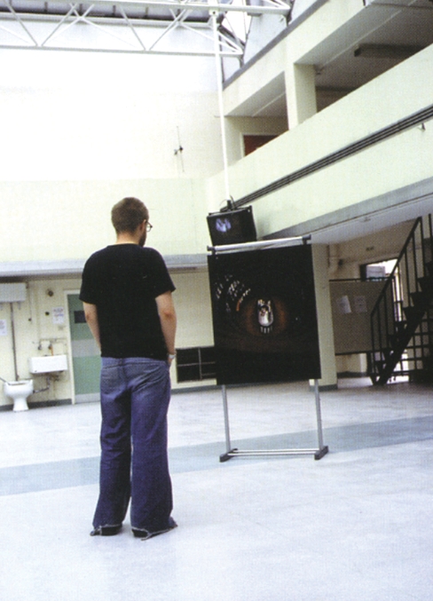
It is this imaginative juxtaposition that I seek within my practice by bringing the past into the present in ways that prompt us to pause and reflect on our preconceptions and assumptions about death, dissection and the disposal of human remains. This can be a daunting task in the realms of anatomy where anatomy museums and medical collections remain largely off-limits to the public. Therefore, perhaps the most important aspect of this particular project was in gaining permission from HM Inspector of Anatomy to exhibit the artworks back in their site of origin, the actual dissecting rooms of Cardiff (Fig. 10) and Guys/King’s College London, for example. In this context the anatomists and their staff acted as ‘gallery guides’ leading the public through the exhibits and giving them the benefit of their firsthand knowledge of the processes of anatomical dissection and the respectful disposal of human remains.
Fig. 10.
Installation shot: Cardiff Dissecting Room, Anatomy Lessons.
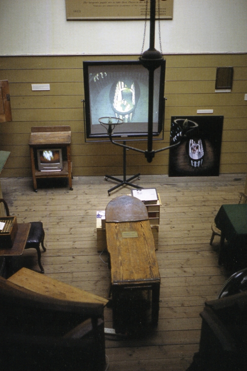
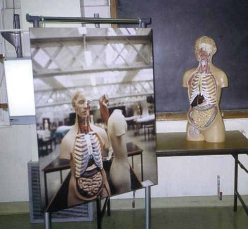
For example, in London members of the public could first visit the Old Operating Theatre Museum (Fig. 11) where elements of the exhibition were installed, then walk directly across the road into the dissecting rooms at Guy’s (Fig. 12).
Fig. 11.
Installation shot: Old Operating Theatre Museum London, Anatomy Lessons.
Fig. 12.
Installation shot: Dissecting Room Guy’s/Kings College London, Anatomy Lessons.
Anecdotal evidence from visitors to the dissecting room exhibition at Guy’s, in the form of a comments book and remarks made to the anatomy staff, suggests that the majority of the public found this experience to be a stimulating and, in at least one case, a cathartic experience as a female tourist from Canada finally found meaning in her Father’s donation of his body for dissection, a decision she had previously been bewildered by.
Mutability
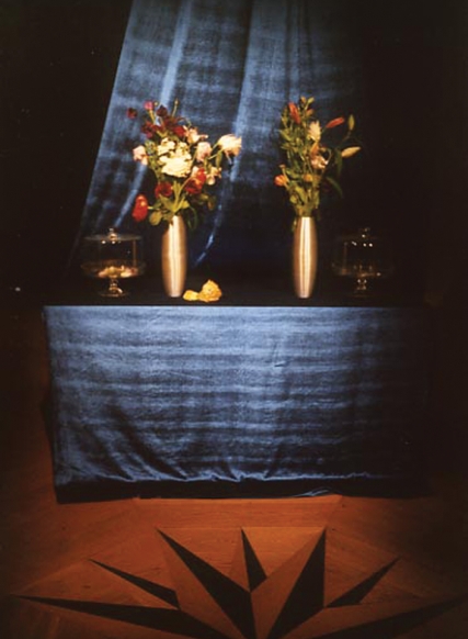
Continuing with the theme of site-specific responses to the anatomical theatre, the ‘Vanitas’ installation (Fig. 13), which took place in the Waag Theatrum Anatomicum in Amsterdam (the site associated with Rembrandt’s Tulp painting), offers an interesting model of how art, medicine and new technology can work in tandem to create new networks of subjectivity and meaning in the anatomical domain. ‘Vanitas’ was a site-specific durational installation that took place in 2005 as the culmination of my artist’s residency with the Waag Society. The Waag is Amsterdam’s oldest secular building and was once home to many of the city’s guilds, the anatomist’s guild being the most notable, but now hosts the Waag Society, a government subsidized ‘new media’ organization. With the exception of rare installations like the ‘Vanitas’ event, the building and its famous anatomy theatre are closed to the public. The Waag is also well known through the work of anatomist Frederik Ruysch (1638–1731) who was the praelector to the Dutch surgeons’ guild in 1667 and whose daughter Rachel was a respected vanitas still-life painter. Ruysch is renowned for his extraordinary tableaux, or dioramas, which incorporated fetal skeletons and bio-botanical backdrops, and were arranged with the vanitas theme of transience and mutability in mind. The still-life genre originated in the Netherlands (particularly Amsterdam and Utrecht) and the vanitas still life, with its emphasis on the all too swift passage of time and the impermanence of the human condition, was symbolized through the use of skulls and flowers; an allegory of death and mutability. ‘Vanitas’ wove together these inter-linked aspects by creating a paradoxical ‘live art’ vanitas ‘still life’. A key element of this installation was the inclusion of the digital film ‘Vanitas: Seed-Head’ (Fig. 14), which depicts three photographic death masks of my partner, myself and our young son, which, through computer animation, appear to ‘morph’ from face to face. I become my son who becomes his father who becomes me in a genetic discourse on the vanitas theme of transience. The deep blue image appears to be suspended in a type of skeletal flower bulb and is in fact an x-ray of my son’s skull taken at the local A & E following a slight head injury. The DVD was projected onto a large screen in the theatrum, located just beneath the domed ceiling of the space, painted with the crests of the various surgeons’ guilds. Two vanitas flower arrangements were also placed within the theatre, one in full bloom, the other still tightly budded, alongside two specimen jars. Over the course of a week a live web broadcast displayed the floral still life alongside the video projection. By the end of the event the petals from the full blooms had filled the jar, symbolizing death and decay. But simultaneously, the flowers in the vase, which were closed at the start of the residency, were now in bloom (a metaphor for new life and the Waag’s present links to creating communal memories).
Fig. 13.
Installation shot: Vanitas, durational, site-specific installation at The Waag, Amsterdam, 2005.
Fig. 14.
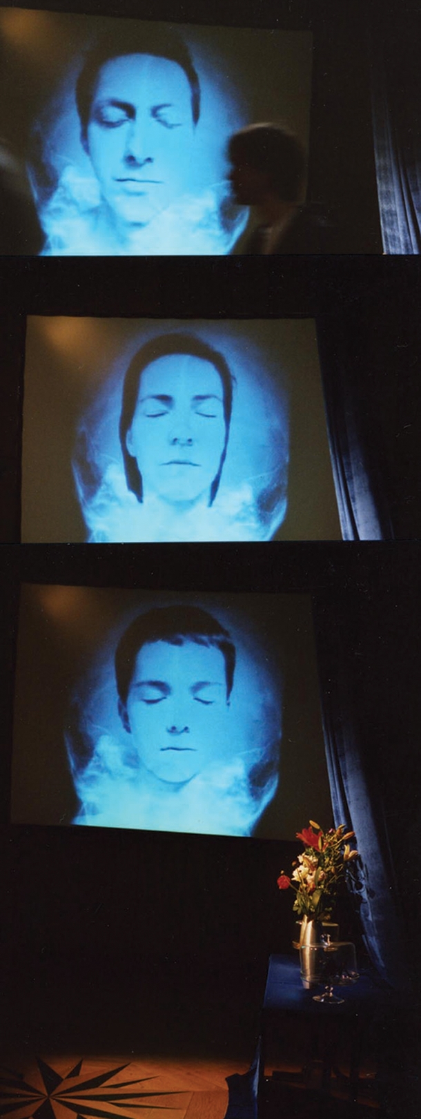
Installation shot: Vanitas: Seed-Head, projected digital film.
The installation was linked to a web-cam and could be accessed live throughout the week. The web-link constructed for the project included a message board where virtual spectators of the Vanitas were encouraged to post an electronic text or image of their own that related to the themes of the event. The public response, in image and text, was to evoke memories of their loved ones who had died and were memorialized, and, interestingly, to respond to the floral symbolism of the vanitas still life with memories of a celebratory nature, rather than dwell solely on the more melancholy aspects of death and mutability. The messages were saved and copied onto a CD and sealed into the other specimen jar.
The electronic memories became an allegory of the 21st century Vanitas where memories are collected, classified, stored and retrieved from the computer’s ‘memory’. The specimen jars, with the preserved flower petals and the ‘preserved’ memories, remain with the Waag as a memento of the project and the website can still be visited on the Waag online archive.
The theme of the vanitas, and of the ‘dance of death’, is the basis of the late Ian Breakwell’s collaborative artwork ‘Death’s Dance Floor’ (1998) made in collaboration with Professor Bernard Moxham and the Cardiff School of Biosciences (Breakwell & Moxham, 1998). Death’s Dance Floor used medical imaging techniques to create a post-modern reading of the allegorical Dance of Death. Breakwell’s lens-based images (photography, medical scans, stained glass, and audio-visual shows) were inspired by Hans Holbein’s popular secular woodcuts of The Dance of Death (1538), which depicted Death as a lively and mischievous figure (in contrast to the melancholic icon of the Grim Reaper). Utilizing medical imaging techniques such as thermography and x-ray, Breakwell created an ironic medicalized self-portrait of himself as simultaneously death’s victorious dancer and death’s skeletal victim. In his commentary on the work, Moxham suggests that the theme of memento mori is implicit in many of Breakwell’s works, particularly the piece ‘Body Mask’:
The work hints at Death, and not merely from the use of the anatomical imagery nor from the self-portrait’s allusion to a memento mori…the very title Body Mask, together with the reference to the skeleton within the depth of the body, also implied that to hide behind a mask need not involve an external deceit but an internal (psychological) deceit. The denying of death to oneself is just such a deceit (Breakwell & Moxham, 1998: p. 38).
The psychological deceit of denying death is bound up with a wider philosophical discourse on the nature of selfhood, consciousness and subjectivity, a discourse I will touch on in the next section.
Art, neuroscience and ‘the mind’s eye’
Like many artists, I am fascinated by Rene Descartes’ search for the soul in the seat of the brain and his notion of ‘the mind’s eye’. Daniel Dennett’s allegorical ‘Cartesian Theatre’ where the mind’s eye turns the anatomical theatre inward and neurons illuminate the search for the self, is a fanciful notion and yet it is a seductive theme to which an artist can respond. Artists interested in neuroscience and neuro-psychology are often drawn to the writing of neurobiologist Semir Zeki (1999), who suggests that the arts are a useful tool for deepening our understanding of the brain. Zeki is one of a growing number of neuroscientists interested in the connections, cultural and synaptic, between creativity and the brain. Given the interdisciplinary nature of the study of the brain it is perhaps not surprising that collaborations across the arts and neuroscience have blossomed over the past decade, as demonstrated in the 2008/2009 ‘Creative Brain Lecture Series’ at Bristol University, which paired leading artists and scientists for a series of public discussions on the brain and creativity.
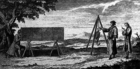
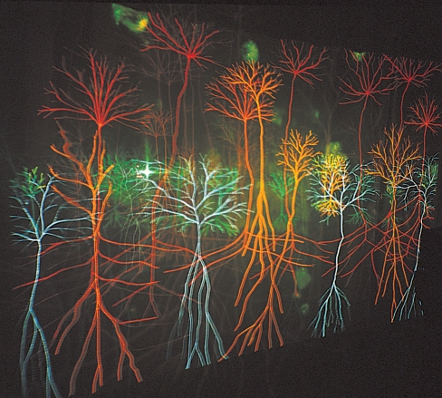
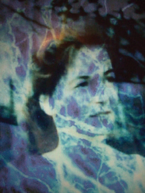
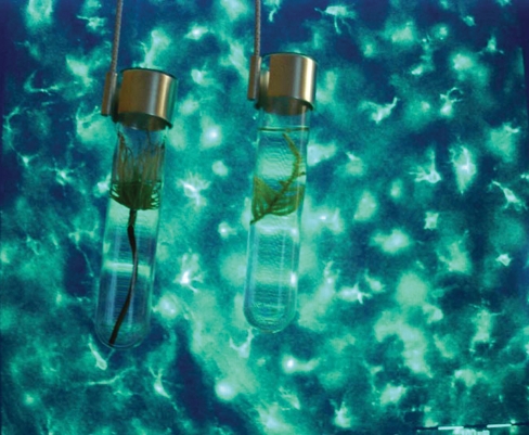
Neuroscience is a very attractive subject for artists, and indeed, for philosophers and scientists alike, as it deals with the ‘big questions’ such as: what is the self (and its corollary, what is ‘other’); what is consciousness; how does memory work (or not work). Another aspect that cannot be ignored is the very real attraction for the artist of access to sophisticated medical imaging technologies such as fMRI and scanning confocal electron microscopy (among a growing range of interesting imaging systems). The impulse for artists to use technology to see ever more detailed descriptions of the body is nothing new, as Gerard Vandergucht’s (1733) ‘A seated male figure looking through a camera obscura’, engraving for William Cheselden’s Osteographia illustrates (Fig. 15). The challenge, I would suggest, is not to be led or seduced by the technology. The artist Susan Aldworth has taken on this challenge and her recent body of work Scribing the Soul (2008) (Fig. 16) demonstrates how medical imaging technology designed to see into the deepest recesses of the brain may be incorporated into moving and personal interpretations on the nature of consciousness, in Aldworth’s case as a direct result of experiencing a brain aneurysm. Aldworth is a serial collaborator with anatomists and neurologists and her work sits happily in both the medical museum and the gallery space. Andrew Carnie has also responded to the notion of the ‘theatre of the mind’ with exquisite artworks on the nature and flux of consciousness, as evinced in his Magic Forest (2002, Fig. 17), a time-based artwork that allegorizes the branch-like structure of dendrites. In one of my own works in progress, See My Thoughts (Fig. 18), I am also seeking to create visual and philosophical insights into the brain by constructing a theatrical space made entirely from structural and chemical brain imagery in which the participant interacts with the space in a similar fashion to that envisaged by Giulio Camillo in his plan for the 16th century Teatro della Memoria. Part of a series of discrete yet inter-related artworks on the meaning of consciousness and memory, this developmental artwork seeks to use scientific imaging from brain scans and scanning confocal electron microscopy to create a multi-layered time-based projection in which the spectator is encouraged to interact with the visualized thought processes on exhibit. The project follows on from Seeds of Memory: art, neuroscience and botany (2006, Fig. 19) funded by an Arts and Humanities Research Council Sci-Art Research Fellowship with the Cardiff Neuroscience Research group, which explored the links between memory and Alzheimer’s disease and the role of plant-based pharmaceuticals in ameliorating dementias (although there are many clinical reservations as to the efficacy of such treatments).
Fig. 15.
‘A seated male figure looking through a camera obscura’, Gerard Vandergucht’s engraving for William Cheselden’s Osteographia,1733, courtesy of The Wellcome Library, London.
Fig. 16.
‘Going Native’ in Scribing the Soul, Susan Aldworth, courtesy of the artist, 2008.
Fig. 17.
Magic Forest, Andrew Carnie, courtesy of the artist, 2002.
Fig. 18.
See My Thoughts, developmental artwork from ‘Theatres of Memory’, Karen Ingham, 2008.
Fig. 19.
Installation shot: ‘Bio-botanical Test Tube Vanitas II’ from Seeds of Memory: art, neuroscience and botany, Karen Ingham, 2006.
This was a very difficult project to develop given the diverse and at times conflicting histories and agendas of the scientific, artistic and botanical communities involved. In this instance the research resulted in something of a one-way process, as ultimately I gained more from the collaboration than my scientific partners. This was in part due to the relative brevity of the project and the pressures on the neuroscience research group, all of whom were at or nearing completion of important research papers: many simply did not have the time to work with an artist in-depth. I make mention of this observation, as I believe it is an important consideration when embarking on collaborations across the arts and biosciences: most projects, if they are to be of lasting mutual value, do require time for reflection and maturation. For obvious reasons, it is sometimes easier to develop longer-term partnerships in the anatomical museum, rather than the actual dissecting room or research laboratory. This was certainly the case in the final collaboration I wish to discuss.
All that remains
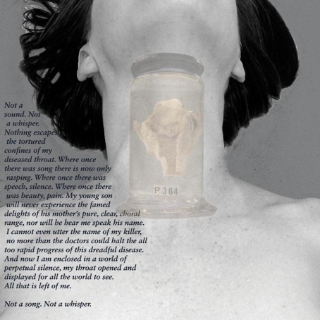
Marina Warner (1985: p. 331) suggests that: ‘Meanings of all kinds flow through the figures of women, and they often do not include who she herself is’. This would certainly seem to be the case in the theatre of the dead. At least we know the name of the cadaver in Tulp’s famous painting, but in the Fabrica Epitome, the image with which I began this review, the identity of the dead woman in the very centre of the picture (the very centre of the universe if we are to believe the Vesalian allegory) remains anonymous. It is this corporeal anonymity, this lack or erasure of what we would now call the patient narrative that is explored in Narrative Remains (Fig. 20). Made in collaboration with the Royal College of Surgeons of England’s Hunterian Museum, and funded by the Wellcome Trust; the project is an exploration of how we consider the postmortem body and the removal and storage of morbid remains. Re-instating the medical narratives of the dead back with their dissected and anonymized body parts creates the opportunity to metaphorically re-present these otherwise forgotten deaths to a public audience. The project is a site-specific response to surgeon-anatomist John Hunter’s anatomical collection and follows the long tradition of narrations by the speaking dead by textually and visually reuniting patient narratives with their displayed organs. Incorporating photo-sculptural vitrines and a digital film, Narrative Remains imagines and re-embodies a semi-fictive first person voice for key historical specimens in the collection.
Fig. 20.
Narrative Remains, Karen Ingham and The Royal College of Surgeons of England Hunterian Museum.
You may wonder how a heart can possibly narrate a story? If we consider the organ as being emblematic of the whole, a metonym so to speak, then it is possible to invest the object with subjective meaning, for example the larynx of Marianne Harland, a woman renowned for the beauty of her singing voice and her musical talents. There was a poignant irony in her death as she lost first her famous voice, and subsequently her life, as a result of tuberculosis. Her oesophagus, larynx and trachea are all that remain; her story now silenced. Co-collaborator and Director of the Hunterian Museum, Simon Chaplin (2009: p. 8,9), summarizes the nature of the project thus:
The emergence of the anatomist as exemplary figure and the demise of the patient as individual have been seen as inextricably linked. In John Hunter – the archetype of the surgeon-scientist – some have seen the origin of an exclusive medical authority that still, today, denies the patient a say – a ‘fearful symmetry’ between Hunter and his successors, as Ruth Richardson so eloquently describes it. Revealing the patient’s voice in John Hunter’s collection, invoking identity to engender emotion, can therefore be seen as a political act. It is a way of highlighting the origins of the clinical gaze in order to deflect or refocus it, and to bring the modern patient back into the picture.
At the time of writing the exhibition has not yet opened and public response to the project will not be known until after the evaluation period. However, it is hoped that the collaboration will engender a revitalized discourse about the nature and meaning of the preservation of human remains. It may also pave the way for future collaborations between the Hunterian Museum and artists seeking to work with this extraordinary space.
Conclusion
This review has focused primarily on the artist’s response to the anatomical body and its spaces, but the aforementioned comment by Professor Bernard Moxham in relation to Ian Breakwell’s art is evidence of the mutual respect and understanding between artist and anatomist.
Moxham is one of many anatomists who have surprised me with their depth of knowledge of the arts and with their willingness to re-introduce the arts into the anatomical domain, not only through external collaboration but also by encouraging their students to engage with the body of dissection in more complex and multi-dimensional ways. I mentioned previously how many artists have moved away from realism and traditional drawing and painting. It is therefore noteworthy that the heart surgeon Francis Wells, featured in Andrew Graham-Dixon’s BBC2 series The Secret of Drawing (2005) uses drawing as a means of better observing, thus understanding, the heart and has on occasion made quick sketches during actual heart surgery so as to gain a clear idea of the logistics of a complex operation. I mention this fact to demonstrate that neither art nor anatomy are exclusive domains and we should be wary of simplification. I would like to conclude by suggesting that science, art and philosophy can no longer afford to be seen as separate disciplines in our quest to know the body and the mind, and thus to know ourselves. It is my hope that this special symposium issue of Journal of Anatomy will stimulate new philosophical and creative insights leading to future collaboration and debate in this rich field of investigation.
Acknowledgments
I would like to thank Professor John Fraher and Professor Gillian Morriss-Kay for their editorial help and suggestions. I would also like to extend my thanks to Professor Bernard Moxham for his continuing support for artists working across art and anatomy collaborations. Several of the projects I discuss in my review could not have been realized without the financial support of The Wellcome Trust Arts Awards and The Arts and Humanities Research Council.
References
- Adams H. In: Elsewhere. MacKinnon K, editor. Swansea: Glynn Vivian Art Gallery; 2003. pp. 6–14. [Google Scholar]
- Andrew Graham-Dixon. BBC2 series: The Secret of Drawing. Broadcast at 20:05 on Saturday 15th October 2005. Available at: http://www.kareningham.org.uk/narrative-remains.html; http://www.kareningham.org.uk/anatomy-lessons.html; http://www.kareningham.org.uk/seedheadlarge.swf. [Google Scholar]
- Bleeker M. Anatomy Live: Performance and the Operating Theatre. Amsterdam: Amsterdam University Press; 2008. [Google Scholar]
- Breakwell I, Moxham B. Death’s Dance Floor. Cardiff: Ffotogallery Publications; 1998. [Google Scholar]
- Chaplin S. ‘Emotion and identity in John Hunter’s Museum’. In: Ingham K, editor. Narrative Remains. London: RCS and CLASI Publications; 2009. pp. 8–15. [Google Scholar]
- Foucault M. The Birth of The Clinic: An Archaeology of Medical Perception. London: Routledge Classics edition; 2003. Original edition, (1963) Naissance de la Clinique First edition, Presses Universitaires de France, Paris. [Google Scholar]
- Horlock M. ‘Between a rock and a soft place’. In: Sladen M, editor. Helen Chadwick: A Retrospective. London: Hatje Cantz Verlag; 2004. pp. 33–46. [Google Scholar]
- Ingham K. Anatomy Lessons. Manchester: Dewi Lewis Publishing; 2004. [Google Scholar]
- Kemp M, Wallace M. Spectacular Bodies: The Art and Science of the Human Body from Leonardo to Now. London and California: Hayward Gallery and the University of California Press; 2000. [Google Scholar]
- Patrizio A. Anatomy Acts: How We Come to Know Ourselves. Edinburgh: Birlinn Press; 2006. [Google Scholar]
- Petherbridge D. The Quick and The Dead. London: Hayward Gallery; 1997. [Google Scholar]
- Pollier-Green P, Van de Velde A, Pollier C. Confronting Mortality with Art & Science: Scientific and Artistic Impressions on What the Certainty of Death Says about Life. Brussels: VubPress; 2007. [Google Scholar]
- Preziosi D. Performing modernity: the art of art history. In: Jones A, Stephenson A, editors. Performing the Body: Performing the Text. London: Routledge; 1999. pp. 29–39. [Google Scholar]
- Richardson R. Death, Dissection and the Destitute. 2nd edn. London: Phoenix Press; 2001. [Google Scholar]
- Sawday J. The Body Emblazoned: Dissection and the Human Body in Renaissance Culture. London: Routledge; 1995. [Google Scholar]
- Sullivan G. Art Practice As Research: Inquiry in the Visual Arts. California and London: Sage Publications; 2005. [Google Scholar]
- Warner M. Monuments and Maidens: Allegory and the Female Form. London: Weidenfeld & Nicolson; 1985. [Google Scholar]
- Zeki S. Inner Vision. Oxford: Oxford University Press; 1999. [Google Scholar]


