Abstract
Venerina (little Venus) is the name given to a wax model representing a pregnant young woman that was created in Florence (Italy) by Clemente Susini (1754–1814) in 1782. It is currently located in the historic Science Museum of the University of Bologna. The model was constructed so as to enable removal of the thoracic and abdominal walls and various organs, exposing the heart, diaphragm and an opened uterus with a well-developed fetus. The woman is small, about 145 cm (4′ 9′) tall and of delicate build; she looks like a teenage girl. We know that Clemente Susini worked directly with the cadaver and copied the anatomical preparation exactly. This artist often represented the true structure using a wax mould; the existence of two other versions of this specimen suggests that this model was made in this way. Therefore, Venerina’s body may be a faithful representation of a young woman who died while pregnant. Observation of the body confirms that the organs are normal, except for the heart and great vessels. The walls of both ventricles are of equal thickness and the ventricles themselves of approximately equal size. The arch of the aorta and the enlarged pulmonary trunk are connected by a short duct about 3.5 mm in diameter. If this structure represents an open arterial duct, we can deduce that the two ventricles worked under the same conditions of blood pressure, hence their equal wall thickness. If the young woman died from this congenital disease, the cause of death has been diagnosed on a wax model of her body after more than two centuries.
Keywords: anatomical wax, arterial duct, Clemente Susini, Venus
Introduction
During the 18th century, Bologna and Florence were the most famous centres for the manufacture of human wax models (Riva et al. 2010; Ballestriero, 2010, this issue). At that time, anatomical modellers used different techniques to represent the different organs of the human body. In Bologna, they started from original human skeletons, to which they added muscles, vessels and nerves made of wax. This wax-modelling technique was very time-consuming. Each preparation was a unique piece, completely distinct from the others. Over a period of time, the anatomists and wax artists of Bologna produced a marvellous collection of human bodies and organs made of wax. The surviving models have been restored and are now exhibited in the Science Museum of the University of Bologna.
The anatomical modellers of Florence used a different technique, making moulds of the anatomical preparations (Lanza et al. 1979). This innovation had two great advantages: (i) the resulting model was an exact representation of the anatomical preparation, showing the actual structure and relations of the different organs; and (ii) it was possible to make several copies of the same anatomical preparation by reusing the mould. The wax representations of bodies and organs realized in Florence were therefore anatomically very accurate. They were also very attractive from an artistic point of view; some of them are reminiscent of the most important sculptures of the Renaissance (Lanza et al. 1979; see also Ballestriero, 2010, this issue). For this reason, many private collectors as well as medical schools in Italy and other European countries bought copies of these wax models from Florence. Several survive and are still exhibited.
In 1802, an anatomist of Bologna bought a copy of the small body of a young woman made in 1782 by Clemente Susini (1754–1814), the most famous Florentine anatomical wax artist, from a private collector. The model is currently located in the historical Science Museum of the University of Bologna. Female wax body representations have long been identified with the name ‘Venus’. The Bologna anatomists called the model described here ‘Venerina’ (little Venus).
Observations
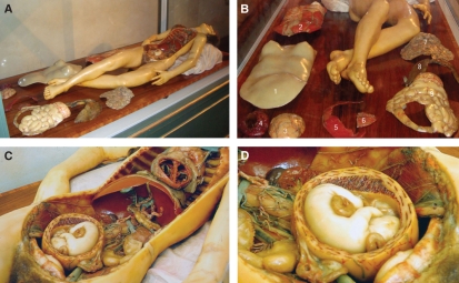
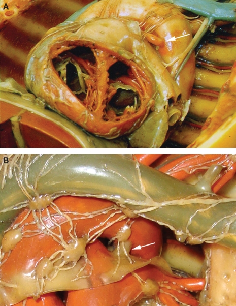
This wax model represents a pregnant young woman. The body represented is very small; it is about 145 cm (4′ 9′) in length and displayed in a relaxed position (Fig. 1A). The realization of this model is very interesting because it consists of many removable parts that can be selectively dismantled from the basic model (Fig. 1B). Indeed, it is possible to remove the thoracic and abdominal walls, the greater omentum and various organs from the body cavities; the pelvis shows an opened pregnant uterus with fetus and placenta (Fig. 1C). The fetus is about 15 cm crown–rump length, from which it has been evaluated that the woman was in the fifth month of her pregnancy (Fig. 1D). Various organs, including the liver and kidneys, were left in situ and are well represented. In the thorax, the lungs have been removed and the heart left in situ; it has been sectioned along a frontal plane so that the cavities of the atria and ventricles can be clearly observed. The inner structure of the heart was represented with extreme care to reveal all of the anatomical features including the atrio-ventricular valves with the papillary muscles and tendinous cords (Fig. 2A).
Fig. 1.
(A) The small body is represented in a relaxed and statuesque pose that relieves the macabre aspect of death. (B) The removable parts of the trunk: (1) skin layer, (2) anterior thoracic wall, (3) wall of the uterus, (4) amniotic sac, (5) anterior part of the diaphragm muscle, (6) sternocostal face of the heart, (7) greater omentum, (8) lungs and (9) small intestine and part of the colon. (C) General view of the thoracic and abdominal cavities; in the pelvis, a pregnant uterus is clearly visible. (D) The uterus contains a well-developed fetus of about 15 cm crown–rump length.
Fig. 2.
(A) The right and left ventricular walls have the same thickness. The arterial duct is easily detectable between the enlarged pulmonary trunk and aortic arch (arrow). (B) A different wax model of a normal heart realized by Clemente Susini. The arterial ligament, partially covered by two lymphatic vessels, is correctly represented in size and colour (arrow) (Museum of Anatomy, Bologna).
Remarkably, the walls of the right and left ventricles have the same thickness. They are both about 5 mm thick, whereas in a normal heart the left ventricle wall is three times thicker than the right (Silver et al. 2001). Considering the great attention that the anatomist Clemente Susini paid to reproducing the structures of the organs, the fact that the ventricular walls have the same thickness is an unusual and interesting feature.
A careful analysis of the visible related structures of the heart reveals two further unusual features: (i) a large pulmonary trunk (> 30 mm) and (ii) the presence of a small duct connecting the arch of the aorta and the pulmonary artery. The duct is about 3.5 mm in diameter and it originates at the level of the division of the pulmonary artery and reaches the aortic arch at the beginning of the descending part (Fig. 2A). This corresponds to the normal insertion of the arterial duct that, in the fetus, connects the two great vessels; after birth it closes and becomes the arterial ligament (Friedman & Fahey, 1993; Kiserud & Acharya, 2004), as described for the first time by the Italian physician Leonardo Botallo (1530–1571) (Péterffy & Péterffy, 2008). The greatest diameter of each ventricular cavity is similar, about 32 mm, and there are no evident differences between the right and left ventricles. The colour of Venerina’s skin does not show any hint of cyanosis and the fingers are well formed with no signs of clubbing, so there is no indication of cyanotic heart disease or hypertension.
Discussion
Over a period covering more than two centuries many museums, libraries and archives have been destroyed, mainly due to the terrible wars occurring in Italy. For this reason, the original moulds used by the wax-workers of Florence have not been preserved and we cannot be certain that this wax body was made according to the usual Florentine techniques. At least three examples of this body exist, located in Florence, Budapest and Bologna. They show slight differences in their structure but it is accepted that they are copies obtained from the same mould, which was reconstructed by observing a true anatomical preparation. In the version in La Specola Museum in Florence (illustrated by Ballestriero, 2010, this issue), the heart has the same features as we have described for the copy in Bologna, with the same thickness of the ventricular walls.
With respect to the Bologna Venerina, we can be confident that the inner structures of the heart were accurately represented. In the light of current scientific knowledge, the fact that the walls of the two ventricles have the same thickness can be explained by the persistence of an open arterial duct. The persistent duct is clearly represented by its size and position, and by the fact that the connection between the two arteries resembles a real duct; it is circular and painted red as are the other arteries of the body. Furthermore, in other anatomical preparations realized by Clemente Susini we can observe that in a normal heart he generally painted the structure of the arterial ligament white or grey and that it was larger at its insertion at the level of the arteries and thinner in its midsection (Fig. 2B). The observed enlargement of the pulmonary trunk completes the morphological spectrum previously described for this congenital disease pathology (Fisher et al. 1986; Lavadenz et al. 1986). The volume of the ventricles evaluated by the Teicholtz formula (Teicholtz et al. 1976) is 45 cm3, which excludes the possibility of a more recently acquired cardiomyopathy.
The presence of an arterial duct of about 3.5 mm is compatible with life and some people with this anomaly have a normal lifestyle without any particular symptoms (Wiyono et al. 2008). However, it has been reported that with this pathology the patient is more vulnerable to illness such as endocarditis (Giroud & Jacobs, 2006). The strong evidence that the Venerina wax model was made from a mould reproducing a true anatomical variant suggests that the small size of the young woman is also anatomically accurate. It is possible that the cardiovascular defect was responsible for a modification in blood circulation and growth rate. Her early death could have been due to endocarditic infection or a more serious cardiovascular insufficiency related to the progression of the pregnancy.
Our conclusion is based on an indirect evaluation of the young woman through examination of the ‘Venerina’ wax model that was made following her death and held in the Science Museum of the University of Bologna for more than 200 years. The physicians and scientists of the late 18th century probably did not know the physiopathology of this disease but, if we accept that the Florentine anatomical preparation was represented exactly as it appeared in reality, we can consider our observations to be the result of a valid scientific study.
Acknowledgments
This work was partially supported by grants from “Ricerca Finalizzata Orientata'' (RFO 2008), University of Bologna.
References
- Ballestriero R. Anatomical models and wax Venuses: art masterpieces or scientific craft works? J Anat. 2010;216:223–234. doi: 10.1111/j.1469-7580.2009.01169.x. [DOI] [PMC free article] [PubMed] [Google Scholar]
- Fisher RG, Moodie DS, Sterba R, et al. Patent ductus arteriosus in adults – long-term follow-up: nonsurgical versus surgical treatment. J Am Coll Cardiol. 1986;8:280–284. doi: 10.1016/s0735-1097(86)80040-7. [DOI] [PubMed] [Google Scholar]
- Friedman AH, Fahey JT. The transition from fetal to neonatal circulation: normal responses and implications for infants with heart disease. Semin Perinatol. 1993;17:106–121. [PubMed] [Google Scholar]
- Giroud JM, Jacobs JP. Fontan’s operation: evolution from a procedure to a process. Cardiol Young. 2006;16:67–71. doi: 10.1017/S1047951105002350. [DOI] [PubMed] [Google Scholar]
- Kiserud T, Acharya G. The fetal circulation. Prenat Diagn. 2004;24:1049–1059. doi: 10.1002/pd.1062. [DOI] [PubMed] [Google Scholar]
- Lanza B, Azzaroli Puccetti ML, Poggesi M, et al. 1979. Le cere Anatomiche della Specola. Arnaud Editore Firenze.
- Lavadenz R, Palmero E, Loma F, et al. Patent ductus arteriosus with pulmonary hypertension. Arq Bras Cardiol. 1986;47:323–327. [PubMed] [Google Scholar]
- Péterffy A, Péterffy M. What is the “Ductus Botallo”? It is an error. Magy Seb. 2008;61:13–16. doi: 10.1556/MaSeb.61.2008.Suppl.5. [DOI] [PubMed] [Google Scholar]
- Riva A, Conti G, Solinas P, et al. The evolution of anatomical illustration and wax modelling in Italy from the 16th to early 19th centuries. J Anat. 2010;216:209–222. doi: 10.1111/j.1469-7580.2009.01157.x. [DOI] [PMC free article] [PubMed] [Google Scholar]
- Silver MD, Gotlieb AI, Schoen FJ. Cardiovascular Pathology. Philadelphia: Churchill Livingstone Ed; 2001. [Google Scholar]
- Teicholtz LE, Kreulen T, Herman MV, et al. Problems in echocardiographic volume determination: echocardiographic-angiographic correlations in presence and absence of asynergy. Am J Cardiol. 1976;37:7–11. doi: 10.1016/0002-9149(76)90491-4. [DOI] [PubMed] [Google Scholar]
- Wiyono SA, Witsenburg M, de Jaegere PP, et al. Patent ductus arteriosus in adults: case report and review illustrating the spectrum of the disease. Neth Heart J. 2008;16:255–259. doi: 10.1007/BF03086157. [DOI] [PMC free article] [PubMed] [Google Scholar]


