Pharmacy and Therapeutics logoLink to Pharmacy and Therapeutics
. 2010 Jan;35(1 Section 2):2–24.

Antihemophilic Factor/von Willebrand Factor Complex (Human), Dried, Pasteurized

PMCID: PMC2816145  PMID: 20182558

INTRODUCTION

Von Willebrand disease (VWD) is the most common inherited bleeding disorder,1 first discovered under the observations of Erik von Willebrand. In 1924, a 5-year-old girl was admitted to a hospital in Finland with mucocutaneous bleeding. Upon further investigation of both her and many of her relatives, von Willebrand reported differences between known hemophilia and the new “hereditary pseudo-hemophilia.”1 In the early 1970s, the medical community began to understand that a new factor (von Willebrand factor, or VWF) was responsible for the hemophilia-like symptoms.2

Although VWD is now recognized, diagnosis and treatment remain complex.3 In a study reviewing women with menorrhagia and known VWD, an average of 16 years elapsed before these women were correctly diagnosed.3 The delay in proper diagnosis can be attributed to physician lack of experience with VWD3 and the lack of a clear and concise diagnostic test to definitively point to VWD. Instead, clinicians are forced to use a variety of tests and surmise the results to make a differential diagnosis. To make matters more difficult, physicians have to consider interlaboratory variation, changes in VWF due to external variables (stress, surgery, exercise, anxiety, crying) and internal variables (systemic inflammation, pregnancy, menstruation, and use of oral contraception).4 After diagnosis, the severity of VWD presents clinicians with treatment challenges that are described herein.

The evolution of treatment of VWD parallels the course of scientific discovery about the disorder itself. Most products on the market to treat VWD in the United States were first indicated for treatment of hemophilia. Humate-P® is a VWF/Factor VIII (FVIII) concentrate, approved in the United States in 1986 first for hemophilia A, then in 1999 for VWD.* Because of its primary ratio of 2.4:1 (VWF:FVIII), Humate-P® is used primarily for treatment of VWD in patients with spontaneous and trauma-induced bleeding episodes, as well as in the prevention of excessive bleeding after surgery.5 A stringent, carefully monitored manufacturing process for Humate-P® has produced a strong safety record, with more than 500 million units infused without any documented reports of viral transmission.5

The introduction of new National Heart, Lung, and Blood Institute (NHLBI) guidelines have renewed awareness of VWD. This Product Profiler reviews the disorder and the evidence base for use of Humate-P® in the treatment of VWD.

Discovery of VWD

VWD is the most common genetically inherited coagulopathy. VWD occurs with a prevalence estimated at 66 to 100 cases per million symptomatic individuals, or 1% to 2% of the general population.1,2 VWD is caused by an anomaly in a blood protein called von Willebrand factor. This glycoprotein (GP) promotes adhesion and aggregation of platelets at wound sites, thereby inducing platelet plug formation. When VWF is defective or deficient, as in VWD, patients may experience easy bruising; frequent or prolonged nosebleeds; heavy or prolonged menstrual bleeding; or prolonged bleeding episodes following even minor injuries, childbirth, surgery, or dental work.1

Recognition of bleeding disorders has an ancient history reaching back to texts of the second century. It was not until the aforementioned Erik von Willebrand, an internist in Helsinki, described a genetic disorder with characteristics different from those of hemophilia. Unlike hemophilia, this disorder affected both sexes with predominantly mucosal bleeding, later determined to be related to loss of platelet adhesion and dependent on a plasma GP other than antihemophilic factor (FVIII). This GP was ultimately identified as VWF and the condition named VWD.6

Early success with fractionated plasma, which inhibited bleeding in hemophilia, did not correct the hemostatic defect in VWD. In 1956, a blood component termed fraction I-O did appear to improve hemostasis in VWD. It also, however, elevated plasma FVIII for 24 hours after administration and carried a small but important risk of transferring blood-borne pathogens. In 1977, Mannucci described positive experience with a vasopressin analogue, desmopressin acetate, that effectively controlled bleeding in most patients with VWD,7 although subsequent reports revealed potentially lethal side effects that limited its use in mild disease and in specific populations.4,8 In the 1980s, advances in the understanding of VWF structure and function, fractionation techniques, and safety yielded the first pasteurized* VWF/FVIII concentrate specifically for treating VWD. Today, it is the mainstay of treatment for VWD8 and nearly universally effective in controlling VWD bleeding episodes when given either episodically (to control discrete bleeding events such as those related to a trauma or monthly menses) or to prevent excessive bleeding during surgery or dental procedures.8

Disease Overview

Inherited as both an autosomal dominant and autosomal recessive disorder, VWD results in prolonged bleeding time despite normal platelet counts in people who carry the disorder. Bleeding from the mucosa, such as in the nasal/oral, gastrointestinal (GI), or reproductive tracts, is the most common clinical symptom. Both sexes and all racial groups are equally affected by VWD.4

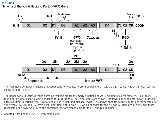
The genetics of VWD present a heterogeneous and diverse expression pattern. The VWF gene is located on chromosome 12 and yields a protein with several regions corresponding to four types of protein domains that comprise the functions of VWF (Figure 1). Genetic mutations within these domains are responsible for various types of VWD (see “classification of VWD,” page 4). An acquired form occurs secondary to other medical disorders, such as autoimmune conditions, some cancers, or cardiovascular disorders,9 but exhibits the same biochemical alterations of inherited VWD.911 This Product Profiler focuses only on hereditary, not acquired, forms of VWD.

FIGURE 1.

FIGURE 1

Schema of the von Willebrand Factor (VWF) Gene

The wide spectrum of gene mutations in VWD, including large deletions, frameshifts from small insertions or deletions, splice-site mutations, nonsense mutations, and missense mutations,9 are similar to defects found in many other diseases. As a result, it is difficult to identify the presence of the disorder on traditional genetic testing, and in most cases VWD is recognized only during a hemostatic challenge.4 To complicate the assessment of VWD further, circulating levels of VWF appear to vary with blood type, with the lowest levels associated with blood type O.4,12 These levels rise with age, at an approximate rate of 6 U/dL per decade.4 During times of metabolic stress, such as during pregnancy or in patients with malignant disease, VWF levels also may rise.4

The importance of appropriate identification and treatment of VWD is underscored by the release of NHLBI guidelines in 2007. Previously, physicians lacked clear guidance regarding the discrepancies between laboratory methods and the relationships between VWF levels and bleeding. The new guidelines address this need and allow physicians to make more informed decisions about the diagnosis and management of VWD. In addition, the guidelines discuss the improvement of clinical diagnostic tools and optimal treatment options for patients suffering from VWD. Because of the limited number of published studies in this arena, many of the recommendations are based on the opinion of the expert panel.9

Classification of VWD

There are three distinct forms of vWD: type 1, type 2, and type 3 (Table 1). Types 1 and 3 involve a partial or complete deficiency of VWF, respectively. Type 2 involves structural or functional defects of the VWF protein.13 In general, the severity of bleeding worsens from type 1 to 3 disease. It should be understood that severity can differ within each type, and all degrees — mild, moderate, and severe forms — can benefit from correction of the deficient or defective VWF.13

TABLE 1.

Classification of von Willebrand Disease

Type 1 Type 2A Type 2B Type 2M Type 2N Type 3
Quantitative deficiency
Qualitative deficiency
Complete deficiency
Inheritance AD AD/AR AD AD/AR AR AR
RCo:VWF (levels range) <30 <30 <30 <30 30–200 Undetectable
FVIII levels (range) 50–200 IU/dL or mildly decreased 50–200 IU/dL or mildly decreased 50–200 IU/dL or mildly decreased 50–200 IU/dL or mildly decreased Significantly decreased <10 IU/dL

AD=autosomal dominant; AR=autosomal recessive

Source: NHLBI 20079

VWF is a relatively large protein made up of a signal peptide (22 amino acids), a large propeptide (741 amino acids), and a mature VWF molecule (2,050 amino acids).2 These different protein regions correspond to four repeating domains that are responsible for the different binding functions of the molecule.2 Monomers of mature VWF are assembled into multimers via disulfide bridges.4 Large complexes are essential for normal platelet adhesion and aggregation under high shear stress conditions, such as those found in arterial capillaries.4

Type 1

Type 1 is the most common form of VWD, accounting for approximately 75% of congenital cases.4 Type 1 disease is marked by a partial quantitative deficiency of functionally normal VWF in platelets or plasma.13 Due to the binding of VWF to FVIII in the plasma, FVIII levels may drop in parallel, warranting replacement of both blood proteins for hematologic normalization.

In type 1 disease, FVIII generally is mildly decreased and VWF levels are low, with no significant decrease in large VWF multimers and only mild bleeding tendency.1,2,13 In many cases, the remaining VWF is able to induce relatively normal platelet adhesion and FVIII binding.9 In cases in which VWF levels range from 30–50 IU/dL, falling just below the normal range (50–200 IU/dL), diagnosis may be difficult; delayed diagnosis can result in unnecessary blood loss, anemia, and, in women, menorrhagia. In cases with VWF levels in the 10–20 IU/dL range (usually related to VWF mutations inhibiting transport of proVWF or promoting rapid VWF clearance from the circulation),15,16 the diagnosis is more apparent.

Type 2

Type 2 composes about 15% to 20% of VWD diagnoses, and is marked by a qualitative defect in the VWF protein. The group is further divided into four subtypes based on pathophysiology:4,17,18

  • Type 2A VWD is recognized by a reduction in high-molecular-weight VWF multimers in the plasma and a decrease in VWF-dependent platelet adhesion.9 Levels of VWF protein and FVIII may be normal or slightly reduced, but VWF function is markedly decreased as determined by ristocetin cofactor (VWF:RCo) activity assay.19 Impaired coagulation may be related to mutations that inhibit normal assembly or secretion of large multimers or increase susceptibility to proteolytic degradation of VWF, and patients may be predisposed to bleeding due to the deficit of large multimers.9

  • Type 2B VWD is associated with proteolytic degradation and depletion of large multimers as a result of mutations that increase VWF-platelet binding.9 VWF also exhibits an increased affinity for the platelet GP complex, GP1bα. The overall effect is spontaneous binding of VWF to platelets, leading to platelet agglutination, thrombocytopenia, and rapid VWF clearance.9,20

  • Type 2M disease is caused by mutation in the GP1b binding domain, which leads to decreased VWF-dependent platelet binding activity despite normal multimer structure.21,22

  • Type 2N is characterized by a full array of normal VWF multimers but reduced VWF-FVIII binding. As a result, FVIII stability and half-life are reduced, and hemostasis is impaired.9 In most of these cases, FVIII is less than 10% of normal; as such, type 2N VWD may be misdiagnosed as an autosomal recessive form of hemophilia.9,23 Only a FVIII-VWF binding assay can distinguish between the two.

Type 3

Patients with type 3 VWD are essentially or completely devoid of VWF and are moderately deficient in FVIII as well, due to its shortened half-life in the absence of VWF. These patients thus have impaired thrombin generation and fibrin formation, and exhibit a severe hemophilia-like bleeding disorder.24 This disorder is rare, representing less than 5% of patients with VWD,18 and can be life-threatening. It is associated with decreased stability and rapid clearance of FVIII, with levels reportedly falling as low as 5% or less of normal.26

Diagnostic and Laboratory Criteria

A positive bleeding history since childhood may be suggestive of VWD.2 In particular, bleeding is prominent and/or easy bruising is seen in one or more of the five following indicators:2,4,27

  • Frequent or prolonged nosebleeds

  • Heavy menstrual bleeding

  • Prolonged bleeding (>5 minutes) or recurrent bleeding during or following childbirth or surgery

  • Prolonged/excessive bleeding or mucous membrane bleeding during dental work

  • Family history of an autosomal dominant or recessive inherited bleeding disorder or easy bruising with indurations, which may also indicate the presence of VWD

Multiple and specialized laboratory evaluations, generally repeated several times over a 1- to 3-month period to compensate for clinical and laboratory variability, are required for a definitive diagnosis of VWD.28 Unfortunately, no single laboratory test firmly establishes a diagnosis of VWD. Instead, an initial hemostatic evaluation is conducted to determine whether there is a coagulation factor deficiency, thrombocytopenia, or other coagulopathy causing the bleeding.9 Typically, laboratory tests include platelet count, complete blood count, partial thromboplastin time (PTT), prothrombin time (PT), and either fibrinogen measure or thrombin time.9

In patients with abnormal results, such as a prolonged PTT that corrects using a 1:1 mixing study, further testing should include evaluation for VWF:Ag, VWF:RCo, and FVIII.9 VWF:Ag measures the amount of protein present in plasma.9 A VWF:RCo assay exposes a plasma specimen to ristocetin, an antibiotic that causes platelet agglutination, indicating reduced VWF activity. This activity is measured via platelet aggregation and is proportional to the reduced levels of VWF in the plasma in subjects with VWD. Decreased concentration of VWF:Ag or decreased activity in VWF:RCo testing is suggestive of type 1 VWD.9 Testing for FVIII helps to determine the ability of VWF to serve as a carrier protein to maintain normal levels of FVIII.9

Additional tests, such as radioimmunoassay or enzymelinked immunoabsorbent assays, can also be used to indicate the amount of VWF antigen in plasma but do not necessarily indicate any structural or functional defects, and thus do not rule out type 2 VWD.9 Structural defects instead must be assessed by means of gel electrophoresis multimer analysis; low- and high-resolution gel electrophoresis techniques are used to distinguish multimeric structure of VWF in plasma and platelets in order to differentiate the variants of VWD.9 A deficiency in FVIII coagulant activity also may be suggestive of VWD due to the protective binding of VWF to FVIII under normal physiologic conditions, and a FVIII binding assay can help to distinguish between disease subtypes.

Closure time (CT) and VWF binding to collagen (VWF: CB) assays also may assist in screening for VWD. CT is a rapid and simple technique for determining VWF-dependent platelet function at high shear stress, and is a good alternative to bleeding time (BT) measures in children or in patients with contraindications. Its limitations are that it appears normal in individuals with type 2N VWD, and it cannot be modified in type 3 patients after administration of a VWF/FVIII concentrate.29 The VWF:CB assay or VWF:CB to VWF:Ag ratio can be used to distinguish types 1 and 2 disease, and should be performed in association with the VWF:RCo test.30,31 Neither assay, however, has been standardized, and therefore is not diagnostic alone and must be performed with other VWD testing.

Several rapid tests for a bleeding disorder, such as activated PTT and BT, may be suggestive of severe type 3 disease, but are not necessarily prolonged in patients with mild disease and therefore are not useful as a global screen for VWD.9 Furthermore, because of the heterogeneity of the disorder and the impact of external influences, repeat testing often is required. For instance, a normal result on a first BT or PT may not rule out VWD; prolongation of response may not be evident until a second or subsequent assessment.

Clinical Manifestations

Mild-to-moderate mucocutaneous bleeding is the most common manifestation of VWD. This is especially evident as easy bruising, prolonged bleeding after injury and during dental work, and prolonged menses and menorrhagia.1 Some cases do not require routine treatment, reflecting the high rate of type 1 VWD relative to other types. This “mild” bleeding can interfere with a rapid and accurate diagnosis for VWD because similar bleeding episodes are observed frequently in the healthy population. In addition, the marker of excess bleeding during menstruation and childbirth in VWD requires careful examination. Menorrhagia may be a sensitive predictor of VWD in women (sensitivity 32–100%), but has limited specificity as similar numbers of healthy women and women with VWD report excessive bleeding during menstruation.32

Excessive and protracted postsurgical bleeding is common in VWD, whereas soft tissue and joint bleeding are observed infrequently and predominantly in persons with type 3 disease or extreme VWF deficiency. Life-threatening bleeding, particularly GI or central nervous system, can occur in persons with type 3 disease and in some individuals with type 2 disease.9 Bleeding severity also may be influenced by concomitant disease or medications, such as worsening among individuals taking aspirin or other nonsteroidal anti-inflammatory drugs.9

Function of VWF

VWF is a complex, multimeric glycoprotein synthesized by endothelial cells and megakaryocytes.1,14,33 Mature VWF proteins are either stored in secretory granules of Weibel-Palade bodies of the endothelial cell or in the alpha-granule of platelets prior to secretion in the event of vessel damage, or circulate as a loosely coiled protein complex.1

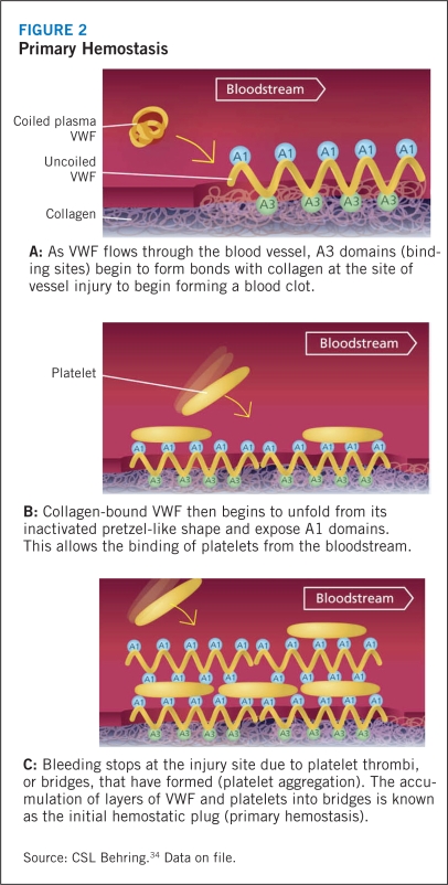
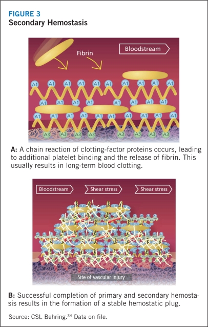
Circulating VWF does not interact with platelets or endothelial cells under normal physiologic conditions. In the event of tissue injury, however, VWF promotes both platelet adhesion and platelet aggregation.1 VWF, particularly the high-molecular-weight multimers, first attach to the subendothelial collagen at the site of injury, where-upon the high shear stress related to rapid blood flow in small vessels and large stenotic arteries activates the subendothelial collagen-bound VWF. This stretches the largest multimers into a filamentous sheet. The GP1bα receptor of circulating platelets adheres to the A1 domain of this reconformed VWF. The platelet adhesion leads to activation and secretion of intracellular proteins in an effort to engulf more platelets in the filament to form a hemostatic plug, a process known as primary hemostasis (Figure 2). Although this soft aggregate plug inhibits bleeding, it is fragile and easily dislodged. In order to secure the plug and complete the thrombus, activation of another platelet receptor protein GPIIb-IIIa35 initiates binding to VWF and fibrinogen, and promotes additional platelet recruitment into the hemostatic plug (Figure 3). During this secondary hemostasis, coagulation factors are activated, fibrin is released, and a stable hemostatic plug is formed.9,4,36

FIGURE 2.

FIGURE 2

Primary Hemostasis

FIGURE 3.

FIGURE 3

Secondary Hemostasis

VWF also influences the hemostatic pathway by its role in binding with FVIII. VWF protects FVIII from proteolytic degradation, prolonging its half-life and localizing it when vascular injury occurs. If VWF levels diminish, FVIII experiences a parallel decrease in concentration leading to faulty coagulation.1

Qualitative or quantitative defects in VWF interfere with normal hemostasis via impaired or delayed platelet adhesion and aggregation. As a result, subsequent platelets may not be recruited at full force or in “normal” time frame to complete the platelet plug. Additional coagulation factor reactions may be stalled, leading to the characteristic bleeding complications of VWD.1

Footnotes

*

Haemate-P® and Humate-P® are the same product. The difference between Haemate-P® and Humate-P® lies in the source of plasma — Humate-P® uses plasma only from U.S. donors. As such, the FDA recognizes the same active ingredients for both, which allows interchangeability of clinical results between the two.

*

The viral inactivation methods used to manufacture Humate-P® are proven safe and effective in eliminating blood-borne pathogens.

Decreased VWF antigen [VWF:Ag], VWF:RCo, and/or FVIII levels, for instance.

P T. 2010 Jan;35(1 Section 2):8–9.

Current Treatment Options and Guidelines

Treatment Guidelines

Treatment of VWD is focused on increasing the availability of VWF (and subsequently FVIII) to correct platelet function through adhesion, aggregation, and hemostatic plug formation.1 Currently, NHLBI recommends three approaches for managing VWD:9

  • Non-replacement therapy enables the release of endogenous VWF by stimulating the endothelial cell with desmopressin, a synthetic derivate of the antidiuretic hormone vasopressin.

  • Replacement therapy replaces missing VWF by delivering safe concentrates of human plasma-derived, viral-inactivated VWF/FVIII.

  • Adjunctive therapy: Other agents, such as antifibrinolytics and oral contraceptives, act to promote hemostasis without altering the VWF concentration at all.

Each of these approaches are described in more detail below. Depending on the type and severity of VWD or the bleeding risk or severity, these approaches can be used singly or in combination.8

The various options available to improve platelet activity or increase VWF and FVIII levels in patients with VWD are outlined below. Humate-P® belongs to the category of replacement therapy.

Nonreplacement Therapy

The synthetic derivative of vasopressin, desmopressin acetate (DDAVP), stimulates the release of VWF from storage sites within the endothelial cells, probably via a cyclic AMP-mediated process. Desmopressin lacks the pressor activity of the parent compound, vasopressin. Available in several formations and strengths, desmopressin used specifically for VWD treatment can be administered via intravenous or subcutaneous injection or nasal spray aspiration (Table 2). DDAVP is the generic scientific name for desmopressin nasal spray formulation (Stimate®), but should not be confused with trademarked DDAVP®, the only brand of desmopressin available for injection.

TABLE 2.

Forms of Desmopressin Acetate

Name Delivery system Concentration Indication
DDAVP®a Injection 4 mcg/mL Mild to moderate classic VWD (Type I) with FVIII levels >5%
Hemophilia A with FVIII coagulant activity levels >5%
Antidiuretic replacement therapy in the management of central (cranial) diabetes insipidus and for the management of the temporary polyuria and polydipsia following head trauma or surgery in the pituitary region
DDAVP® Nasal spray 0.1 mg/mL Antidiuretic replacement therapy in the management of central cranial diabetes insipidus and for the management of the temporary polyuria and polydipsia following head trauma or surgery in the pituitary region
Stimate® Nasal spray 1.5 mg/mLb Mild to moderate classic VWD (Type I) with FVIII levels > 5%b
Hemophilia A with FVIII coagulantactivity levels >5%
a

DDAVP is a trademark of Sanofi-Aventis. Generic forms of desmopressin acetate 0.004 mg/mL for injection exist.

b

Stimate 1.5 mg/mL is a high-concentrate formulation of this product. A low-concentrate form is used to treat diabetes and bed wetting, and is not indicated for VWD.

Sources: Stimate® [prescribing information],25 DDAVP® [prescribing information]37

The intranasal formulation of desmopressin contains 150 ug per metered nasal puff (0.1 mL of a 1.5 mg/mL solution), given as one (<50 kg person) or two (≥50 kg person) puffs. After nasal administration of desmopressin, absorption is variable and if possible patients should undergo a pretreatment dosing trial, in the nonbleeding state, to be certain that both VWF and FVIII rise adequately.9

Desmopressin also appears to promote secretion of tissue plasminogen activator, although this protein is rapidly inactivated before it can induce fibrinolysis or bleeding.9 Minor side effects, such as headache, flushing, or transient hyper- or hypotension, are common but rarely treatment limiting.9 Some reports of hyponatremia leading to seizures and death, however, have led to its contraindication for less serious diagnoses, such as bedwetting and minor bleeds from VWD,38 and infrequent cases of myocardial infarction suggest caution when using desmopressin in specific populations, including those at high risk for cardiovascular or cerebrovascular disease and the elderly.9 A risk for water retention and increased urinary osmolality may warrant fluid and electrolyte monitoring while receiving desmopressin.9

The National Hemophilia Foundation’s Medical and Scientific Advisory Council (MASAC) recommends use of desmopressin in patients with type 1 VWD, and in patients with types 2A, 2M, and 2N disease if they have been shown to be responsive to DDAVP.39 It may be cautiously considered in patients with type 2B VWD.9

A low-concentration desmopressin spray is reserved for the treatment of diabetes insipidus, whereas treatment for VWD requires the high-concentration formulation.

Replacement Therapy

Replacement of functional VWF and FVIII can be achieved via administration of plasma-derived concentrates containing both of these proteins (VWF/FVIII). There are three products currently approved in the United States for VWD; however, only Humate-P® is appropriate for all types of VWD and all surgical procedures. These products are indicated for episodic treatment and prevention of surgical bleeding in all types of VWD.4,40 FVIII alone cannot be used to treat congenital VWD.

The available VWF/FVIII concentrates have differing ratios of VWF to FVIII and different dosing regimens, and therefore should not be considered equivalent.9,41,42 The main goal of treatment in VWD is to replace the missing VWF. However, elevations in FVIII also have been found to increase the comorbidity risk for thromboembolic events.43 In fact, the risk for venous thromboembolism increased by 10% for every 10 IU/dL of FVIII administered.43 For these reasons, caution must be exercised in evaluating the VWF to FVIII ratio of a product.4 Humate-P® contains more VWF than FVIII, with a target ratio of VWF:RCo to FVIII activity of 2.4 to 1.0, which allows adjustment of VWF levels without raising FVIII to unacceptable levels.5,43 In addition, the concentration of high-molecular-weight VWF multimers, which contribute the most potent hemostatic activity, vary among products.9 These differences should be considered when selecting a treatment.

NHLBI guidelines suggest use of a VWF concentrate as replacement therapy for patients with significant bleeding events or those who need surgery and who have types 2 and 3 VWD, as well as in patients who have type 1 VWD who are unresponsive to DDAVP or require longer duration of therapy, or where DDAVP is contraindicated.9 The dose and duration of therapy depend on the intensity of the hemostatic challenge and the expected duration of treatment. VWF/FVIII concentrates are dosed primarily based on VWF:RCo units, with secondary consideration of FVIII units. Published results of the use of these products have generally reported good to excellent control of bleeding in 99% of surgeries and 97% of bleeding episodes.1,24,44–48

Similarly, MASAC recommends use of FVIII concentrate with high-molecular-weight VWF multimers that has been virally attenuated to eliminate transmission of HIV and hepatitis A, B, and C in patients with types 2B and 3 VWD and in other VWD patients with inadequate response to DDAVP.39

Adverse reactions are generally rare, but can include anaphylaxis, urticaria, chest tightness, rash, or edema.9 In rare occurrences, alloantibodies or inhibitors to VWF will occur. VWF/FVIII concentrates should be used in caution in patients with history of or known risk factors for thrombosis, and during extended therapy (i.e., ≥3 days), plasma FVIII levels should be monitored.9

Adjunctive Therapy

Antifibrinolytic agents. Antifibrinolytic agents currently used in the United States are aminocaproic and oral tranexamic acids.9 These agents inhibit the conversion of plasminogen to plasmin and, as a result, reduces the fibrinolysis that causes a clot to dissolve.9 When combined with desmopressin or VWF/FVIII concentrates as indicated, they are particularly effective in controlling bleeding in the oral cavity or GI or genitourinary tracts.9

Side effects of antifibrinolytic agents include nausea and vomiting, but they also less frequently can cause serious thrombotic complications.9

Oral contraceptives. Differences of opinions exist regarding the mechanism of action for oral contraceptives in VWD patients.1,9 Oral contraceptives have no direct effect on VWF/FVIII levels but may be useful in reducing the severity of menorrhagia in women with VWD.1 Efficacy rates vary from 24% to 73% in published reports.49,50 Estrogen has been shown to increase plasma VWF and FVIII, but current oral contraceptives do not contain sufficient estrogen levels to be effective as a treatment for bleeding episodes.1

Cryoprecipitate. Because it is not virally inactivated, MASAC recommends that cryo precipitate from human plasma not be used except in circumstances where any of the above-mentioned products are unavailable in a life-or limb-threatening emergency.39

P T. 2010 Jan;35(1 Section 2):10–11.

Indications, Chemistry, and Pharmacology

Indications5

Antihemophilic factor/von Willebrand factor (VWF) complex (human), dried, pasteurized (Humate-P®), is indicated in adult patients for treatment and prevention of bleeding in hemophilia A (classical hemophilia). Humate-P® is also indicated in adult and pediatric patients with von Willebrand disease (VWD) for (1) treatment of spontaneous and trauma-induced bleeding episodes and (2) prevention of excessive bleeding during and after surgery. This applies to patients with severe VWD, as well as patients with mild to moderate VWD where use of desmopressin is known or suspected to be inadequate.

Chemistry5

Humate-P® is a stable, purified, sterile, lyophilized concentrate of antihemophilic factor (human) and von Willebrand factor (human). It is administered intravenously in the treatment of patients with VWD.

Humate-P® is purified from the cold insoluble fraction of pooled human fresh-frozen plasma. It contains the human glycoproteins that play a role in hemostasis in a highly purified and concentrated formulation, and has a high degree of purity with a low amount of non-factor proteins. Fibrinogen is less than or equal to 0.2 mg/mL. This formulation has a higher factor potency than cryoprecipitate preparations.

Each vial of Humate-P® contains the labeled amount of FVIII activity in international units (IU). In addition, each vial contains the labeled amount of VWF:RCo activity expressed in IU. (An IU FVIII or IU VWF:RCo, as defined by the World Health Organization standard, is exactly equal to the level of FVIII or VWF:RCo found in one 1.0 mL of fresh-pooled human plasma.)

Upon reconstitution with the volume of the sterile diluent provided (Sterile Diluent for Humate-P®), each mL of Humate-P® contains 40 to 80 IU FVIII activity; 72 to 224 IU VWF:RCo activity (which correlates to a VWF:RCo-to-FVIII activity average ratio of 2.4, which is used to calculate the nominal values of VWF:RCo activity and is the average VWF:RCo activity); 15 to 33 mg of glycine; 3.5 to 9.3 mg of sodium citrate; 2.0 to 5.3 mg of sodium chloride; 8 to 16 mg of albumin (human); 2 to 14 mg of other proteins; and 10 to 20 mg of total proteins.

Humate-P® has been demonstrated in several studies to contain the high-molecular-weight multimers of VWF.5155 This component is considered important for correcting the coagulation defect in patients with VWD. When administered to patients with VWD types 1, 2, or 3, bleeding time decreased. This effect was correlated with the presence of multimeric composition of VWF similar to that found in normal plasma.

Humate-P® contains anti-A and anti-B blood group isoagglutins. This product is prepared from pooled human plasma collected from U.S.-licensed facilities in the United States.

All source plasma used in the manufacture of this product was tested by U.S. Food and Drug Administration-licensed nucleic acid tests (NAT) for hepatitis C virus and HIV-1 and found to be nonreactive (negative). An investigational NAT for hepatitis B virus (HBV) also was performed on all source plasma used in the manufacture of this product, and was nonreactive (negative). An investigational NAT for HBV was also performed on all source plasma used in the manufacture of this product and found to be nonreactive (negative). The HBV test is intended to detect low levels of viral material; however, the significance of a nonreactive (negative) result has not been established.

Virus Reduction Capacity5

The manufacturing procedure for Humate-P®` includes multiple processing steps that reduce the risk of virus transmission. The virus reduction capacity of the manufacturing process was evaluated in a series of in vitro spiking experiments: the steps were: cryoprecipitation; Al(OH) adsorption, glycine precipitation, and NaCl precipitation, studied in combination; and pasteurization in aqueous solution at 60° C for 10 hours. Total mean cumulative virus reductions ranged from 6.0 to ≥ 11.3 log10 (Table 3).

TABLE 3.

Mean Virus Reduction Factors

Virus studied Cryoprecipitation (log10) Al(OH)2 adsorption/glycine precipitation/NaCl precipitation (log10) Pasteurization (log10) Total cumulative (log10)
Enveloped viruses
HIV-1 ND 3.6 ≥6.4 ≥10.0
BVDV ND 2.4 ≥8.9 ≥11.3
PRV 1.6 3.7 4.6 9.9
WNV ND ND ≥7.8 NA
Non-enveloped viruses
HAV 1.5 2.4 4.2 8.1
CPV 1.5 3.4 1.1 6.0
B19V ND ND ≥3.9* NA

B19V=parvovirus B19; BVDV=bovine viral diarrhea virus, model for HIV types 1 and 2; CPV=canine parvovirus, model for parvovirus B19; HAV=Hepatitis A virus; NA=not applicable; ND=not determined; PRV=pseudorabies virus, model for large enveloped DNA viruses (e.g., herpes virus); WNV=West Nile virus.

* The virus evaluation studies for parvovirus B19 employed a novel experimental infectivity assay utilizing a clone of the cell line UT7 that contains erythropoietic progenitor cells; (Residual) virus titer was determined using an immunofluorescence-based detection method.

Source: Humate-P® [prescribing information]5

Pharmacology5

The antihemophilic factor/von Willebrand factor complex consists of two different noncovalently bound proteins (FVIII and VWF). FVIII is an essential cofactor in activation of factor X, leading ultimately to formation of thrombin and fibrin. The VWF promotes platelet aggregation and platelet adhesion on damaged vascular endothelium. It also serves as a stabilizing carrier protein for the procoagulant protein FVIII. The activity of VWF is measured as VWF:RCo.

Pharmacokinetics in von Willebrand Disease5

Pharmacokinetic studies of Humate-P® have been performed with cohorts of subjects in the nonbleeding state. Wide intersubject variability was observed in pharmacokinetic values obtained from these studies.

The pharmacokinetics of Humate-P® were evaluated in 41 subjects in a prospective U.S. study in the nonbleeding state prior to a surgical procedure. Subjects received 60 IU VWF:RCo/kg body weight of Humate-P®. Sixteen subjects had type 1 VWD, two had type 2A, four had type 2B, six had type 2M, and 13 had type 3. The median terminal half-life of VWF:RCo was 11 hours (range: 3.5–33.6 hours), excluding five subjects with a half-life exceeding the blood sampling time of 24–48 hours. The median clearance and volume of distribution at steady state were 3.1 mL/hr/kg (range: 1.0–16.6 mL/hr/kg) and 53 mL/kg (range: 29–141 mL/kg), respectively. The median in vivo recovery for VWF:RCo activity was 2.4 IU/dL per IU/kg (range: 1.1–4.2). High-molecular-weight multimers were measured in 13 subjects with type 3 disease; 11 had absent or barely detectable multimers at baseline. Of those 11 subjects, all had some high-molecular-weight multimers present 24 hours after infusion of Humate-P®.

Pharmacokinetics also were evaluated in 28 subjects in a European study in the nonbleeding state prior to a surgical procedure. Subjects received 80 IU VWF:RCo/kg body weight of Humate-P®. Ten subjects had type 1 disease, 10 had type 2A, one had type 2M, and seven had type 3. The median terminal half-life of VWF:RCo was 10 hours (range: 2.8–28.3 hours), excluding one subject with a half-life exceeding the blood sampling time of 48 hours. The median clearance and volume of distribution at steady state were 4.8 mL/hr/kg (range: 2.1–53 mL/hr/kg) and 59 mL/kg (range: 32–290 mL/kg), respectively. The median in vivo recovery for VWF:RCo activity was 1.9 IU/dL per IU/kg (range: 0.6–4.5). Infusion of Humate-P® corrected the defect of the multimer pattern in subjects with types 2A and 3 disease. High-molecular-weight multimers were detectable until at least 8 hours after infusion.

Based on this evaluation, it appears that age, sex, and types of VWD have no impact on the pharmacokinetics of VWF:RCo.

P T. 2010 Jan;35(1 Section 2):12–15.

Summary of Key Published Clinical Trials


Humate-P® has extensive clinical experience in the treatment of VWD. In a series of prospective and retrospective trials, Humate-P® has been shown to provide effective hemostatic control for VWD bleeding episodes in the vast majority of adult and pediatric patients treated. Three key trials described below outline the biochemistry, hemostatic activity, and clinical application of Humate-P® in the treatment of VWD.

Efficacy and Safety of Humate-P® in Children and Adults With von Willebrand Disease46

In this retrospective review of data on Humate-P®/Haemate-P® use in Canada, the authors examined the efficacy and safety of this VWF/FVIII complex therapy when dosed in VWF:RCo units for the treatment of VWD. Efficacy was assessed by a predetermined rating system as:

  • Excellent (hemostasis achieved/complete cessation of bleeding)

  • Good (slight oozing/partial but adequate bleeding control that did not require additional products)

  • Poor (moderate bleeding/moderate control that required additional products for unplanned treatment)

  • None (severe uncontrolled bleeding).

An excellent/good rating was used if patients experienced an effective reduction in observed bleeds. A poor/none rating was used if there was an inability to reduce the number of bleedings in prophylactic patients.

Adverse events (AEs) were considered to be any change from baseline in a patient’s health status that occurred within 24 hours of Humate-P®/Haemate-P® administration. AE recording excluded any events directly related to surgery.

The study population included 97 patients with VWD types 1, 2, or 3 or other (2N, 2M, or acquired). There were 437 bleeding events (73 surgery, 344 bleeding, and 20 prophylaxis) considered in the final analysis. A group of 93 “other” events — which included minor biopsies, test doses, and childbirth — were excluded from analysis because their heterogeneity diminished the value of information gained.

Results. Overall, 97% of events treated with Humate-P®/Haemate-P® achieved an excellent or good clinical result (Table 4). In a pediatric patient subanalysis, results revealed excellent/good outcomes in 100% of treatment events in infants age 1 month to <2 years; 95% of children age 2 to <12 years; and 94% of children age 12 to <16 years. Overall, the majority of patients were treated for 10 days or less, although some patients, mostly treated for prophylaxis, were treated for longer durations (11–20 days or more).

TABLE 4.

Efficacy of Humate-P® in Surgery and Bleeding or Prophylactic Use

VWD type 1 2A 2B 3 Other Total

No. of patients 32 5 18 28 14 97
Surgeries (n) 26 6 14 21 6 73
Outcomes
   Excellent/good (%) 100 100 93 100 100 99
   Poor/none (%) 7 1
Bleeding episodes (n) 32 17 60 208 27 344
Outcomes
   Excellent/good (%) 100 100 10 95 93 97
   Poor/none (%) 5 7 3
Prophylaxis (n of uses) 20 20
Outcomes
   Excellent/good (%) 100 100
   Poor/none (%) 0 0

Source: Lillicrap 200246

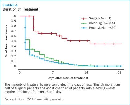
The median doses of Humate-P®/Haemate-P® varied according to bleeding events but were similar across VWD types. The median doses were 69.1 IU VWF:RCo/kg for surgical events (range: 11.9–222.8 IU), 55.3 IU VWF:RCo/kg for acute bleeding episodes (range: 17.1–227.5 IU), and 41.6 IU VWF:RCo/kg for prophylaxis (range: 34.6–81.0 IU). Most treatments required no more than 2 to 3 days of therapy, and only about 55% of surgical events and 35% of bleeding events required treatment after the first post-event day (Figure 4).

FIGURE 4.

FIGURE 4

Duration of Treatment

Laboratory assessment of in vivo recovery of VWF:RCo activity in 17 patients not bleeding at the time of Humate-P®/Haemate-P® administration indicated a median measure of 1.35 IU/mL. Thirteen of these patients had physician-rated excellent/good hemostatic efficacy ratings; the median recovery of VWF:RCo activity in these patients was 1.23 IU/mL. The other four patients, who were not evaluated for hemostatic efficacy, had an in vivo recovery of 1.82 IU/mL.

Probable treatment-related AEs were noted in four patients and none were considered to be serious. There were no reported cases of clinically observed thrombosis;however, one patient discontinued treatment due to an AE from an accidental traumatic injury that resulted in death.

This analysis established the efficacy and safety of Humate-P®/Haemate-P® when dosed according to VWF: RCo units to improve hemostatic activity during surgery, bleeding events, or as prophylaxis for dental or surgical interventions in patients with VWD. It was effective in pediatric and adult patients and across all subtypes of VWD.

High-Molecular-Weight VWF Correlates With Increased Coagulant Activity33

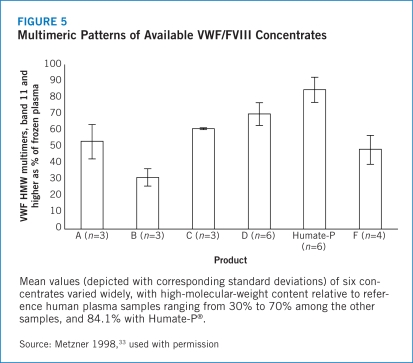
The relative content of high-molecular-weight multimers of VWF influences the hemostatic activity of VWF/FVIII concentrates used in the treatment of VWD, with an increased presence of high-molecular-weight variants associated with improved hemostatic function.56,57 To quantify the multimeric structure of VWF content, Metzner used an SDS-agarose gel electrophoresis method to quantify high-molecular-weight VWF in six VWF/FVIII concentrates, with particular attention to Humate-P®.33

Results. This analysis revealed a high consistency of multimeric band patterns among samples of Humate-P®. It also showed a multimeric band pattern very similar to that seen with the reference human plasma, with only a slight decrease in large multimer content. In fact, after analysis of 47 batches of Humate-P®, the average high-molecular-weight VWF content was 84.1% of that reflected in corresponding bands of reference human plasma (minimum 70%, maximum 95%; SD, 6.3%). Humate-P® high-molecular-weight VWF concentrations more closely corresponded with reference human plasma samples than any of the other commercially available VWF/FVIII concentrates (Figure 5).

FIGURE 5.

FIGURE 5

Multimeric Patterns of Available VWF/FVIII Concentrates

Similarly, Humate-P® measures of VWF:RCo activity relative to VWF:Ag concentration, as determined on Laurell rocket electrophoresis, closely approximated that of human plasma, with relative ratios of 0.8 to 0.9 compared with 1.0 for the reference plasma.

The other VWF/FVIII products exhibited lower specific activities of VWF, in the range of 0.3 to about 0.8. The data, furthermore, indicated a linear correlation between the high-molecular-weight VWF content of a concentrate and the specific activity of VWF for that product.

This study established Humate-P® as closer to human plasma in both high-molecular-weight VWF content and in specific activity of VWF than other VWF/FVIII complex products evaluated. This is important because high-molecular-weight VWF increases platelet agglutination to a greater degree than medium- and low-molecular-weight multimers, and improves overall hemostatic function in patients with VWD.

Humate-P® Dosing According to Ristocetin Cofactor Assay24

VWF:RCo activity was long used to diagnose and follow treatment activity in patients with VWD, but not until 2002 was this tool applied to guide dosing with VWF/FVIII concentrates.40,46 Based on successful outcomes in these earlier trials, Gill conducted a prospective, multicenter, open-label, non-randomized study to evaluate the efficacy and safety of Humate-P® via VWF:RCo-based dosing. Patients enrolled in this study were not candidates for desmopressin therapy and had urgent bleeding episodes or emergency surgery. Only the experience in patients who had urgent bleeds is reported here.

Humate-P® (average ratio of IU VWF:RCo per IU FVIII:C of ~2.5:1.0) was administered intravenously (see dosing, Table 5A). VWF:RCo activity was maintained at >50 IU/dL (50%) for the first 3 days and FVIII:C was sustained at levels in keeping with the usual clinical practice of the investigator. Patient closure to study was 3 days after the investigator deemed no further benefit from therapy. Hemostatic efficacy was rated daily and overall (at completion of study) as:

  • Excellent/good (hemostasis similar to that of individual without bleeding disorder)

  • Fair/poor (less hemostasis than individual without bleeding disorder)

  • None (severe bleeding due to VWD despite Humate-P®).

TABLE 5A.

Humate-P® Dosing According to Ristocetin Cofactor Assay/Surgery

  • Loading dose: 60–80 IU VWF:RCo/kg body weight

  • Maintenance doses: 40–60 IU VWF:RCo/kg every 8–12 hours for 3 days

  • As needed additional treatment: 40–60 IU VWF:RCo/kg daily, for up to 7 days (longer only in unusual circumstances)

Efficacy also was assessed by the number of study medication infusions required, number of days treated, and occurrence of visible hemorrhages. Bleeding was characterized as severe (refractory epistaxis, gastrointestinal hemorrhage, central nervous system trauma, other traumatic hemorrhage), moderate (moderate epistaxis, oral bleeding, menorrhagia), or mild.

Results. Among 33 patients enrolled at 19 centers, 53 serious bleeding events were treated — 48 with complete follow-up and five discontinued. Slightly more than half of the bleeding events occurred in patients with type 3 VWD and events predominantly occurred in the nose, oral cavity, or GI tract.

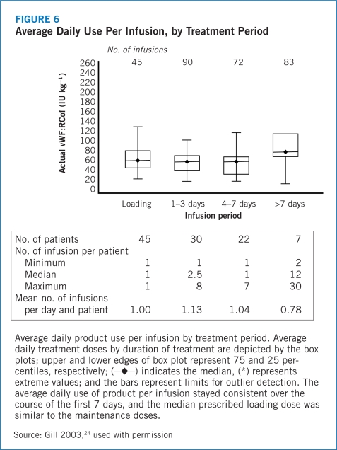
Median loading dose of Humate-P® was 67.0 IU/kg−1 VWF:RCo and the median daily maintenance dose was 74.0 IU/kg VWF:RCo (Table 5B) given in a median of two doses per event. The median treatment duration was 3 days. Patients receiving treatment for more than 7 days tended to use higher dosages but with less frequent administration (Figure 6). The type of VWD did not have a significant impact on the daily dose of Humate-P® required, with wide ranges in dosage and number of days of treatment across types.

TABLE 5B.

Intensity of Humate-P® Treatment Based on Severity of Bleeding Event

Bleeding severity
Severe Moderate Mild Overall
Patients (N=33)*
Events (n) 16 34 3 53
Median loading dose (IU VWF:RCo/kg−1) 87.3 66.7 57.4 67.0
Median maintenance dose (IU VWF:RCo/kg−1) 100.1 68.0 67.8 74.0
Median duration of treatment days 5 3 1 3
Median no. of infusions 4 2 1 2
*

Nine patients were enrolled more than once for multiple events.

Source: Gill 200324

FIGURE 6.

FIGURE 6

Average Daily Use Per Infusion, by Treatment Period

Higher dosing in both loading and maintenance phases appeared to correlate positively with severity of the bleeding episode, as did the number of infusions and days on treatment. Similarly, the intensity of therapy related to the organ affected, with patients with GI or neurological bleeds receiving the highest loading doses and those with genitourinary events getting higher maintenance doses than those affecting other sites. More infusions were given to patients with orthopedic or neurological events (12 and 2 infusions, respectively), and they were treated for longer periods (5 and 6 days, respectively) than were individuals with endocrine, ear, nose and throat, or dental bleeding (1, 12, 4 infusions, and 1 day, respectively).

There were wide variations in plasma VWF:RCo activity recovery, based on samples obtained 15 to 60 minutes after the first infusion in 7 patients with urgent bleeding. The median peak and trough recoveries were 1.5 and 0.6 IU/dL per IU kg−1 VWF:RCo infused. There were no episodes of thrombosis in these nonsurgical patients, likely related to the low ratio of FVIII to VWF:RCo in Humate-P®.

The efficacy analysis revealed an excellent overall hemostatic effect, with Humate-P® dosed according to VWF:RCo activity; 98% of treated bleeding episodes were rated as excellent/good. There was one episode of fair/poor or temporary ineffectiveness, but the patient was managed according to the investigator’s judgment, and resolved to be effectively managed thereafter with Humate-P®. There were no unexpected treatment-related adverse events; the only two episodes associated with serious adverse events were not considered related to study medication. None required treatment discontinuation.

In summary, this study showed that treating urgent bleeding episodes with Humate-P® at doses determined by VWF:RCo assay is an effective and safe method to correct hemostatic activity during acute bleeding emergencies in patients with VWD. This approach appears to ensure effective levels of both FVIII and VWF activity without elevating FVIII sufficiently to predispose to thrombosis. Humate-P® worked across all VWD types and in patients of all disease severities, with dosing intensity and duration correlated with severity and site of bleeding.

Footnotes

The FVIII content in Humate-P® is listed in the product label as FVIII:C.

P T. 2010 Jan;35(1 Section 2):16–17.

Efficacy & Safety

Efficacy5

The hemostatic activity and safety of Humate-P® has been studied in one retrospective review of use in the control of bleeding and in two clinical studies in subjects undergoing surgery. The retrospective review was based on Canadian subjects treated under an Emergency Drug Release Program. Humate-P® was administered to 97 subjects for 530 treatment courses: 73 for surgery, 344 for bleeding, and 20 for prophylaxis. The 99 “other” uses involved dental procedures, diagnostic procedures, prophylaxis prior to procedures, or a test dose. Efficacy ratings of excellent/good were yielded in 100% of bleeding episodes treated in type 1, 2A, and 2B subjects, and in 95% of type 3 subjects; poor (or no) response was observed in the remaining 5% of subjects.

In a U.S. clinical study, Humate-P® was given to 35 subjects (21 women and 14 men) with VWD undergoing surgery, including 28 major procedures (e.g., orthopedic joint replacement, intracranial surgery, multiple tooth extractions, laparoscopic cholecystectomy), four minor procedures (e.g., placement of intravenous access device), and three oral surgeries (removal of fewer than three teeth). The first 15 subjects received a loading dose of 1.5 times the “full dose” (dose predicted to achieve a peak VWF:RCo level of 100 IU/dL), and the next 20 subjects were dosed based on individual pharmacokinetic assessments and target peak VWF:RCo levels of 80 to 100 IU/dL for major surgery and 50 to 60 IU/dL for minor or oral surgery, respectively. Maintenance doses were 0.5 times full dose to start, then adjusted based on regular measurements of trough VWF:RCo and FVIII:C levels. The median duration of treatment was 1 day (range: 1–2 days) for oral surgery, 5 days (range: 3–7 days) for minor surgery, and 5.5 days (range: 2–26 days) for major surgery. A European clinical study also used Humate-P® to prophylactically treat 27 subjects (18 women and nine men) with VWD undergoing surgery. Dosing was individualized based on a pharmacokinetic assessment performed before surgery. The median duration of treatment was 3.5 days (range: 1–17 days) for minor surgery and 9 days (range: 1–17 days) for major surgery.

In both studies, hemostatic efficacy at the end of surgery was found to be “effective” (excellent/good), including in 32 (91.4%) of U.S. participants and in 25 (96%) of European subjects for whom data were available. The hemostatic efficacy was classified by investigators as excellent/good for all surgical subjects in the both the U.S. and European settings. Humate-P® also was effective in preventing excessive bleeding during and after surgery. In the U.S. study, the median actual estimated blood loss did not exceed the median expected blood loss, regardless of the type of surgery. Four subjects received transfusions, three due to adverse events and one due to pre-existing anemia. In the European study, one subject received transfusions to treat pre-existing anemia.

The outcomes of studies to determine the clinical efficacy of Humate P® are described in more detail in the Clinical Trials section of this publication.

Safety5

Contraindications. Humate-P® is contraindicated in individuals with a history of anaphylactic or severe systemic response to antihemophilic factor or von Willebrand factor preparations. It also is contraindicated in individuals with a known hypersensitivity to any of its components.

Warnings. Thromboembolic events have been reported in VWD patients receiving antihemophilic factor/von Willebrand factor complex replacement therapy, especially in the setting of known risk factors for thrombosis. Early reports might indicate a higher incidence in women. In addition, endogenous high levels of FVIII also have been associated with thrombosis but no causal relationship has been established. In all VWD patients in situations of high thrombotic risk receiving coagulation factor replacement therapy, caution should be exercised and antithrombotic measures should be considered.

Humate-P® is made from human plasma. Products made from human plasma may contain infectious agents, such as viruses, that can cause disease. Because Humate-P® is made from human blood, it may carry a risk of transmitting infectious agents, e.g., viruses, and theoretically, the Creutzfeldt-Jakob disease agent. The risk that such products will transmit an infectious agent has been reduced by screening plasma donors for prior exposure to certain viruses, by testing for the presence of certain current viral infections, and by inactivating and/or removing certain viruses during manufacture. Stringent procedures, utilized at plasma collection centers, plasma testing laboratories, and fractionation facilities are designed to reduce the risk of virus transmission. The primary virus reduction step of the Humate-P® manufacturing process is the heat treatment of the purified, stabilized aqueous solution at 60°C for 10 hours (i.e., pasteurization). In addition, the purification procedure, which includes several precipitation steps and an adsorption step, used in the manufacture of Humate-P® also provides virus reduction capacity. Despite these measures, such products may still potentially contain human pathogenic agents, including those not yet known or identified. Thus the risk of transmission of infectious agents cannot be totally eliminated. Any infections thought by a physician possibly to have been transmitted by this product should be reported by the physician or other healthcare provider to CSL Behring at (866) 915-6958 (from the United States and Canada). The physician should discuss the risks and benefits of this product with the patient.

Precautions. It is important to determine that the coagulation disorder is caused by FVIII or VWF deficiency, since no benefit in treating other deficiencies can be expected.

Thromboembolic events have been reported in VWD patients receiving coagulation factor replacement therapy, especially in the setting of known risk factors for thrombosis.

As a precaution, the administration equipment and any unused Humate-P® should be discarded after use.

Animal reproduction studies have not been conducted with antihemophilic factor/von Willebrand factor (human). It also is not known whether Humate-P® can cause fetal harm when administered to a pregnant woman or can affect reproduction capacity. Humate-P® should be given to a pregnant woman only if clearly needed.

The safety and effectiveness of Humate-P® for the treatment of von Willebrand disease was demonstrated in 26 pediatric subjects, including infants, children, and adolescents, but has not been evaluated in neonates. The safety of Humate-P® for the prevention of excessive bleeding during and after surgery was demonstrated in eight pediatric subjects (ages 3–15) with VWD. Of the 34 pediatric subjects studied for both treatment of VWD and prevention of excessive bleeding during and after surgery, four were infants (1 month to under 2 years of age), 23 were children (2–12 years), and 7 were adolescents (13–15 years). As in adults, pediatric patients should be dosed based upon weight (kg) in accordance with information in the Dosage and Administration section of the full prescribing information.

Clinical studies of Humate-P® did not include sufficient numbers of subjects aged 65 and over to determine whether they respond differently from younger subjects. As for all patients, dosing for geriatric patients should be appropriate to their overall situation.

Adverse reactions. The most serious adverse reaction observed in patients receiving Humate-P® is anaphylaxis. Thromboembolic events also have been observed in patients receiving Humate-P® for the treatment of VWD. Reports of thromboembolic events in VWD patients with other thrombotic risk factors receiving coagulation factor replacement therapy have been obtained from spontaneous reports, published literature, and a European clinical study. Early reports might indicate a higher incidence in women. In some cases, inhibitors to coagulation factors may occur. However, no inhibitor formation was observed in any of the clinical trials.

Although few adverse reactions have been reported in clinical studies and in the postmarketing setting in patients receiving Humate-P® for treatment of hemophilia A and VWD, the most commonly reported are allergic-anaphylactic reactions (including urticaria, chest tightness, rash, pruritus, edema, and shock). For patients undergoing surgery, the most common adverse reactions are postoperative wound or injection-site bleeding.

P T. 2010 Jan;35(1 Section 2):18–20.

P&T Committee Considerations

Thromboembolic Risk

The risks of treating any patient with a bleeding disorder may result in thromboembolic events, especially those patients with more than one risk factor. Patients should be monitored frequently to avoid the accumulation of thromboembolic-causing factors.

Humate-P® contains a high ratio of VWF:RCo to FVIII,5 which may minimize the risk that adjustment of VWF in VWD is ac companied by an unacceptable increase in FVIII.

Insurance/Reimbursement58

Because the majority of Humate-P® utilization is for VWD, the product is sold and reimbursed on the basis of VWF:RCo international units (IUs). Each box of Humate-P® is labeled, however, with VWF:RCo and FVIII units of the specific lot, as dosing guidelines are based on replacing the defective or missing proteins. Dosing for hemophilia A is dosed in FVIII units, while dosing for VWD will be in VWF:RCo units.

Some physicians may prescribe in FVIII units for VWD patients because laboratory testing of FVIII is more readily available than for VWF:RCo. Healthcare professionals should carefully review prescriptions for accuracy. A potential for improper dosing exists if the prescription is not based on either VWF:RCo or FVIII units for dosing accuracy in VWD.

How Supplied5

Humate-P® is supplied in a single-dose vial containing the labeled amount of VWF:RCo and FVIII activity expressed in IU. Each package contains a vial of Humate-P, a vial of diluent containing sterile water (meets USP chemistry requirements Sterile Water for Injection, except for pH; ph 4.5–8.5), a Mix2Vial™ filter transfer set, and two alcohol swabs. The components used in the packaging for Humate-P® are latex-free.5

NDC number VWF:RCo/vial (IU) FVIII/vial (IU) Dosage Diluent (mL)
63833-615-02 600 250 Low 5
63833-616-02 1,200 500 Mid 10
63833-617-02 2,400 1,000 High 15

Dosage & Administration5

Humate-P® is for intravenous administration only. Each vial of Humate-P® contains the labeled amount of FVIII:C activity in IU (target 2.4 to 1.0 VWF:RCo to FVIII) for the treatment of hemophilia A. Additionally, each vial of Humate-P® also contains VWF:RCo activity in IU for the treatment of VWD. Studies performed to determine the effective dosing strategies were performed on patients with hereditary VWD, not acquired VWD.

Episodic treatment of bleeding for VWD. The dosage should be adjusted according to the extent and location of bleeding. As a rule, 40–80 IU VWF:RCo (corresponding to 17 to 33 IU FVIII in Humate-P®) per kg body weight are given every 8 to 12 hours. Repeat doses are administered for as long as needed, based on repeat monitoring of appropriate clinical and laboratory measures. Expected levels of VWF:RCo are based on an expected in vivo recovery of 2.0 IU/dL rise per IU/kg VWF:RCo administered. The administration of Humate-P® 1 IU of FVIII per kg body weight can be expected to lead to a rise in circulating VWF:RCo of approximately 5 IU/dL. See the full prescribing information for specific dosing guidelines.

Prevention of excessive bleeding during and after surgery in VWD. The full prescribing information provides guidelines for calculating the initial loading dose in addition to maintenance doses, which are 50% of the loading dose, of Humate-P® for patients undergoing surgery. In the case of emergency surgery, administer a loading dose of 50 to 60 IU/kg and, subsequently, closely monitor the patient’s trough coagulation factor levels. When possible, it is recommended that the incremental in vivo recovery be measured and that baseline plasma VWF:RCo and FVIII:C be assessed in all patients prior to surgery.

In presurgical dosing, IVR is measured as follows:

  1. Measure baseline plasma VWF:RCo.

  2. Infuse 60 IU VWF:RCo/kg product intravenously at time 0.

  3. At time +30 minutes, measure plasma VWF:RCo. IVR = (Plasma VWF:RCo time +30 min – Plasma VWF:RCo baseline) / 60 IU kg

Calculation of the loading dose requires four values: the target peak plasma VWF:RCo level, the baseline VWF:RCo level, body weight (BW) in kilograms, and IVR. When individual recovery values are not available, a standardized loading dose can be used based on an assumed VWF:RCo IVR of 2.0 IU/dL per IU/kg of VWF:RCo product administered. Table 6 provides guidelines for calculating the loading dose for adult and pediatric patients.

TABLE 6.

VWF:RCo and FVIII:C Loading Dose Recommendations for the Prevention of Excessive Bleeding During and After Surgery

Type of surgery VWF:RCo target peak plasma level FVIII:C target peak plasma level Calculation of loading dose (to be administered 1 to 2 hours before surgery)
Major 100 IU/dL 80–100 IU/dL Δ*VWF:RCo x BW (kg)/IVR#=IU VWF:RCo required
If the incremental IVR is not available, assume an IVR of 2 IU/dL per IU/kg and calculate the loading dose as follows:
(100 – baseline plasma VWF:RCo) x BW (kg) / 2.0
In the case of emergency surgery, administer a dose of 50–60 IU/kg
Minor / oral§ 50–60 IU/dL 40–50 IU/dL Δ*VWF:RCo x BW (kg)/IVR#=IU VWF:RCo required

BW=body weight.

* Δ=Target peak plasma VWF:RCo – baseline plasma VWF:RCo

#

IVR=Incremental recovery as measured in the patient

§

Oral surgery is defined as removal of fewer than three teeth, if the teeth are non-molars and have no bony involvement. Removal of more than one impacted wisdom tooth is considered major surgery due to the expected difficulty of the surgery and the expected blood loss, particularly in subjects with type 2A or type 3 VWD. Removal of more than two teeth is considered major surgery in all patients.

For example, the loading dose of Humate-P® required, assuming a target VWF:RCo level of 100 IU/dL, baseline VWF:RCo level 20 IU/dL, an IVR of 2.0 (IU/dL)/(IU/kg), Δ of 80 IU/dL, and a body weight of 70 kg, would be calculated as follows:
80IU/dL ×70kg2(IU/dL)/(IU/kg)=2,800IU VMF:RCo required

Source: Humate-P® prescribing information5

Attaining a target peak FVIII:C plasma level of 80 to 100 IU FVIII:C/dL for major surgery and 40 to 50 IU FVIII:C/dL for minor surgery or oral surgery might require additional dosing with Humate-P®. Because the ratio of VWF:RCo to FVIII:C activity in Humate-P® is 2.4 to 1, any additional dosing will increase VWF:RCo proportionally more than FVIII:C. Assuming an incremental IVR of 2.0 IU VWF:RCo/dL per IU/kg infused, additional dosing to increase FVIII:C in plasma also will increase plasma VWF:RCo by approximately 5 IU/dL for each IU/kg of FVIII administered. The initial maintenance dose for the prevention of excessive bleeding during and after surgery should be half the loading dose, irrespective of additional dosing required to meet FVIII:C targets.

Table 7 provides recommendations for target trough plasma levels (based on type of surgery and number of days following surgery) and minimum duration of treatment for subsequent maintenance doses. These recommendations apply to both adult and pediatric patients.

TABLE 7.

VWF:RCo and FVIII:C Target Trough Plasma Level and Minimum Duration of Treatment Recommendations for Subsequent Maintenance Doses for the Prevention of Excessive Bleeding During and After Surgery

Type of surgery VWF:RCo target trough plasma levels* FVIII:C target trough plasma levels* Minimum duration of treatment
Up to 3 days following surgery After day 3 Up to 3 days following surgery After day 3
Major >50 IU/dL >30 IU/dL >50 IU/dL >30 IU/dL 72 hours
Minor ≥ 30 IU/dL >30 IU/dL 48 hours
Oral# ≥ 30 IU/dL >30 IU/dL 8–12 hours§

* Trough levels for either coagulation factor should not exceed 100 IU/dL

# Oral surgery is defined as removal of fewer than three teeth, if the teeth are non-molars and have no bony involvement. Removal of more than one impacted wisdom tooth is considered major surgery due to the expected difficulty of the surgery and the expected blood loss, particularly in subjects with type 2A or type 3 VWD. Removal of more than two teeth is considered major surgery in all patients.

§ At least one maintenance dose following surgery based on individual pharmacokinetic values.

Source: Humate-P® prescribing information5

Based on individual pharmacokinetic-derived half-lives, the frequency of maintenance doses is generally every 8 or 12 hours; patients with shorter half-lives may require dosing every 6 hours. In the absence of pharmacokinetic data, it is recommended that Humate-P® be administered initially every 8 hours with further adjustments determined by monitoring trough coagulation factor levels. When hemostatic levels are judged insufficient or trough levels are outside the recommended range, consider modifying the administration interval and/or the dose.

It is advisable to monitor trough VWF:RCo and FVIII:C levels at least once daily in order to adjust Humate-P® dosing as needed to avoid excessive accumulation of coagulation factors. The duration of treatment generally depends on the type of surgery performed, but must be assessed for individual patients based on their hemostatic response.

Storage5

When stored up to 25ºC (up to 77ºF), Humate-P® is stable up to the expiration date printed on its label, approximately 24 months after production. Do not freeze.

P T. 2010 Jan;35(1 Section 2):2–24.

Conclusion


Humate-P has been available in the United States for more than 20 years with an initial indication for hemophilia A. Today, however, its primary use is for treating patients with VWD. Humate-P® has been shown to be a safe and effective plasma-derived anti-hemophilic VWF/FVIII concentrate used to treat all patients with VWD for episodic bleeding episodes or for the prevention of bleeding associated with surgery. In clinical studies, Humate-P® has consistently delivered improved hemostatic activity with few side effects and little risk for thromboembolic complications.

The effect of VWF has been clearly linked to the proportion of high-molecular-weight multimers, and Humate-P® has been shown to provide a better approximation of the content in normal human plasma than any other concentrate studied.33 In addition, the VWF/FVIII products that have been used for the treatment of bleeding in VWD carry varying ratios of VWF to FVIII, with low ratios raising FVIII levels more relative to VWF than high ratios. The ratio of VWF to FVIII in Humate-P is about 2.4:1.0,5 compared with 0.4:1.0 to 1.0:1.0 for other VWF/FVIII concentrates. NHLBI recommendations for diagnosis, treatment, and management strategies may be reviewed.

Humate-P® is indicated in both adult and pediatric patients and is effective in all types of VWD. It contains more VWF:RCo per FVIII IU than other VWF/FVIII concentrates, which improves its ability to efficiently replace the deficient or dysfunctional VWF in patients with VWD.

Glossary

Agglutination:

A reaction in which particles (as red blood cells or bacteria) suspended in a liquid collect into clumps and which occurs especially as a serological response to a specific antibody.59

Alloantibodies:

An antibody produced following introduction of an alloantigen into the system of an individual of a species lacking that particular antigen — called also isoantibody.59

Antihemophilic factor:

(Factor VIII, or FVIII) A glycoprotein clotting factor of blood plasma that is essential for blood clotting and is absent or inactive in hemophilia.59

Closure:

The drawing together of edges or parts to form a united integument wound closure by suture immediately after laceration.59

Fibrin:

A white insoluble fibrous protein formed from fibrinogen by the action of thrombin especially in the clotting of blood.59

FVIII:C:

Factor VIII coagulant activity.9

Glycoprotein:

A conjugated protein in which the nonprotein group is a carbohydrate — called also glucoprotein.59

Hemophilia:

A bleeding disorder in which a specific clotting factor protein, namely factor VIII or IX, is missing or does not function normally.60

Hemostasis:

The process by which the body stops bleeding. It is the stoppage of blood flow through a blood vessel or an organ of the body.60

Inhibitor:

An agent that slows or interferes with a chemical reaction, or a substance that reduces the activity of another substance (as an enzyme).59

Megakaryocyte:

A large cell that has a lobulated nucleus, is found especially in the bone marrow, and is the source of blood platelets.59

Multimers:

A protein molecule made up of more than one polypeptide chain.61

Partial thromboplastin time (PTT)

A measure of the coagulation factors of the intrinsic pathway of coagulation in plasma; now largely superseded by the test of activated partial thromboplastin t.61

Primary hemostasis:

The vascular contraction, platelet adhesion, and formation of a soft aggregate plug.62

Proteolytic:

Of, relating to, or producing proteolysis (hydrolysis of proteins or peptides with formation of simpler and soluble products, such as in digestion or degradation).59

Prothrombin time:

Time required for prothrombin to induce blood-plasma clotting under standardized conditions, between 11.5 and 12 seconds for normal human blood.59

Ristocetin:

Either of two antibiotics or a mixture of both produced by an actinomycete of the genus Nocardia (N. lurida).59

Ristocetin cofactor VWF:RCo:

Binding activity of VWF that causes binding of VWF to platelets in the presence of ristocetin with consequent agglutination.9

Secondary hemostasis:

Stabilization of the soft clot and maintenance of vasoconstriction initiated during primary hemostasis.62

Thrombin:

A proteolytic enzyme formed from prothrombin that facilitates the clotting of blood by catalyzing conversion of fibrinogen to fibrin.59

Thrombin time:

Time required for plasma fibrinogen to form thrombin: exogenous thrombin is added to citrated plasma and the time to clot formation is measured; it is prolonged with abnormalities of fibrinogen and in the presence of heparin or of fibrin/fibrinogen degradation products.61

Thrombocytopenia:

Persistent decrease in the number of blood platelets that often is associated with hemorrhagic conditions — called also thrombopenia.59

Thromboplastin:

A complex enzyme that is found in brain, lung, and other tissues and especially in blood platelets and that functions in the conversion of prothrombin to thrombin in the clotting of blood — called also thrombokinase.59

von Willebrand disease:

A bleeding disorder in which von Willebrand factor, a blood protein, is either missing or does not function properly. Von Willebrand disease is the most common bleeding disorder as it can be inherited by both men and women equally.60

von Willebrand factor:

A protein secreted especially by endothelial cells that circulates in blood plasma as a large variable aggregation consisting usually of repeating dimers, that mediates platelet adhesion to collagen in subendothelial tissue at injury sites, that is often found complexed to factor VIII in plasma where it serves to protect it from degradation, and that is deficient or defective in individuals affected with von Willebrand disease —called also VW factor.59

VWF antigen VWF:Ag:

VWF protein as measured by protein assays; does not imply functional ability.9

VWF binding to collagen (VWF:CB):

Ability of VWF to bind to collagen.9

References

  • 1.Federici AB, Mannucci PM. Management of inherited von Willebrand disease in 2007. Ann Med. 2007;39:346–358. doi: 10.1080/07853890701513738. [DOI] [PubMed] [Google Scholar]
  • 2.Castaman G, Federici AB, Rodeghiero F, Mannucci PM. Von Willebrand’s disease in the year 2003: towards the complete identification of gene defects for correct diagnosis and treatment. Haematologica. 2003;88:94–108. [PubMed] [Google Scholar]
  • 3.Dilley A. Bleeding disorders in women: The CDC program. Hemaware. 2003;8:47–49. [Google Scholar]
  • 4.Totonchi A, Eshraghi Y, Beck D, et al. Von Willebrand disease: screening, diagnosis, and management. Asthetic Surg J. 2008;28:189–194. doi: 10.1016/j.asj.2007.12.002. [DOI] [PubMed] [Google Scholar]
  • 5.Humate-P® [prescribing information] Kankakee, Ill: CSL Behring; Oct, 2007. [Google Scholar]
  • 6.Tosetto A, Castaman G, Rodeghiero F. Evidence-based diagnosis of type 1 vonWillebrand disease: a Bayes theorem approach. Blood. 2008;111:3998–4003. doi: 10.1182/blood-2007-08-105940. [DOI] [PubMed] [Google Scholar]
  • 7.Mannucci PM, Pareti FI, Ruggeri ZM, Capitanio A. 1-Deamino-8-D-Arginine Vasopression: A new pharmacological approach to the management of haemophilia and von Willebrands’s disease. Lancet. 1977;309:869–872. doi: 10.1016/s0140-6736(77)91197-7. [DOI] [PubMed] [Google Scholar]
  • 8.Mannucci PM. Treatment of von Willebrand’s disease. N Engl J Med. 2004;351:683–694. doi: 10.1056/NEJMra040403. [DOI] [PubMed] [Google Scholar]
  • 9.National Heart, Lung, and Blood Institute The Diagnosis, Evaluation and Management of von Willebrand disease Bethesda, Md: National Institutes of Health; publication no. 08–5832December2007 [Google Scholar]
  • 10.Budde U, Scharf RE, Franke P, et al. Elevated platelet count as a cause of abnormal von Willebrand factor multimer distribution in plasma. Blood. 1993;82:1749–1757. [PubMed] [Google Scholar]
  • 11.van Genderen PJ, Budde U, Michiels JJ, et al. The reduction of large von Willebrand factor multimers in plasma in essential thrombocythaemia is related to the platelet count. Br J Haematol. 1996;93:962–965. doi: 10.1046/j.1365-2141.1996.d01-1729.x. [DOI] [PubMed] [Google Scholar]
  • 12.Gill JC, Endres-Brooks J, Bauer PJ, et al. The effect of ABO blood group on the diagnoses of von Willebrand disease. Blood. 1987;69:1691–1695. [PubMed] [Google Scholar]
  • 13.Sadler JE, Budde U, Eikenboom CJ, et al. Update on the pathophysiology and classification of von Willebrand disease: a report of the subcommittee on von Willebrand factor. J Thromb Haemost. 2006;5:647–649. doi: 10.1111/j.1538-7836.2006.02146.x. [DOI] [PubMed] [Google Scholar]
  • 14.Ruggeri ZM. Structure of von Willebrand factor and its function in platelet adhesion and thrombus formation. Best Pract Res Clin Haematol. 2001;14:257–279. doi: 10.1053/beha.2001.0133. [DOI] [PubMed] [Google Scholar]
  • 15.Goodeve A, Eikenboom J, Castaman G, et al. Phenotype and genotype of a cohort of families historically diagnosed with type 1 von Willebrand disease in the European study, Molecular and Clinical Markers for the Diagnosis and Management of Type 1 von Willebrand Disease (MCMDM-1VWD) Blood. 2007;109:112–121. doi: 10.1182/blood-2006-05-020784. [DOI] [PubMed] [Google Scholar]
  • 16.James PD, Notley C, Hegadorn C, et al. The mutational spectrum of type 1 von Willebrand disease: results from a Canadian cohort study. Blood. 2007;109:145–154. doi: 10.1182/blood-2006-05-021105.. [DOI] [PubMed] [Google Scholar]
  • 17.Federici AB. Diagnosis of inherited von Willebrand disease: a clinical perspective. Semin Thromb Hemost. 2006;32:555–565. doi: 10.1055/s-2006-949661. [DOI] [PubMed] [Google Scholar]
  • 18.Geil JD.Von Willebrand disease. emedicine.medscape. com. «http://emedicine.medscape.com/article/959825-overview». Accessed Nov. 15, 2009.
  • 19.Ruggeri ZM, Pareti FI, Mannucci PM, et al. Heightened interaction between platelets and factor VIII/von Willebrand factor in a new subtype of von Willebrand’s disease. N Engl J Med. 1980;302:1047–1051. doi: 10.1056/NEJM198005083021902. [DOI] [PubMed] [Google Scholar]
  • 20.Zimmerman TS, Dent JA, Ruggeri ZM, Nannini LH. Subunit composition of plasma von Willebrand factor. Cleavage is present in normal individuals, increased in IIA and IIB von Willebrand disease, but minimal in variants with aberrant structure of individual oligomers (types IIC, IID, and IIE) J Clin Invest. 1986;77:947–951. doi: 10.1172/JCI112394. [DOI] [PMC free article] [PubMed] [Google Scholar]
  • 21.Mancuso DJ, Kroner PA, Christopherson PA, et al. Type 2M:Milwaukee-1 von Willebrand disease: an in-frame deletion in the Cys509-Cys695 loop of the von Willebrand factor A1 domain causes deficient binding of von Willebrand factor to platelets. Blood. 1996;88:2559–2568. [PubMed] [Google Scholar]
  • 22.Hilbert L, Jenkins PV, Gaucher C, et al. Type 2M VWD resulting from a lysine deletion within a four lysine residue repeat in the A1 loop of von Willebrand factor. Thromb Haemost. 2000;84:188–194. [PubMed] [Google Scholar]
  • 23.Mazurier C, Dieval J, Jorieux S, et al. A new von Willebrand factor (VWF) defect in a patients with factor VIII (FVIII) deficiency but with normal levels and multimeric patterns of both plasma and platelet VWF. Characterization of abnormal VWF/FVIII interaction. Blood. 1990;75:20–26. [PubMed] [Google Scholar]
  • 24.Gill JC, Ewenstein BM, Thompson AR, et al. Successful treatment of urgent bleeding in von Willebrand disease with factor VIII/VWF concentrate (Humate-P) to measure potency and to guide therapy. Haemophilia. 2003;9:688–695. doi: 10.1046/j.1351-8216.2003.00816.x. [DOI] [PubMed] [Google Scholar]
  • 25.Stimate® [prescribing information] King of Prussia, Pa: CSL Behring; Sep, 2007. [Google Scholar]
  • 26.Franchini M. Advances in the diagnosis and management of von Willebrand disease. Hematology. 2006;11:219–225. doi: 10.1080/10245330600841311. [DOI] [PubMed] [Google Scholar]
  • 27.de Diego JI, Prim MP, Rodriguez E, et al. Von Willebrand disease as a cause of unanticipated bleeding following adeno-tonsillectomy. Int J Pediatr Otorhinolaryngol. 1999;49:185–188. doi: 10.1016/s0165-5876(99)00195-0. [DOI] [PubMed] [Google Scholar]
  • 28.Rick M. Diagnosis and management of von Willebrand’s syndrome. Med Clin NA. 1994;78:609–623. doi: 10.1016/s0025-7125(16)30149-3. [DOI] [PubMed] [Google Scholar]
  • 29.Cattaneo M, Federici AB, Lecchi A, et al. Evaluation of the PFA-100 System in the diagnosis and therapeutic monitoring of patients with von Willebrand Disease. Thromb Haemost. 1999;82:35–39. [PubMed] [Google Scholar]
  • 30.Favaloro EJ. Collagen binding assay for von Willebrand factor (VWF:CBA): detection of von Willebrand disease (VWD), and discrimination of VWD subtypes, depends on collagen source. Thromb Haemost. 2000;83:127–135. [PubMed] [Google Scholar]
  • 31.Federici AB, Canciani MT, Forza I, Cozzi G. Ristocetin cofactor and collagen binding activity normalized to antigen levels for a rapid diagnosis of type 2 von Willebrand disease: single center comparison of four different assays. Thromb Haemost. 2000;84:1127–1128. [PubMed] [Google Scholar]
  • 32.Kirtava A, Drews C, Lally C, et al. Medical, reproductive and psychosocial experiences of women diagnosed with von Willebrand’s disease receiving care in haemophilia treatment centres: a case-control study. Haemophilia. 2003;9:292–297. doi: 10.1046/j.1365-2516.2003.00756.x. [DOI] [PubMed] [Google Scholar]
  • 33.Metzner HJ, Hermentin P, Cuesta-Linker T, et al. Characterization of factor VIII/von Willebrand factor concentrates using a modified method of von Willebrand factor multimer analysis. Haemophilia. 1998;4(suppl 3):25–32. doi: 10.1046/j.1365-2516.1998.0040s3025.x. [DOI] [PubMed] [Google Scholar]
  • 34.CSL Behring. Data on file.
  • 35.De Marco L, Girolami A, Zimmerman TS, Ruggeri ZM. Von Willebrand factor interaction with the GP IIb/IIIa complex. Its role in platelet function as demonstrated in patients with congenital afibrinogenemia. J Clin Invest. 1986;77:1272–1277. doi: 10.1172/JCI112430. [DOI] [PMC free article] [PubMed] [Google Scholar]
  • 36.De Marco L, Girolami A, Russell S, Ruggeri ZM. Interaction of asialo von Willebrand factor with GP Ib induces fibrinogen binding to GP IIb/IIIa complex and mediates platelet aggregation. J Clin Invest. 1985;75:1198–1203. doi: 10.1172/JCI111816. [DOI] [PMC free article] [PubMed] [Google Scholar]
  • 37.DDAVP® [prescribing information] Bridgewater, N.J.: Sanofi Aventis; Jul, 2007. [Google Scholar]
  • 38.U.S. Food and Drug Administration Desmopressin Acetate (marketed as DDAVP Nasal Spray, DDAVP Rhinal Tube, DDAVP, DDVP, and Stimate Nasal Spray): Safety Update Aug. 72008. «http://www.fda.gov/Safety/Med-Watch/SafetyInformation/SafetyAlertsforHumanMedicalProducts/ucm079928.htm». Accessed Nov. 15, 2009.
  • 39.National Hemophilia Foundation Medical and Scientific Advisory Council (MASAC)MASAC Recommendation #186. Nov. 15, 2008. «http://www.hemophilia.org/NHFWeb/MainPgs/MainNHF.aspx?menuid=57&contentid=689». Accessed Nov. 15, 2009.
  • 40.Mannucci PM, Chediak J, Hanna W, et al. Treatment of von Willebrand disease with a high-purity VIII/von Willebrand factor concentrate: a prospective, multicenter study. Blood. 2002;99:450–456. doi: 10.1182/blood.v99.2.450. [DOI] [PubMed] [Google Scholar]
  • 41.Agrawal YP, Dzik W. The VWF content of factor VIII concentrates. Transfusion. 2001;41:153–154. doi: 10.1046/j.1537-2995.2001.41010153.x. [DOI] [PubMed] [Google Scholar]
  • 42.Chang AC, Rick ME, Ross PL, Weinstein MJ. Summary of workshop on potency and dosage of von Willebrand factor concentrates. Haemophilia. 1998;4(suppl 3):1–6. doi: 10.1046/j.1365-2516.1998.0040s3001.x. [DOI] [PubMed] [Google Scholar]
  • 43.Kraaijenhagen RA, Anker PS, Koopman MM, et al. High plasma concentration of factor VIIIc is a major risk factor for venous thromboembolism. Thrombos Haemost. 2000;83:5–9. [PubMed] [Google Scholar]
  • 44.Michiels JJ, Berneman ZN, van der Planken M, et al. Bleeding prophylaxis for major surgery in patients with type 2 von Willebrand disease with an intermediate purity factor VIII-von Willebrand factor concentrate (Haemate-P) Blood Coagul Fibrinolysis. 2004;15:323–330. doi: 10.1097/00001721-200406000-00006. [DOI] [PubMed] [Google Scholar]
  • 45.Thompson AR, Gill JC, Ewenstein BM, et al. Successful treatment for patients with von Willebrand disease undergoing urgent surgery using factor VIII/VWF concentrate (Humate-P) Haemophilia. 2004;10:42–51. doi: 10.1046/j.1351-8216.2003.00809.x. [DOI] [PubMed] [Google Scholar]
  • 46.Lillicrap D, Poon MC, Walker I, et al. Efficacy and safety of the factor VIII/von Willebrand factor concentrate, haemate-P/humate-P: ristocetin cofactor unit dosing in patients with von Willebrand disease. Thromb Haemost. 2002;87:224–230. [PubMed] [Google Scholar]
  • 47.Nitu-Whalley IC, Griffioen A, Harrington C, Lee CA. Retrospective review of the management of elective surgery with desmopressin and clotting factor concentrates in patients with von Willebrand disease. Am J Hematol. 2001;66:280–284. doi: 10.1002/ajh.1058. [DOI] [PubMed] [Google Scholar]
  • 48.Lubetsky A, Schulman S, Varon D, et al. Safety and efficacy of continuous infusion of a combined factor VIII-von Willebrand factor (VWF) concentrate (Haemate-P) in patients with von Willebrand disease. Thromb Haemost. 1999;81:229–233. [PubMed] [Google Scholar]
  • 49.Foster PA. The reproductive health of women with von Willebrand disease unresponsive to DDAVP. Results of an international survey. Thromb Haemost. 1995;74:784. [PubMed] [Google Scholar]
  • 50.Kouides PA, Phatak PD, Burkart P, et al. Gynaecological and obstetrical morbidity in women with type 1 von Willebrand disease: results of a patient survey. Haemophilia. 2000;6:643. doi: 10.1046/j.1365-2516.2000.00447.x. [DOI] [PubMed] [Google Scholar]
  • 51.Berntorp E, Nilsson IM. Biochemical and in vivo properties of commercial virus-inactivated factor VIII concentrates. Eur J Haematol. 1988;40:205–214. doi: 10.1111/j.1600-0609.1988.tb00825.x. [DOI] [PubMed] [Google Scholar]
  • 52.Berntorp E, Nilsson IM. Use of a high-purity factor VIII concentrate (Hemate P) in von Willebrand’s disease. Vox Sang. 1989;56:212–217. doi: 10.1111/j.1423-0410.1989.tb02031.x. [DOI] [PubMed] [Google Scholar]
  • 53.Mannucci PM, Tenconi PM, Castaman G, Rodeghiero F. Comparison of four virus-inactivated plasma concentrates for treatment of severe von Willebrand disease: A cross-over randomized trial. Blood. 1992;79:3130–3137. [PubMed] [Google Scholar]
  • 54.Berntorp E. Plasma product treatment in various types of von Willebrand’s disease. Haemostasis. 1994;24:289–297. doi: 10.1159/000217116. [DOI] [PubMed] [Google Scholar]
  • 55.Scharrer I, Vigh T, Aygören-Pürsün E. Experience with Haemate P in von Willebrand’s disease in adults. Haemostasis. 1994;24:298–303. doi: 10.1159/000217117. [DOI] [PubMed] [Google Scholar]
  • 56.Sixma JJ, Sakariassen KS, Beeser-Visser NH, et al. Adhesion of platelets to human artery subendothelium: effect of factor VIII-von Willebrand factor of various multimeric composition. Blood. 1984;63:128–139. [PubMed] [Google Scholar]
  • 57.Zimmerman TS, Ruggeri ZM. Von Willebrand’s disease. Clin Haematol. 1983;12:175–200. [PubMed] [Google Scholar]
  • 58.CSL Behring. Data on file.
  • 59.Medical Dictionary«http://medlineplus.nlm.nih.gov/medlineplus/mplusdictionary.html». Accessed Nov. 15, 2009.
  • 60.National Hemophilia Foundation Glossary«http://www.hemophilia.org/NHFWeb/MainPgs/MainNHF.aspx?menuid=39&contentid=28». Accessed Nov. 15, 2009.
  • 61.Dorland’s Medical Dictionary Merck Medicus Web site«http://www.merckmedicus.com/pp/us/hcp/thcp_dorlands_content_split.jsp?pg=/ppdocs/us/common/dorlands/drlnd/misc/dmd-a-b-000.htm». Accessed Nov. 15, 2009.
  • 62.Karolinska Institute of Medicine. Laboratory Medicine«http://www.rnceus.com/coag/coaghemo.html». Accessed Nov. 15, 2009.

Articles from Pharmacy and Therapeutics are provided here courtesy of MediMedia, USA

RESOURCES