Abstract
Hybrid carcinoma of the salivary gland is a very rare entity that has been described only in the parotid and palate. The occurrence of a hybrid carcinoma of maxillary sinus has not been reported. The diagnosis of hybrid carcinoma is important particularly when the components of tumor have different biologic behaviors. Diagnosis and treatment require a high index of suspicion, especially when the tumor is an epithelial-myoepithelial carcinoma, pathological effort to look for a more aggressive accompanying tumor, and proper oncologic treatment. We describe a case of 26-yr-old woman with a hybrid carcinoma composed of epithelial-myoepithelial carcinoma with an adenoid cystic carcinoma component (cribriform pattern) in the right maxillary sinus with a brief review of the relevant literature.
Keywords: Hybrid; Carcinoma; Maxillary Sinus Neoplasms; Carcinoma, Adenoid Cystic
INTRODUCTION
Multiple tumors are rare but possible in the salivary gland. Their combinations according to the histological classification of the tumors, localization, and origin (origin in independent topographical areas or in the same tissue) are diverse. Hybrid tumor of the salivary gland is a single lesion that arises at a single site but is composed of two or more distinct tumor entities, each of which conforms to a defined tumor category (1).
The limited number of reported cases of hybrid cancer makes prognostic assessment and clinical management of these lesions difficult.
In this report, we describe a case of hybrid carcinoma occurring in the maxillary sinus of a 26-yr-old woman.
CASE REPORT
A 26-yr-old woman was referred to the authors because of a 3-month history of bloody discharge from the right nasal cavity in June, 1993. On physical examination, a reddish ulcerated mass in the right middle meatus was found. She had received an operation for a pleomorphic adenoma in the same region 2 yr before and recently, the lesion was suspected to have recurred on punch biopsy at another hospital.
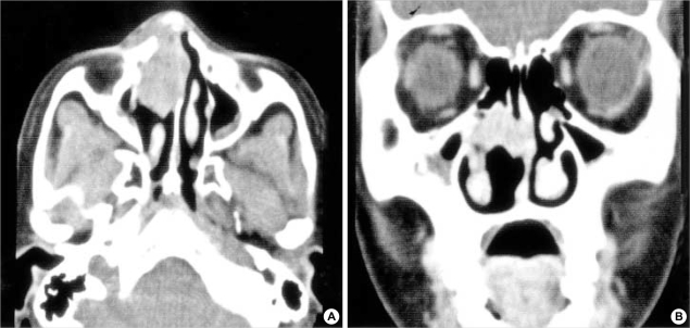
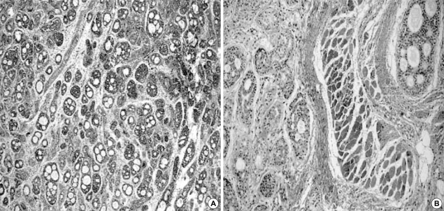
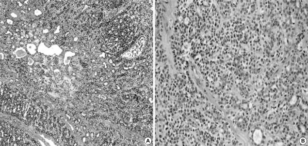
A paranasal sinus computed tomography scan showed a slightly enhanced, soft tissue mass filling the right anterior nasal cavity and maxillary sinus, extending to the nasal septum and the right nasal dorsum (Fig. 1). The tumor was diagnosed as epithelial-myoepithelial carcinoma through incisional biopsy. Right medial maxillectomy with resection of the nasal septum was performed, and the pathologic diagnosis was hybrid carcinoma composed of epithelial-myoepithelial carcinoma with an adenoid cystic carcinoma component (cribriform pattern) (Fig. 2, 3). A total of 5,400 cGy of postoperative radiation was added as an adjuvant therapy.
Fig. 1.
A CT scan showing a slightly enhanced, soft tissue mass filling the right anterior nasal cavity and the maxillary sinus, extending to the nasal septum and the right nasal dorsum.
Fig. 2.
An area of typical adenoid cystic carcinoma with a cribriform pattern (H-E stain, ×40 (A), ×200 (B)).
Fig. 3.
An area of epithelial-myoepithelial carcinoma composed of ductular structures surrounded by clear myoepithelial cells (H-E stain, ×40 (A), ×200 (B)).
The postoperative course was uneventful for 7 yr; however, the patient revisited because of tumor recurrence with extension to the right orbit in May, 2000. The right orbit was not included in the surgical specimen at that time of revision surgery according to the patient's decision. The pathologic diagnosis was adenoid cystic carcinoma (solid pattern) rather than epithelial-myoepithelial carcinoma. Adjuvant radiation therapy was given to the patient again with 4,000 cGy. As expected, the lesion invaded the intracranial space and showed metastases to multiple bones 8 months after the second radiation therapy. Total maxillectomy with palliative chemotherapy was the only choice for the patient. After all, the patient died of the disease in October, 2001.
DISCUSSION
A hybrid tumor is composed of two different tumors arising in the same topographical site and invariably produces a single mass, both clinically and macroscopically (1, 2). Hybrid carcinoma is a rare neoplasm, accounting for less than 0.1% of all registered tumors in salivary glands (1). Up to now, a few cases of hybrid tumor of salivary glands have been described exclusively in the parotid gland, submandibular gland, and palate (1, 3, 4). This is the first case of hybrid carcinoma arising from the maxillary sinus. The limited number of reported cases of hybrid carcinoma makes prognostic assessment and clinical management of this lesion difficult.
Since multiple (different or identical; unilateral or bilateral) tumors can occur independently in salivary glands, synchronously or metachronously, it is important to differentiate hybrid carcinoma from other multiple tumors of salivary gland such as collision tumors, biphasically differentiated tumors, and so on. The collision tumors represent the meeting of two malignant neoplasms arising at independent topographic sites. The majority of these tumors represent collision between carcinomas with sarcomas or lymphomas and rarely between two types of carcinomas (5, 6). Biphasically differentiated tumors represent regular, repetitive mixtures of two cellular patterns with a corresponding term in the tumor classification, such as epithelial-myoepithelial carcinoma, adenoid-cystic carcinoma, mucoepidermoid carcinoma, and carcinosarcoma (2).
Since the two different compartments of hybrid carcinoma show different characteristics as regards to cellular differentiation and proliferative activity, their recognition is important to establish a treatment strategy. Most series of epithelial-myoepithelial carcinoma have indicated a low-grade malignant behavior (7, 8). Therefore recognition of other component that is more aggressive and has a worse prognosis, just as in our case, has therapeutic and prognostic ramifications. A thorough sampling of the salivary gland tumors can help identify the possible presence of a higher-grade component. This finding will aid in differential diagnosis, determining the aggressiveness and metastatic potential of the tumor, prognosis, and treatment.
There have been some controversial issues regarding the criteria for hybrid carcinomas because of shared foci of phenotypic differentiation within salivary tumors. Grenko et al. (9). have described three adenoid cystic carcinomas and two epithelial-myoepithelial carcinomas that focally shared both histological features. They suggested that these two entities shared common differentiation pathways and did not categorize these examples as hybrid carcinomas. On the other hand, Seifert and Donath (1), Croitoru et al. (3), Nagao et al. (10), and Chetty (11) suggested that completely divergent differentiation may lead to the appearance of two distinct tumor entities (hybrid tumor), and also suggested that when each carcinoma component occupied more than 30% of the tumor mass and could be separated from the other component, the case should be considered as hybrid carcinomas.
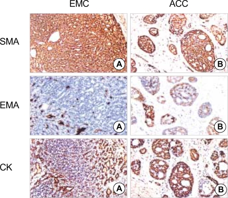
In our case, the cellular composition of the two tumor entities was confirmed by using immunohistochemical stains and by clinical course of the patient. Now, we speculate, however, that the treatment for the recurrent tumor was not adequate and the anatomical complexity of the maxillary region prohibited obtaining an adequate resection margin. Since the pathologic diagnosis of our case was adenoid cystic carcinoma rather than epithelial-myoepithelial carcinoma, the more aggressive tumor component might have a greater potential of recurrence.
Occasionally only one component of the hybrid carcinoma that is more aggressive may has the potential to metastases (3). In our case, the adenoid cystic carcinoma might have metastasized to multiple bones and invaded to the intracranial space.
Di Palma (12) suggested that multidirectional differentiation may be mediated through intercalated duct hyperplasia and may account for the simultaneous occurrence of more than one tumor, especially in association with epithelial-myoepitheial carcinoma.
In an epithelial-myoepithelial carcinoma in the salivary gland, a meticulous search for other accompanying tumors that may be more aggressive is mandatory to establish an optimal management strategy.
Fig. 4.
Immunohistochemical staining for smooth muscle actin (SMA) shows cytoplasmic staining on the myoepithelial cells of epithelial-myoepithelial carcinoma (EMC)(A) and adenoid cystic carcinoma (ACC)(B). Ductal epithelial cells are highlighted by immunostaining for epithelial membrane antigen (EMA) and low molecular weight cytokeratin (CK) in both EMC and ACC (ABC stain, ×400).
References
- 1.Seifert G, Donath K. Hybrid tumours of salivary glands: definition and classification of five rare cases. Eur J Cancer B. 1996;32B:251–259. doi: 10.1016/0964-1955(95)00059-3. [DOI] [PubMed] [Google Scholar]
- 2.Seifert G, Donath K. Multiple tumours of the salivary glands: terminology and nomenclature. Eur J Cancer Oral Oncol. 1996;32B:3–7. doi: 10.1016/0964-1955(95)00063-1. [DOI] [PubMed] [Google Scholar]
- 3.Croitoru CM, Suarez PA, Luna MA. Hybrid carcinomas of salivary glands: report of 4 cases and review of the literature. Arch Pathol Lab Med. 1999;123:698–702. doi: 10.5858/1999-123-0698-HCOSG. [DOI] [PubMed] [Google Scholar]
- 4.Snyder ML, Paulino AF. Hybrid carcinoma of the salivary gland: salivary duct adenocarcinoma adenoid cystic carcinoma. Histopathology. 1999;35:380–383. doi: 10.1046/j.1365-2559.1999.00761.x. [DOI] [PubMed] [Google Scholar]
- 5.Spagnolo DV, Heenan PJ. Collision carcinoma at the esophagogastric junction: report of two cases. Cancer. 1980;46:2702–2708. doi: 10.1002/1097-0142(19801215)46:12<2702::aid-cncr2820461228>3.0.co;2-m. [DOI] [PubMed] [Google Scholar]
- 6.Wanke M. Collision tumour of the cardia: case report. Virchows Arch Pathol Anat. 1972;357:81–86. doi: 10.1007/BF00548218. [DOI] [PubMed] [Google Scholar]
- 7.Batsakis JG, El-Naggar AK, Luna MA. Epithelial-myoepithelial carcinoma of salivary glands. Ann Otol Rhinol Laryngol. 1992;101:540–542. doi: 10.1177/000348949210100617. [DOI] [PubMed] [Google Scholar]
- 8.Tralongo V, Daniele E. Epithelial-myoepithelial carcinoma of the salivary glands: a review of literature. Anticancer Res. 1998;18:603–608. [PubMed] [Google Scholar]
- 9.Grenko RT, Abendroth CS, Davis AT, Levin RJ, Dardick I. Hybrid tumors or salivary gland tumors sharing common differentiation pathways? Reexamining adenoid cystic and epithelial-myoepithelial carcinomas. Oral Surg Oral Med Oral Pathol Oral Radiol Endod. 1998;86:188–195. doi: 10.1016/s1079-2104(98)90124-x. [DOI] [PubMed] [Google Scholar]
- 10.Nagao T, Sugano I, Ishida Y, Asoh A, Munakata S, Yamazaki K, Konno A, Iwaya K, Shimizu T, Serizawa H, Ebihara Y. Hybrid carcinomas of the salivary glands: report of nine cases with a clinicopathologic, immunohistochemical, and p53 gene alteration analysis. Mod Pathol. 2002;15:724–733. doi: 10.1097/01.MP.0000018977.18942.FD. [DOI] [PubMed] [Google Scholar]
- 11.Chetty R. Intercalated duct hyperplasia: possible relationship to epithelial-myoepithelial carcinoma and hybrid tumours of salivary gland. Histopathology. 2000;37:260–263. doi: 10.1046/j.1365-2559.2000.00976.x. [DOI] [PubMed] [Google Scholar]
- 12.Di Palma S. Epithelial-myoepithelial carcinoma with coexisting multifocal intercalated duct hyperplasia of the parotid gland. Histopathology. 1994;25:494–496. doi: 10.1111/j.1365-2559.1994.tb00014.x. [DOI] [PubMed] [Google Scholar]


