Abstract
While leukemia-originating stem cells are critical in the initiation and maintenance of leukemias, the existence of similar cell populations that may generate B-cell lymphoma upon mutation remains uncertain. Here we propose that committed lymphoid progenitor/precursor cells with an active V-D-J recombination program are the initiating cells of follicular lymphoma and mantle cell lymphoma when targeted by immunoglobulin (IG)- gene translocations in the bone marrow. However, these pre-malignant lymphoma-initiating cells cannot drive complete malignant transformation, requiring additional cooperating mutations in specific stem-cell programs to be converted into the lymphoma-originating cells able to generate and sustain lymphoma development. Conversely, diffuse large B-cell lymphoma and sporadic Burkitt’s lymphoma derive from B lymphocytes that acquire translocations through IG-hyper-mutation or class-switching errors within the germinal center. Although secondary reprogramming mutations are generally required, some cells such as centroblasts or memory B cells that have certain stem cell-like features, or lymphocytes with MYC rearrangements that deregulate self-renewal pathways, may bypass this need and directly function as the lymphoma-originating cells. An alternative model supports an aberrant epigenetic modification of gene sets as the first occurring hit, which either leads to retaining stem-cell features in hematopoietic stem or progenitor cells, or reprograms stemness into more committed lymphocytes, followed by secondary chromosomal translocations that eventually drive lymphoma development. Isolation and characterization of the cells that are at the origin of the different B-cell non-Hodgkin’s lymphomas will provide critical insights into the disease pathogenesis and will represent a step towards the development of more effective therapies.
Keywords: cancer stem cells, B-cell non-Hodgkin’s lymphoma, lymphoma-initiating vs. lymphoma-originating cells, lymphoid plasticity
Introduction
Stem cells are primarily characterized by the properties of unlimited self-renewal, which maintains and expands the undifferentiated cell pool over the lifetime of the host, and multi-lineage differentiation, which produces progeny of diverse mature phenotypes to generate and regenerate tissues.1 These stem cell attributes are tightly regulated in normal development, yet their alteration may lead to many human diseases including cancer. In fact, because stem cells and some cancer cells share self-renewal and differentiation capacities, it was suggested that tumors were derived from mutated stem cells, the so-called cancer stem cells.1–3 Although this hypothesis was postulated in early reports,4–6 definite proof of their existence came from recent studies in leukemia, where among the complete tumor cell population only a small subset of cells could initiate, regenerate and maintain the leukemia after transplantation into immunocompromised mice.7,8 Using similar functional approaches, a variety of cancer stem cells have been identified in an increasing number of epithelial tumors, including breast, prostate, pancreatic, and head and neck carcinomas, all of which were distinguished by the expression of the cell-surface glycoprotein CD44.9 Another cell surface marker, the CD133 glycoprotein, defined the tumor-initiating cells of brain and colon carcinomas.9–11 The concept of cancer stem cells is not only changing our current understanding of cancer biology, but may also have profound consequences on cancer diagnostics and therapeutics. For example, gene expression profiles associated with the stemness/differentiation states of tumors might be used as molecular predictors of therapeutic outcome.12,13 In addition, studies in glioblastoma and breast cancers support the view that cancer stem cells are more resistant to radiotherapy and chemotherapy-induced apoptosis, which allows these cells to survive and generate tumor relapse.14,15 A prototypical example of a stem cell disorder is chronic myeloid leukemia (CML), where the tyro-sine kinase inhibitor imatinib has replaced IFNa as the standard therapy.16 However, clinical trials have not shown the expected cure rates with the use of imatinib, as a consequence of this compound being active against differentiated CML progenitors while presenting limited activity against quiescent CML stem cells.17 Consequently, many patients in apparent complete molecular remission relapsed when the drug was discontinued.17,18 These data suggest that CML stem cells would constitute a reservoir responsible for disease relapse, a phenomenon which may also occur in other cancers.
Despite these outstanding discoveries in leukemias and solid tumors, the existence of lymphoma-originating cells with stem-cell properties that may similarly generate lymphoma upon mutation remains a controversial and largely unexplored issue.19
Indeed, a recent study identified rare clonal B lymphocytes expressing CD20, the memory B-cell antigen CD27, and the stem cell marker aldehyde dehydrogenase (ALDH) that may be responsible for the generation and maintenance of the predominant Hodgkin and Reed-Sternberg cells in classical Hodgkin’s lymphoma.20 These data point to these B lymphocytes as the originating cells for Hodgkin’s lymphoma, and opens a debate on whether this is also the case for the different B-cell non-Hodgkin’s lymphomas.20,21 Consequently, here we review the role of the diverse hematopoietic and lymphoid cell populations as the putative cells of origin for the B-cell lymphoma subgroups, highlighting the similarities and differences with other known stem cell-derived cancer models.
Chromosomal translocations as the first occurring genetic hit in B-cell lymphomas
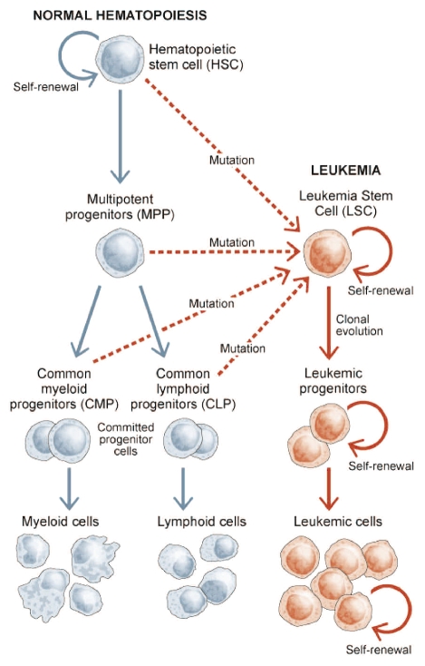
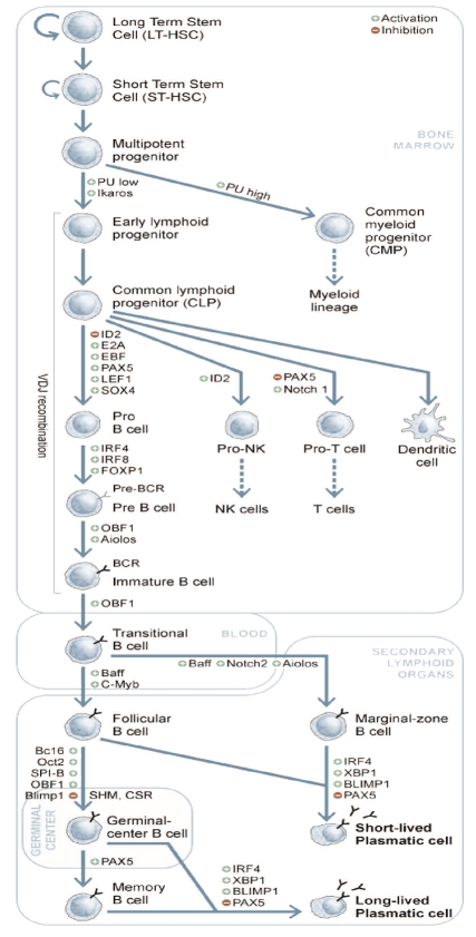
The different acute myeloid leukemia (AML) subgroups were shown to be derived from a common leukemia stem cell (LSC) that shares a CD34+CD38− phenotype with normal hematopoietic stem cells (HSCs).8,22 The similarities between HSCs and LSCs strongly suggest that HSCs can be the source of LSCs when targeted by oncogenic translocations. Instead, more committed progenitors may also be transformed into LSCs through the accumulation of appropriate mutations that restore the critical stem-cell abilities of self-renewal and multi-differentiation (Figure 1). But are lymphomas, a group of disorders also characterized by oncogenic chromosomal translocations, derived from hematopoietic stem cells or from committed lymphoid progenitors? The study of normal lymphocyte lineage development together with the analysis of the molecular structure of the chromosomal translocations involving immunoglobulin (IG) genes can help to assign the putative lymphoma-initiating cells to specific developmental stages (Figure 2).23–28 Errors in any of the physiological IG gene rearrangements may result in aberrant chromosomal translocations involving IGH (or more rarely IGL or IGK) genes, which usually become juxtaposed to a variety of oncogenes, and are characteristic of the different lymphoma subtypes.27 Direct proof of the involvement of the V-D-J recombination process in the generation of lymphoma translocations has been provided, as the DNA of the BCL2 major translocation breakpoint acquires an altered structure that is cut by RAG nucleases, which regulate V-D-J recombination.29 Molecular analysis of translocation breakpoints provides insights into their timing of occurrence during B-cell differentiation. In the t(14;18)(q32;q21) involving BCL2 in follicular lymphoma (FL), in the t(11;14)(q13;q32) targeting CCND1 in mantle cell lymphoma (MCL), and in the t(14;18)(q32;q21) deregulating MALT1 gene in mucosa-associated lymphoid tissue (MALT), the gene rearrangements usually involve the nonfunctional IG gene at the 5′ end of J heavy-chain (JH) gene segments, pointing to mistakes occurring at the DH to JH stage in bone marrow lymphoid progenitors (ELPs and CLPs) or in B-cell precursors (pro-B and pre-B cells).23,27,30 However, one-third of BCL2-IGH gene rearrangements in FL occur at later V-D-J recombination stages (primarily VH to DHJH), involving pre-B or immature B cells.31,32 In any case, FL cells invariably show somatic hypermutation (SHM) of both IGH alleles, suggesting that the immortalized B cells with BCL2 overexpression must have continued their normal differentiation path including the transit through the germinal center. In contrast, most MCL cases show unmutated IGH genes, reflecting that B cells with acquired CCND1 translocations did not enter the germinal center.
Figure 1.
Leukemia-originating stem cell model. Functional studies in acute myeloid and chronic leukemias have led to the identification and characterization of the leukemia-originating stem cells (LSCs), which share features with normal hematopoietic stem cells (HSCs). LSCs derive either from HSCs, multipotent progenitors (MPPs), committed progenitor cells or even more differentiated cells that accumulate mutations that reprogram the necessary stem-cell features to generate and regenerate tumors.
Figure 2.
Schematic representation of the normal lymphoid cell development. HSCs generate multipotent progenitors (MPPs) that are committed to form either common myeloid progenitors (CMPs) or early lymphoid progenitors (ELPs). ELPs differentiate into common lymphoid progenitors (CLPs), which are restricted to lymphoid development, giving rise to B cells, T cells, dendritic cells and natural killer cells. Expression of the B-cell marker B220 by a subset of CLPs (known as pro-B cells) indicates their entry into the B-cell differentiation pathway. However, the process of IG gene remodeling is initiated earlier by ELPs, with the assembly of genes for the variable regions of the IG heavy (IGH) and light (IGL) chains in a process called V-D-J gene recombination. This event occurs prior to contact with antigen, and the molecular anatomy of the productive V-D-J rearrangements suggests that nuclear terminal deoxynucleotidyl transferase (Tdt) is involved. Immature B cells then leave the bone marrow microenvironment and populate the peripheral lymphoid organs. It is here that these naïve B cells contact antigen and lead to the formation of the germinal centers of secondary lymphoid organs, where some of them continue BCR modification to increase affinity for the immunizing antigen. This secondary phase of BCR remodeling requires two different mechanisms unique to the germinal center, IG class-switch recombination (CSR) and somatic hyper-mutation (SHM). During CSR, B cells change the isotype of the expressed BCR, while SHM introduces nucleotide changes, deletions and duplications at a very high frequency in both productive and non-productive IG genes, resulting in the generation of antibody variants. These mechanisms require the activation-induced cytidine deaminase (AICDA). Finally, cells expressing favorable antibody mutants are positively selected, vigorously expanded and released into the periphery as IG-secreting plasma cells or long-lived memory B cells. B-cell ontogeny-determining transcription factors are shown, indicating whether they are activated (+) or inhibited (−) during each cellular step.
A set of molecularly different chromosomal translocations are promoted by the process of SHM.26 In normal germinal-center lymphocytes, this mechanism generates point mutations, deletions and duplications in the variable regions of the IG genes that are intimately associated with DNA cleavage and can be recombinogenic.33 Aberrant hypermutation activity can also target BCL6 and other oncogenes such as PIM1, MYC, RhoH and PAX5 in germinal center-derived lymphomas and in Hodgkin’s lymphoma.34–36 Mutations are distributed in the 5′ untranslated or coding sequences, regions that are commonly disrupted by chromosomal translocations, consistent with a role for SHM in generating these translocations by DNA double-strand breaks. Accordingly, BCL6 fuses its 5′ untranslated sequences with either IG genes or with a large variety of partner genes in DLBCL.37 A third class of chromosomal translocations occurs in IG switch sequences through an illegitimate class-switch recombination (CSR) process in the germinal center. As an example, in most sporadic Burkitt’s lymphomas with t(8;14)(q24;q32), the MYC oncogene fuses to IGH μ switch (Sμ) region, whereas endemic Burkitt’s lymphomas display breakpoints in the JH region.38,39 Similar rearrangements targeting IGH Sμ sequences are observed in DLBCL with t(3;14)(q27;q32) involving the 5′ unstranslated region of BCL6, and in multiple myeloma with translocations of IRF4, c-MAF, FGFR3 and MMSET oncogenes.26,27,37,40 Most tumors with CSR-related translocations show concomitant hypermutation of IG genes, consistent with cell germinal center transit. However, a few cases of low grade B-cell lymphoma with CSR-related translocations, including the t(14;19)(q32;q13) involving BCL3 gene or the t(2;14)(p13;q32) targeting BCL11A, displayed non-mutated IGH gene sequences.41,42 An explanation for these cases is that the translocation occurred in activated B cells in the course of T-cell independent immune responses outside the germinal center. Lastly, rare cases of FL display CSR-related BCL2-IGH rearrangements, indicating a late germinal center origin.43
Importantly, in FL and in some germinal center-derived DLBCL cells, the presence of intraclonal IGH nucleotide variation indicates that the translocation t(14;18)(q32;q21) must have occurred in pre-germinal or early germinal center B lymphocytes (possibly centroblasts in the dark zone of the germinal center), after which divergences in the SHM and CSR processes gave rise to different clones with heterogeneous IG sequences and multiple isotype transcripts.44,45 Moreover, these lymphomas usually present variation in the pattern of IG hypermutation during disease progression.46 Conversely, ongoing SHM of IG genes is rarely observed in DLBCL with activated B-cell phenotype, but instead, aberrant CSR and IGH switch translocations involving different oncogenes are common.47 These data indicate that activated B-cell DLBCL may be derived from B lymphocytes (possibly centrocytes) in transit through the light zone of the germinal center, where CSR but not SHM take place normally.44,47
Evidence to support that B-cell lymphomas originate from common lymphoid progenitor or precursor cells
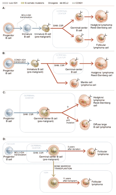
Initial support for the existence of clonogenic cells that may be the origin of lymphoma was gleaned from the molecular analysis of composite lymphomas.48 The study of patients presenting with both Hodgkin’s and non-Hodgkin’s lymphoma demonstrated the same clonal IGH gene rearrangements in the Reed-Sternberg cells and in non-Hodgkin’s lymphoma cells; however, the IGH somatic hypermutation pattern in each lymphoma was different.49,50 These data suggested that lymphomas were derived from a common B-cell precursor cell that acquired the BCL2 gene rearrangement and then diverged to generate clonally distinct tumors (Figure 3A). One patient simultaneously developed MCL and Hodgkin’s lymphoma, both with identical CCND1 translocation. However, MCL cells showed unmutated IGH genotype whereas SHM was observed in Hodgkin’s lymphoma cells.50 These data further support the occurrence of the CCND1 translocations in MCL in pre-germinal center B cells (Figure 3B).
Figure 3.
B-cell progenitors or germinal-center B lymphocytes as the lymphoma-initiating originating cells in composite lymphomas. (A) Composite lymphoma with Hodgkin’s and follicular lymphoma, both carrying identical BCL2-IGH chromosomal translocation and different IG somatic hypermutation patterns, indicating that the translocation occurred in a cell before entering the germinal center. (B) Composite lymphoma with mantle cell lymphoma (MCL) and Hodgkin’s lymphoma, both with identical CCND1-IGH chromosomal translocation. However, MCL did not show somatic hypermutation of IG genes, pointing to a pre-germinal center cell as the initiating cell for both lymphomas. (C) In a Hodgkin’s and non-Hodgkin’s (diffuse large B-cell lymphoma, DLBCL) composite lymphoma, both tumors shared a common IG somatic hypermutation pattern, but differed on the P53 gene mutation status, which was only observed in the DLBCL cells. This finding suggests a germinal-center B lymphocyte as the cell suffering the first genetic event. (D) Transmission of follicular lymphoma by bone marrow transplantation. Both the donor and recipient presented identical BCL2-IGH rearrangement and IG hypermutation pattern, suggesting that the cell that transmitted lymphoma, the lymphoma-originating cell, was a germinal center B lymphocyte, although the chromosomal translocation resulting in the BCL2-IGH fusion occurred in a pre-germinal center lymphocyte.
Conversely, germinal-center B cells may also be the initiating cells of some composite lymphomas. A patient developed DLBCL and Hodgkin’s lymphoma, both with identically hypermutated IGH genes. However, a P53 gene bi-allelic mutation was detected only in DLBCL cells (Figure 3C). These data suggest that a late germinal center B-cell initiated both lymphomas, and that subsequent P53 inactivation led a fraction of cells towards development of DLBCL.50 The transmission of an FL by bone marrow transplantation (BMT) further supports the existence of a germinal center lymphoma-originating cell.51 In 1992, a 32-year old man developed AML, for which he received allogeneic BMT from his father, after which he remained in remission. Three years later the patient’s father was diagnosed with FL. Although he received therapy, he died of the disease. In 2003, eleven years after the diagnosis of AML, the son developed FL, and despite a good initial clinical response, his disease slowly progressed.52 Both lymphoma biopsies in father and son revealed identical BCL2-IGH gene rearrangements. In addition, both tumors had identical DH-JH allele-types and shared SHM patterns that defined a putative common precursor cell with 91% homology to the VH germline sequence of IGH. Although the first genetic hit (the BCL2-IGH chromosomal translocation) must have occurred in a precursor B lymphocyte (the lymphoma-initiating cell), the genetic similarity between the father’s and the son’s lymphomas points to a germinal-center B lymphocyte as the lymphoma-originating cell for both subjects’ tumors (Figure 3D).
Further compelling evidence arguing in favor of the existence of a common lymphoma-initiating cell in FL comes from cytogenetic and genotyping studies of sequential biopsies of patients undergoing histological transformation from FL to DLBCL (t-DLBCL). Classical karyotyping and comparative genomic hybridization studies of paired specimens revealed that the transformed biopsy shares some, but not all, of the alterations evident in the diagnostic FL sample.53,54 This finding suggests subclone selection or divergent outgrowth of a common malignant ancestral cell in contrast to clonal evolution resulting from stochastic genetic events, only some of which provide a selective advantage. Moreover, recent studies have reported that transformation of FL to t-DLBCL occurs over approximately 15–17 years following a diagnosis of FL and seems to have a constant rate of 3% per year.55,56 These results suggest that a sizable proportion of the patients with FL may not be at risk to transform. If clonal selection and evolution were the primary drivers of this process, one might expect the rate of histological transformation to change over time. The observed steady-state risk of transformation might be better modeled if a precursor cell pool with limited proliferative capacity existed and was at-risk for stochastic genetic events, some of which provide a growth advantage.
Plasticity of lymphoid cells: generation of the lymphoma-originating cells through the second hit
Most of the oncogenes targeted by translocations in B-cell lymphomas are regulators of lymphocyte proliferation, apoptosis, development and differentiation pathways, but do not have a defined role in stem-cell self-renewal programs.27 If we accept that the translocations in FL and MCL target either B-cell progenitors or precursors within the bone marrow that do not conserve stem-cell properties, these pre-malignant lymphoma-initiating cells need to restore the necessary stemness to generate and maintain the tumor, thus becoming the lymphoma-originating cells. This cellular reprogramming must be achieved through mutations in genes controling stemness and/or pluripotency pathways. In germinal center-derived lymphomas, the initiating translocations seem to target more mature lymphocytes within the germinal center, and in most cases these cells also need to accumulate secondary reprogramming mutations. However, there may be exceptions to this rule, such as (i) the translocations occurring in centroblasts or in centrocytes, which are cells that can have certain stem-cell properties; (ii) translocations/mutations in memory B cells, which have self-renewal capacity to maintain life-long immunological memory; and (iii) the translocations involving the MYC oncogene, which deregulate self-renewal pathways in the targeted lymphocytes (see below).57–60 In these cells, the occurrence of secondary mutations may not be necessary, as they can directly become the lymphoma-originating cells able to produce lymphoma. Currently, there is compelling evidence for the enormous plasticity of B cells in physiological and pathological conditions, and some of the gene pathways that regulate these stem cell properties are beginning to be characterized.61–65
PAX5 and CEBPα/β transcription factors
Mouse and human somatic cells can be reprogrammed into pluripotent stem cells by introducing a combination of factors (OCT3/4, SOX2, MYC, KLF4, NANOG or LIN28) under certain cell culture conditions.66–68 This exceptional plasticity is also observed in lymphocytes, where expression of CEBPα/β transcription factors was sufficient to reprogram differentiated B cells into macrophages by inhibiting PAX5.62 The most convincing demonstration of the plasticity of mature B cells was provided by Cobaleda and colleagues by generating conditional PAX5 deletion mutants in mice that allowed mature B cells from peripheral lymphoid organs to dedifferentiate back to early uncommitted progenitors.65 In addition, these mice lacking PAX5 in mature B cells developed progenitor cell-derived B-cell lymphomas.65 More recently, reprogramming of terminally differentiated mouse mature B lymphocytes to a pluripotent state was achieved by expression of OCT4, SOX2, KLF4 and MYC plus either ectopic expression of CEBPα/β or specific knock-down of PAX5.64 These reports underscore that the reprogramming capacity of lymphoid cells possibly represents a critical and necessary feature of lymphomagenesis.64,65 However, genetic or epigenetic mutations of PAX5 or CEBPα/β genes are not commonly found in lymphoma biopsies, and thus their implication in human B-cell lymphomas is not completely clear. Conversely, in B-cell precursor acute lymphoblastic leukemia (BCP-ALL), inactivating mutations of PAX5 are frequent, suggesting that these rearrangements may contribute to the cellular differentiation arrest observed in this disorder.69 Indeed, five members of the CEBP family of transcription factors can be over-expressed through IG-related chromosomal translocations in a subset of BCP-ALL.70 In these cases, CEBP proteins may have inhibited PAX5 aberrantly, thereby promoting B-cell arrest.62,71
MYC oncogene
MYC is mutated and deregulated in a large proportion of B-cell lymphomas of different subgroups through various genetic mechanisms.27,35,38,39 Unlike other lymphoma oncogenes, forced expression of MYC in murine lymphoid cells is sufficient to generate B-cell leukemia and lymphoma.72 In addition, MYC activation is necessary to develop lymphoma in BCL2 or CCND1 transgenic mice.73,74 These oncogenic features may in part rely on the role of MYC as a regulator of the balance between self-renewal and differentiation in HSCs.57 Accordingly, MYC is among the few key players inducing pluripotency in somatic and embryonic cells and mature lymphocytes.64,66–68 A recent study revealed that tumor growth in a mouse model of Eμ-MYC lymphoma need not be driven by rare cancer stem cells but rather, and in contrast to most leukemias, a large population of tumor cells was able to initiate and sustain lymphoma development.75 One possible explanation of these data might be that the MYC oncogene confers stem-cell properties to a large number of lymphoma cells within the tumor.
The Polycomb protein BMI1
Proteins belonging to the Polycomb group (PcG) are epigenetic silencers that regulate lymphopoiesis by forming multimeric complexes that bind and remodel chromatin, thus regulating gene activity.76 Some PcG genes have been implicated in lymphomagenesis.77 Forced expression of the PcG protein BMI1 in lymphocytes was associated with lymphoma generation, whereas BMI1 gene is amplified and over-expressed in human MCL and in other B-cell lymphomas.77–79 BMI1 has essential roles in regulating proliferation and self-renewal of both normal and leukemic stem cells.80 These findings underscore a potential critical characteristic of mutated BMI1 in lymphomagenesis by reprogramming pre-malignant lymphocytes bearing different chromosomal rearrangements into lymphoma-originating cells with de novo stem cell-like properties.
Lessons from mouse models of leukemia and lymphoma
A number of reports have demonstrated the transformation potential of known leukemia oncogenes in the different mouse hematopoietic cell compartments. Short-lived myeloid progenitors transduced with MLL-ENL, MOZ-TIF2 and MLL-AF9 oncogenic fusions generated AML with similar latencies and characteristics to those observed in long-term HSC models.81–84 As normal myeloid progenitors do not conserve stem-cell features, those oncogenic rearrangements must have restored self-renewal and pluripotent abilities necessary for leukemic transformation, as was demonstrated by Krivtsov and colleagues in an MLL-AF9 transgenic model.83 In BCP-ALL, oncogenic fusions may similarly awaken stem-cell properties in committed B lymphocytes. In one report, the common genetic rearrangements observed in patients with BCP-ALL were modeled in mice (including p190-BCR-ABL and TEL-AML1 gene fusions), concluding that these leukemias were derived from committed B-cell progenitor cells rather than from HSCs.85 However, the analysis of human BCP-ALL samples did not confirm these findings but revealed that the blasts representative of all of the different maturational lymphoid stages were able to reconstitute and re-establish the complete leukemic phenotype in vivo.86 Further studies in monochorionic twins with BCP-ALL carrying a TEL-AML1 rearrangement led to the isolation of a CD34+CD19+CD38− cell population able to initiate a pre-leukemic state that eventually transformed to frank BCP-ALL.87 Analysis of TEL-AML1-transduced human cord blood cells suggested that this rearrangement may function as a first-hit mutation that also endows these pre-leukemic cells with altered self-renewal and survival properties, sufficient for leukemia development.87
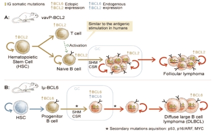
In contrast to leukemia, many engineered mice expressing specific lymphoma oncogenes have failed to generate the expected human-like lymphomas in vivo. Mice in which the human CCND1 oncogene was juxtaposed to the IGμ-chain gene (Eμ-CCND1), which restricts the expression of the oncogene in the lymphoid compartment,73 did not develop malignancy spontaneously, requiring the cooperation of additional activating mutations to drive lymphoma formation.74 The Eμ-MYC transgenic mice developed lymphoma with histopathological features of human lymphoblastic lymphoma rather than Burkitt’s lymphoma, lacking for instance the typical germinal center-associated IGH somatic hypermutations.72,88 Similarly, the Eμ-BCL2 mice developed high-grade B-cell lymphoma after a significant latency period, but not human-like FL.89 Failure of these models to recapitulate human disease may rely on the inappropriate selection of the cell compartment in which the oncogenes were expressed and/or on their inability to restore self-renewal and pluripotent states in lymphoid progenitors or in more mature B cells. Supporting these hypotheses, the vavP-BCL2 transgenic mice, which present constitutive BCL2 overexpression from HSCs to mature B cells, developed lymphoma that was preceded by germinal center hyperplasia, thus mimicking human FL.90 Unlike Eμ-BCL2 transgenes, vavP-BCL2 mice showed higher levels of BLC2 in T cells, causing enlargement of the peripheral T-cell compartment, which was critical to generate abundant germinal centers where B cells underwent IG hypermutation and class switching, eventually leading to FL (Figure 4A). These data in mice are compatible with the portrait of human FL, where micro-environmental cells that surround tumor cells, including several T-cell subsets, appear crucial for lymphoma formation and maintenance.91 These findings theoretically provide support for either a hematopoietic stem cell or a lymphoid progenitor cell as the putative FL-initiating cells, as they are able to permit BCL2 expression in both B- and T-cell lineages. However, hematopoietic stem cells do not seem to have the functional V-D-J machinery subsidiary to suffer mechanistic errors leading to chromosomal breaks.23,24 Conversely, lymphoid progenitor cells may have started V-D-J recombination and are prone to suffer translocations, thus representing the ideal FL-initiating cells. However, these cells have lost self-renewal and multi-differentiation potentials, therefore needing additional reprogramming mutations to restore stem-cell features. If these secondary genetic alterations do not occur, BCL2-expressing lymphocytes may circulate for several years without giving rise to FL.92
Figure 4.
Cells of origin for B-cell lymphomas according to mouse models of disease. (A) The vavP-BCL2 mice show BCL2 expression in all hematopoietic cell populations, and promoted follicular lymphoma development that was preceded by germinal center hyperplasia, thus mimicking human FL development. (B) In the Iμ-BCL6 mice, BCL6 is expressed in mature B cells. Development of DLBCL is dependent on the acquisition of secondary mutations able to dedifferentiate back pre-malignant B cells into lymphoma-originating stem cells.
Engineered mice with full-length murine BCL6 placed downstream of the IG heavy chain (Iμ) promoter (Iμ-BCL6 mice) led to an increase in germinal center formation followed by the generation of DLBCL between 15 and 20 months of age.93 These tumors were developed through expression of BCL6 in mature B-lymphocytes, resembling the cellular and genetic features of human germinal center-derived DLBCL. Because of the long latency period, the cells carrying the initiating BCL6 alteration possibly required late secondary mutations to drive malignant transformation (Figure 4B). Remarkably, crossing Iμ-BCL6 mice with mice lacking AID, the enzyme required for both SHM and CSR, prevented development of BCL6-driven DLBCL, suggesting that AID is required for germinal center-derived lymphomas.94 Another interesting model is an AID-dependent conditional MYC transgene (the Vk*MYC mice), which allowed MYC activation in germinal center B lymphocytes.95 Despite theoretically resembling the dynamics of human Burkitt’s lymphoma, these animals develop multiple myeloma. MYC activation was caused by AID-dependent SHM, because crossing Vk*MYC mice with AID knockout mice failed to activate the SHM/CSR machinery and thus to generate myeloma.95 Indeed, AID is required for MYC-IGH chromosomal translocations in vivo.96 Moreover, loss of AID in Eμ-MYC mice changed the lymphoma phenotype from B-cell to pre-B cell tumors, further suggesting that AID activity contributes to mature B-cell but not to pre-B cell lymphoma development.97 Why the selective deregulation of MYC in germinal center B cells in the Vk*MYC mice failed (again) to reproduce the human disease phenotype is not fully understood. However, this model reinforces the concept that the single deregulation of MYC in centroblasts is sufficient to directly induce malignant transformation.
Epigenetic modifications in B-cell lymphomagenesis
It is widely accepted that cancers are heterogeneous disorders caused by a series of clonally selected genetic and epigenetic changes in tumor genes. However, some cancers may originate from a polyclonal epigenetic alteration of stem or progenitor cells within specific tissues, initially producing a pre-neoplastic epigenetically aberrant state.98 Recent studies have provided additional evidence for an instructive mechanism behind aberrant DNA methylation in cancer, which might indicate that specific DNA sequences are predisposed to acquire epigenetic aberrations.99,100 Indeed, a highly significant proportion of genes that are hypermethylated in solid tumors were already repressed at the embryonic stem cell stage by PcG marks.100 These findings support the cancer stem cell theory by stating that aberrant epigenetic changes of PcG-target genes might represent an initial event in tumorigenesis.101 Moreover, the genome-wide methylation pattern observed in a mouse model of MYC-induced lymphomas was driven by the genetic configuration of tumor cells, providing experimental evidence for a causal role of DNA hypermethylation in tumor progression.102
Martin-Subero et al. have recently studied the DNA methylation profiles of different morphological, genetic and transcriptional subtypes of lymphomas, including Burkitt’s lymphoma, activated B-cell DLBCL, and germinal center-like DLBCL.103 Hierarchical clustering of microarray-based DNA methylation data indicated that DNA methylation patterns in lymphoma cells were not strictly associated with morphological, genetic or transcriptional features. Using supervised analyses, however, they identified a small group of genes that appeared to be de novo methylated in a lymphoma subtype-specific manner. Strikingly, a different set of genes was methylated in all lymphoma subtypes and unmethylated in hematopoietic controls. This latter group of genes was mostly unmethylated in adult and embryonic stem cells and highly enriched for PcG targets in embryonic stem cells.
These findings could on one hand suggest that mature aggressive B-cell lymphomas with different genetic backgrounds might derive from lymphoma precursor cells with similar stem cell-like features that have acquired aberrant methylation of PcG target genes.103 Alternatively, an epigenetic footprint of stemness could have been acquired during lymphomagenesis by epigenetic remodeling. Consequently, as an alternative hypothesis to the development of lymphoma by linear acquisition of chromosomal translocations in lymphoid precursor or progenitor cells followed by reprogramming mutations, we may postulate that epigenetically aberrant lymphoma-initiating stem or progenitor cells that retain stem-cell features are susceptible to malignant transformation when acquiring, for instance, BCL2 or BCL6 gene rearrangements, during their transit through the bone marrow or the germinal center.
Lymphoma-originating cells: a therapeutic perspective
Most conventional chemotherapy kills rapidly proliferating tumor cells whereas cancer stem cells may remain viable during treatment because of their relative quiescence. At the end of treatment, cancer stem cells may regenerate the tumor entirely, providing one possible explanation of why tumors can relapse in patients in apparent complete clinical remission.1,9,104 Consequently, destruction of the stem cell population capable of initiating and maintaining tumors has become a major goal in the treatment of human malignancies. FL is characterized by the t(14;18)(q32;q21) resulting in overexpression of BCL2 protein. Most patients with FL initially respond to chemotherapy, but they typically experience relapses and/or transformation into high-grade therapy-resistant lymphoma.105 Thus, it is still considered an incurable disease using conventional treatment approaches. In mice, forced expression of the anti-apoptotic BCL2 protein in HSCs makes them more resistant to radiotherapy.106 In human biopsies, Dogan et al. identified an interfollicular neoplastic B-cell population surrounding the malignant follicles in the majority of FLs. While both follicular and inter-follicular cell populations shared clonal identity, the latter showed low-grade cytological features, lower proliferation activity and downregulation of activation markers such as CD19, CD38 and CD95, suggesting that these more quiescent cells may partially account for FL therapeutic resistance.107 Based on these reports, we may postulate that the yet unidentified FL-originating cells express high levels of BCL2 and are slowly proliferating, making them resistant to standard chemo/radiotherapy, whereas most other FL cells are killed following treatment. At the conclusion of a treatment regimen, the quiescent cell population regenerates the lymphoma, requiring further treatments followed by subsequent relapses that are accompanied by novel stochastic genetic changes that ultimately lead to transformation to DLBCL in those patients destined to transform. Therefore, identification of the FL-originating cells seems crucial to better define future treatment strategies. For instance, one promising therapy in FL is ABT-737, a small-molecule inhibitor of BCL2, which is effective in solid tumors.108 However, whether this and other similar anti-BCL2 compounds will be active against the FL-originating cells or will otherwise encounter selective resistance (mimicking imatinib therapy in CML) remains unclear. Another issue is whether the FL negative for BCL2 expression will respond to these compounds.109 A different example is the monoclonal antibody against CD20 (rituximab), which has shown clinical benefit for both induction and maintenance therapy in FL clinical trials.105 Whether rituximab given in combination with chemotherapy will eventually achieve cure in FL or will simply prolong complete remissions is uncertain, but may in part depend on whether the FL-originating cells express CD20 on the cell surface in a configuration and antigen density that allows this targeted therapy to kill them.
Stem cells reside in a special microenvironment termed the niche, where they interact via adhesion molecules and exchange molecular signals that maintain the specific features of stem cells. Based on these findings, it has been shown that a cancer stem cell niche exists, being involved in cancer stem cell regulation, tumor invasion, and metastasis.110 Although there has not been a formal demonstration of the existence of a niche for the putative lymphoma-originating cells, these niches may serve as reservoirs that protect these cells from current therapies.
Conclusions
B-cell non-Hodgkin’s lymphomas, in contrast to most leukemias, are derived from either committed lymphoid progenitor/precursor cells or from more mature B-lymphocytes that acquire chromosomal translocations through errors in the IG gene remodeling processes during normal B-cell differentiation. These lymphoma-initiating cells harboring characteristic chromosomal translocations are pre-malignant cells unable to form tumors, needing to acquire stem-cell features through secondary reprogramming mutations to become the lymphoma-originating cells fully capable of generating and maintaining lymphomas. While this scenario applies to FL and MCL, diffuse large B-cell lymphoma and Burkitt’s lymphoma are derived from more mature germinal center-derived lymphocytes. Although reprogramming mutations are also required in most cases, centroblasts or memory B cells that have certain stem cell-like properties and are targeted by translocations or lymphocytes that suffer MYC rearrangements that deregulate self-renewal pathways may not need these secondary mutations, directly functioning as lymphoma-originating cells able to drive malignancy. An alternative hypothesis states that the first hit consists of an aberrant epigenetic state of stem or progenitor cells that either retain stem cell-like features or reprogram stemness into more mature cells, followed by chromosomal instability and subsequent genetic aberrations that eventually lead to lymphoma formation. Detailed isolation and molecular characterization of the specific cells of origin for each B-cell lymphoma entity are crucial steps not only to better understand lymphomagenesis but also to make meaningful improvements in the treatment and eventual cure of these patients.
Acknowledgments
the authors wish to thank Heber Longas for excellent graphical assistance. Funding: the authors’ own work in the field has been supported by the Spanish Ministry of Science and Innovation, the Navarra Government (Education and Health Councils) and the UTE-CIMA project (JAM-C, LF and FP); the National Cancer Institute of Canada (NCIC), Terry Fox Foundation Program Project grant # 016003 (RDG); and the Deutsche Krebshilfe, the Wilhelm Sander Stiftung, the BMFB, and the KinderKrebsInitative Buchholz Holm-Seppensen (RS).
Footnotes
Authorship and Disclosures
All authors designed the manuscript and wrote the paper.
RS received lecture fees from Abbott/Vysis and scientifically collaborates with Illumina. The other authors declare no competing financial interests.
References
- 1.Reya T, Morrison SJ, Clarke MF, Weissman IL. Stem cells, cancer, and cancer stem cells. Nature. 2001;414(6859):105–11. doi: 10.1038/35102167. [DOI] [PubMed] [Google Scholar]
- 2.Pardal R, Molofsky AV, He S, Morrison SJ. Stem cell self-renewal and cancer cell proliferation are regulated by common networks that balance the activation of proto-oncogenes and tumor suppressors. Cold Spring Harb Symp Quant Biol. 2005;70:177–85. doi: 10.1101/sqb.2005.70.057. [DOI] [PubMed] [Google Scholar]
- 3.Clarke MF, Fuller M. Stem cells and cancer: two faces of eve. Cell. 2006;124(6):1111–5. doi: 10.1016/j.cell.2006.03.011. [DOI] [PubMed] [Google Scholar]
- 4.Bruce WR, Van Der Gaag H. A Quantitative assay for the number of murine lymphoma cells capable of proliferation in vivo. Nature. 1963;199:79–80. doi: 10.1038/199079a0. [DOI] [PubMed] [Google Scholar]
- 5.Park CH, Bergsagel DE, McCulloch EA. Mouse myeloma tumor stem cells: a primary cell culture assay. J Natl Cancer Inst. 1971;46(2):411–22. [PubMed] [Google Scholar]
- 6.Hamburger AW, Salmon SE. Primary bioassay of human tumor stem cells. Science. 1977;197(4302):461–3. doi: 10.1126/science.560061. [DOI] [PubMed] [Google Scholar]
- 7.Lapidot T, Sirard C, Vormoor J, Murdoch B, Hoang T, Caceres-Cortes J, et al. A cell initiating human acute myeloid leukaemia after transplantation into SCID mice. Nature. 1994;367(6464):645–8. doi: 10.1038/367645a0. [DOI] [PubMed] [Google Scholar]
- 8.Bonnet D, Dick JE. Human acute myeloid leukemia is organized as a hierarchy that originates from a primitive hematopoietic cell. Nat Med. 1997;3(7):730–7. doi: 10.1038/nm0797-730. [DOI] [PubMed] [Google Scholar]
- 9.Al-Hajj M, Clarke MF. Self-renewal and solid tumor stem cells. Oncogene. 2004;23(43):7274–82. doi: 10.1038/sj.onc.1207947. [DOI] [PubMed] [Google Scholar]
- 10.Singh SK, Hawkins C, Clarke ID, Squire JA, Bayani J, Hide T, et al. Identification of human brain tumour initiating cells. Nature. 2004;432(7015):396–401. doi: 10.1038/nature03128. [DOI] [PubMed] [Google Scholar]
- 11.Ricci-Vitiani L, Lombardi DG, Pilozzi E, Biffoni M, Todaro M, Peschle C, et al. Identification and expansion of human colon-cancer-initiating cells. Nature. 2007;445(7123):111–5. doi: 10.1038/nature05384. [DOI] [PubMed] [Google Scholar]
- 12.Liu R, Wang X, Chen GY, Dalerba P, Gurney A, Hoey T, et al. The prognostic role of a gene signature from tumorigenic breast-cancer cells. N Engl J Med. 2007;356(3):217–26. doi: 10.1056/NEJMoa063994. [DOI] [PubMed] [Google Scholar]
- 13.Ben-Porath I, Thomson MW, Carey VJ, Ge R, Bell GW, Regev A, et al. An embryonic stem cell-like gene expression signature in poorly differentiated aggressive human tumors. Nat Genet. 2008;40(5):499–507. doi: 10.1038/ng.127. [DOI] [PMC free article] [PubMed] [Google Scholar]
- 14.Bao S, Wu Q, McLendon RE, Hao Y, Shi Q, Hjelmeland AB, et al. Glioma stem cells promote radioresistance by preferential activation of the DNA damage response. Nature. 2006;444(7120):756–60. doi: 10.1038/nature05236. [DOI] [PubMed] [Google Scholar]
- 15.Glinsky GV, Berezovska O, Glinskii AB. Microarray analysis identifies a death-from-cancer signature predicting therapy failure in patients with multiple types of cancer. J Clin Invest. 2005;115(6):1503–21. doi: 10.1172/JCI23412. [DOI] [PMC free article] [PubMed] [Google Scholar]
- 16.Druker BJ, Tamura S, Buchdunger E, Ohno S, Segal GM, Fanning S, et al. Effects of a selective inhibitor of the Abl tyrosine kinase on the growth of Bcr-Abl positive cells. Nat Med. 1996;2(5):561–6. doi: 10.1038/nm0596-561. [DOI] [PubMed] [Google Scholar]
- 17.Graham SM, Jorgensen HG, Allan E, Pearson C, Alcorn MJ, Richmond L, et al. Primitive, quiescent, Philadelphia-positive stem cells from patients with chronic myeloid leukemia are insensitive to STI571 in vitro. Blood. 2002;99(1):319–25. doi: 10.1182/blood.v99.1.319. [DOI] [PubMed] [Google Scholar]
- 18.Michor F, Hughes TP, Iwasa Y, Branford S, Shah NP, Sawyers CL, et al. Dynamics of chronic myeloid leukaemia. Nature. 2005;435(7046):1267–70. doi: 10.1038/nature03669. [DOI] [PubMed] [Google Scholar]
- 19.Boman BM, Wicha MS. Cancer stem cells: a step toward the cure. J Clin Oncol. 2008;26(17):2795–9. doi: 10.1200/JCO.2008.17.7436. [DOI] [PubMed] [Google Scholar]
- 20.Jones RJ, Gocke CD, Kasamon YL, Miller CB, Perkins B, Barber JP, et al. Circulating clonotypic B cells in classic Hodgkin lymphoma. Blood. 2009;113(23):5920–6. doi: 10.1182/blood-2008-11-189688. [DOI] [PMC free article] [PubMed] [Google Scholar]
- 21.Gascoyne RD. Stem cells in Hodgkin lymphoma? Blood. 2009;113(23):5694. doi: 10.1182/blood-2009-03-208512. [DOI] [PubMed] [Google Scholar]
- 22.Hope KJ, Jin L, Dick JE. Acute myeloid leukemia originates from a hierarchy of leukemic stem cell classes that differ in self-renewal capacity. Nat Immunol. 2004;5(7):738–43. doi: 10.1038/ni1080. [DOI] [PubMed] [Google Scholar]
- 23.Busslinger M. Transcriptional control of early B cell development. Annu Rev Immunol. 2004;22:55–79. doi: 10.1146/annurev.immunol.22.012703.104807. [DOI] [PubMed] [Google Scholar]
- 24.Matthias P, Rolink AG. Transcriptional networks in developing and mature B cells. Nat Rev Immunol. 2005;5(6):497–508. doi: 10.1038/nri1633. [DOI] [PubMed] [Google Scholar]
- 25.Medina KL, Singh H. Genetic networks that regulate B lymphopoiesis. Curr Opin Hematol. 2005;12(3):203–9. doi: 10.1097/01.moh.0000160735.67596.a0. [DOI] [PubMed] [Google Scholar]
- 26.Kuppers R, Dalla-Favera R. Mechanisms of chromosomal translocations in B cell lymphomas. Oncogene. 2001;20(40):5580–94. doi: 10.1038/sj.onc.1204640. [DOI] [PubMed] [Google Scholar]
- 27.Willis TG, Dyer MJ. The role of immunoglobulin translocations in the pathogenesis of B-cell malignancies. Blood. 2000;96(3):808–22. [PubMed] [Google Scholar]
- 28.Muramatsu M, Kinoshita K, Fagarasan S, Yamada S, Shinkai Y, Honjo T. Class switch recombination and hypermutation require activation-induced cytidine deaminase (AID), a potential RNA editing enzyme. Cell. 2000;102(5):553–63. doi: 10.1016/s0092-8674(00)00078-7. [DOI] [PubMed] [Google Scholar]
- 29.Raghavan SC, Swanson PC, Wu X, Hsieh CL, Lieber MR. A non-B-DNA structure at the Bcl-2 major breakpoint region is cleaved by the RAG complex. Nature. 2004;428(6978):88–93. doi: 10.1038/nature02355. [DOI] [PubMed] [Google Scholar]
- 30.Sanchez-Izquierdo D, Buchonnet G, Siebert R, Gascoyne RD, Climent J, Karran L, et al. MALT1 is deregulated by both chromosomal translocation and amplification in B-cell non-Hodgkin lymphoma. Blood. 2003;101(11):4539–46. doi: 10.1182/blood-2002-10-3236. [DOI] [PubMed] [Google Scholar]
- 31.Stamatopoulos K, Kosmas C, Belessi C, Papadaki T, Afendaki S, Anagnostou D, et al. t(14;18) chromosomal translocation in follicular lymphoma: an event occurring with almost equal frequency both at the D to J(H) and at later stages in the rearrangement process of the immunoglobulin heavy chain gene locus. Br J Haematol. 1997;99(4):866–72. doi: 10.1046/j.1365-2141.1997.4853290.x. [DOI] [PubMed] [Google Scholar]
- 32.Jager U, Bocskor S, Le T, Mitterbauer G, Bolz I, Chott A, et al. Follicular lymphomas’ BCL-2/IgH junctions contain templated nucleotide insertions: novel insights into the mechanism of t(14;18) translocation. Blood. 2000;95(11):3520–9. [PubMed] [Google Scholar]
- 33.Peled JU, Kuang FL, Iglesias-Ussel MD, Roa S, Kalis SL, Goodman MF, et al. The biochemistry of somatic hypermutation. Annu Rev Immunol. 2008;26:481–511. doi: 10.1146/annurev.immunol.26.021607.090236. [DOI] [PubMed] [Google Scholar]
- 34.Shen HM, Peters A, Baron B, Zhu X, Storb U. Mutation of BCL-6 gene in normal B cells by the process of somatic hypermutation of Ig genes. Science. 1998;280(5370):1750–2. doi: 10.1126/science.280.5370.1750. [DOI] [PubMed] [Google Scholar]
- 35.Pasqualucci L, Neumeister P, Goossens T, Nanjangud G, Chaganti RS, Kuppers R, et al. Hypermutation of multiple proto-oncogenes in B-cell diffuse large-cell lymphomas. Nature. 2001;412(6844):341–6. doi: 10.1038/35085588. [DOI] [PubMed] [Google Scholar]
- 36.Liso A, Capello D, Marafioti T, Tiacci E, Cerri M, Distler V, et al. Aberrant somatic hypermutation in tumor cells of nodular-lymphocyte-predominant and classic Hodgkin lymphoma. Blood. 2006;108(3):1013–20. doi: 10.1182/blood-2005-10-3949. [DOI] [PubMed] [Google Scholar]
- 37.Ci W, Polo JM, Melnick A. B-cell lymphoma 6 and the molecular pathogenesis of diffuse large B-cell lymphoma. Curr Opin Hematol. 2008;15(4):381–90. doi: 10.1097/MOH.0b013e328302c7df. [DOI] [PMC free article] [PubMed] [Google Scholar]
- 38.Haluska FG, Finver S, Tsujimoto Y, Croce CM. The t(8; 14) chromosomal translocation occurring in B-cell malignancies results from mistakes in V-D-J joining. Nature. 1986;324(6093):158–61. doi: 10.1038/324158a0. [DOI] [PubMed] [Google Scholar]
- 39.Neri A, Barriga F, Knowles DM, Magrath IT, Dalla-Favera R. Different regions of the immunoglobulin heavy-chain locus are involved in chromosomal translocations in distinct pathogenetic forms of Burkitt lymphoma. Proc Natl Acad Sci USA. 1988;85(8):2748–52. doi: 10.1073/pnas.85.8.2748. [DOI] [PMC free article] [PubMed] [Google Scholar]
- 40.Kuehl WM, Bergsagel PL. Multiple myeloma: evolving genetic events and host interactions. Nat Rev Cancer. 2002;2(3):175–87. doi: 10.1038/nrc746. [DOI] [PubMed] [Google Scholar]
- 41.Kuppers R, Sonoki T, Satterwhite E, Gesk S, Harder L, Oscier DG, et al. Lack of somatic hypermutation of IG V(H) genes in lymphoid malignancies with t(2;14)(p13;q32) translocation involving the BCL11A gene. Leukemia. 2002;16(5):937–9. doi: 10.1038/sj.leu.2402480. [DOI] [PubMed] [Google Scholar]
- 42.Martin-Subero JI, Ibbotson R, Klapper W, Michaux L, Callet-Bauchu E, Berger F, et al. A comprehensive genetic and histopathologic analysis identifies two subgroups of B-cell malignancies carrying a t(14;19)(q32;q13) or variant BCL3-translocation. Leukemia. 2007;21(7):1532–44. doi: 10.1038/sj.leu.2404695. [DOI] [PubMed] [Google Scholar]
- 43.Fenton JA, Vaandrager JW, Aarts WM, Bende RJ, Heering K, van Dijk M, et al. Follicular lymphoma with a novel t(14;18) breakpoint involving the immunoglobulin heavy chain switch mu region indicates an origin from germinal center B cells. Blood. 2002;99(2):716–8. doi: 10.1182/blood.v99.2.716. [DOI] [PubMed] [Google Scholar]
- 44.Ottensmeier CH, Thompsett AR, Zhu D, Wilkins BS, Sweetenham JW, Stevenson FK. Analysis of VH genes in follicular and diffuse lymphoma shows ongoing somatic mutation and multiple isotype transcripts in early disease with changes during disease progression. Blood. 1998;91(11):4292–9. [PubMed] [Google Scholar]
- 45.Lossos IS, Alizadeh AA, Eisen MB, Chan WC, Brown PO, Botstein D, et al. Ongoing immunoglobulin somatic mutation in germinal center B cell-like but not in activated B cell-like diffuse large cell lymphomas. Proc Natl Acad Sci USA. 2000;97(18):10209–13. doi: 10.1073/pnas.180316097. [DOI] [PMC free article] [PubMed] [Google Scholar]
- 46.Oeschger S, Brauninger A, Kuppers R, Hansmann ML. Tumor cell dissemination in follicular lymphoma. Blood. 2002;99(6):2192–8. doi: 10.1182/blood.v99.6.2192. [DOI] [PubMed] [Google Scholar]
- 47.Lenz G, Nagel I, Siebert R, Roschke AV, Sanger W, Wright GW, et al. Aberrant immunoglobulin class switch recombination and switch translocations in activated B cell-like diffuse large B cell lymphoma. J Exp Med. 2007;204(3):633–43. doi: 10.1084/jem.20062041. [DOI] [PMC free article] [PubMed] [Google Scholar]
- 48.Cleary ML, Galili N, Trela M, Levy R, Sklar J. Single cell origin of bigenotypic and biphenotypic B cell proliferations in human follicular lymphomas. J Exp Med. 1988;167(2):582–97. doi: 10.1084/jem.167.2.582. [DOI] [PMC free article] [PubMed] [Google Scholar]
- 49.Brauninger A, Hansmann ML, Strickler JG, Dummer R, Burg G, Rajewsky K, et al. Identification of common germinal-center B-cell precursors in two patients with both Hodgkin’s disease and non-Hodgkin’s lymphoma. N Engl J Med. 1999;340(16):1239–47. doi: 10.1056/NEJM199904223401604. [DOI] [PubMed] [Google Scholar]
- 50.Schmitz R, Renne C, Rosenquist R, Tinguely M, Distler V, Menestrina F, et al. Insights into the multistep transformation process of lymphomas: IgH-associated translocations and tumor suppressor gene mutations in clonally related composite Hodgkin’s and non-Hodgkin’s lymphomas. Leukemia. 2005;19(8):1452–8. doi: 10.1038/sj.leu.2403841. [DOI] [PubMed] [Google Scholar]
- 51.Hart J, Turner AR, Larratt L, Russell J, Franko B, Frantz C, et al. Transmission of a follicular lymphoma by allogeneic bone marrow transplantation--evidence to support the existence of lymphoma progenitor cells. Br J Haematol. 2007;136(1):166–7. doi: 10.1111/j.1365-2141.2006.06398.x. [DOI] [PubMed] [Google Scholar]
- 52.Carlotti E, Wrench D, Matthews J, Iqbal S, Davies A, Norton A, et al. Transformation of follicular lymphoma to diffuse large B-cell lymphoma may occur by divergent evolution from a common progenitor cell or by direct evolution from the follicular lymphoma clone. Blood. 2009;113(15):3553–7. doi: 10.1182/blood-2008-08-174839. [DOI] [PubMed] [Google Scholar]
- 53.Martinez-Climent JA, Alizadeh AA, Segraves R, Blesa D, Rubio-Moscardo F, Albertson DG, et al. Transformation of follicular lymphoma to diffuse large cell lymphoma is associated with a heterogeneous set of DNA copy number and gene expression alterations. Blood. 2003;101(8):3109–17. doi: 10.1182/blood-2002-07-2119. [DOI] [PubMed] [Google Scholar]
- 54.Fitzgibbon J, Iqbal S, Davies A, O’Shea D, Carlotti E, Chaplin T, et al. Genome-wide detection of recurring sites of uniparental disomy in follicular and transformed follicular lymphoma. Leukemia. 2007;21(7):1514–20. doi: 10.1038/sj.leu.2404696. [DOI] [PubMed] [Google Scholar]
- 55.Montoto S, Davies AJ, Matthews J, Calaminici M, Norton AJ, Amess J, et al. Risk and clinical implications of transformation of follicular lymphoma to diffuse large B-cell lymphoma. J Clin Oncol. 2007;25(17):2426–33. doi: 10.1200/JCO.2006.09.3260. [DOI] [PubMed] [Google Scholar]
- 56.Al-Tourah AJ, Gill KK, Chhanabhai M, Hoskins PJ, Klasa RJ, Savage KJ, et al. Population-based analysis of incidence and outcome of transformed non-Hodgkin’s lymphoma. J Clin Oncol. 2008;26(32):5165–9. doi: 10.1200/JCO.2008.16.0283. [DOI] [PubMed] [Google Scholar]
- 57.Wilson A, Murphy MJ, Oskarsson T, Kaloulis K, Bettess MD, Oser GM, et al. c-Myc controls the balance between hematopoietic stem cell self-renewal and differentiation. Genes Dev. 2004;18(22):2747–63. doi: 10.1101/gad.313104. [DOI] [PMC free article] [PubMed] [Google Scholar]
- 58.Wong DJ, Liu H, Ridky TW, Cassarino D, Segal E, Chang HY. Module map of stem cell genes guides creation of epithelial cancer stem cells. Cell Stem Cell. 2008;2(4):333–44. doi: 10.1016/j.stem.2008.02.009. [DOI] [PMC free article] [PubMed] [Google Scholar]
- 59.Luckey CJ, Bhattacharya D, Goldrath AW, Weissman IL, Benoist C, Mathis D. Memory T and memory B cells share a transcriptional program of self-renewal with long-term hematopoietic stem cells. Proc Natl Acad Sci USA. 2006;103(9):3304–9. doi: 10.1073/pnas.0511137103. [DOI] [PMC free article] [PubMed] [Google Scholar]
- 60.Fearon DT, Manders P, Wagner SD. Arrested differentiation, the self-renewing memory lymphocyte, and vaccination. Science. 2001;293(5528):248–50. doi: 10.1126/science.1062589. [DOI] [PubMed] [Google Scholar]
- 61.Streubel B, Chott A, Huber D, Exner M, Jager U, Wagner O, et al. Lymphoma-specific genetic aberrations in microvascular endothelial cells in B-cell lymphomas. N Engl J Med. 2004;351(3):250–9. doi: 10.1056/NEJMoa033153. [DOI] [PubMed] [Google Scholar]
- 62.Xie H, Ye M, Feng R, Graf T. Stepwise reprogramming of B cells into macrophages. Cell. 2004;117(5):663–76. doi: 10.1016/s0092-8674(04)00419-2. [DOI] [PubMed] [Google Scholar]
- 63.Mathas S, Janz M, Hummel F, Hummel M, Wollert-Wulf B, Lusatis S, et al. Intrinsic inhibition of transcription factor E2A by HLH proteins ABF-1 and Id2 mediates reprogramming of neoplastic B cells in Hodgkin lymphoma. Nat Immunol. 2006;7(2):207–15. doi: 10.1038/ni1285. [DOI] [PubMed] [Google Scholar]
- 64.Hanna J, Markoulaki S, Schorderet P, Carey BW, Beard C, Wernig M, et al. Direct reprogramming of terminally differentiated mature B lymphocytes to pluripotency. Cell. 2008;133(2):250–64. doi: 10.1016/j.cell.2008.03.028. [DOI] [PMC free article] [PubMed] [Google Scholar]
- 65.Cobaleda C, Jochum W, Busslinger M. Conversion of mature B cells into T cells by dedifferentiation to uncommitted progenitors. Nature. 2007;449(7161):473–7. doi: 10.1038/nature06159. [DOI] [PubMed] [Google Scholar]
- 66.Takahashi K, Yamanaka S. Induction of pluripotent stem cells from mouse embryonic and adult fibroblast cultures by defined factors. Cell. 2006;126(4):663–76. doi: 10.1016/j.cell.2006.07.024. [DOI] [PubMed] [Google Scholar]
- 67.Wernig M, Meissner A, Foreman R, Brambrink T, Ku M, Hochedlinger K, et al. In vitro reprogramming of fibroblasts into a pluripotent ES-cell-like state. Nature. 2007;448(7151):318–24. doi: 10.1038/nature05944. [DOI] [PubMed] [Google Scholar]
- 68.Yu J, Vodyanik MA, Smuga-Otto K, Antosiewicz-Bourget J, Frane JL, Tian S, et al. Induced pluripotent stem cell lines derived from human somatic cells. Science. 2007;318(5858):1917–20. doi: 10.1126/science.1151526. [DOI] [PubMed] [Google Scholar]
- 69.Mullighan CG, Goorha S, Radtke I, Miller CB, Coustan-Smith E, Dalton JD, et al. Genome-wide analysis of genetic alterations in acute lymphoblastic leukaemia. Nature. 2007;446(7137):758–64. doi: 10.1038/nature05690. [DOI] [PubMed] [Google Scholar]
- 70.Akasaka T, Balasas T, Russell LJ, Sugimoto KJ, Majid A, Walewska R, et al. Five members of the CEBP transcription factor family are targeted by recurrent IGH translocations in B-cell precursor acute lymphoblastic leukemia (BCP-ALL) Blood. 2007;109(8):3451–61. doi: 10.1182/blood-2006-08-041012. [DOI] [PubMed] [Google Scholar]
- 71.Mikkola I, Heavey B, Horcher M, Busslinger M. Reversion of B cell commitment upon loss of Pax5 expression. Science. 2002;297(5578):110–3. doi: 10.1126/science.1067518. [DOI] [PubMed] [Google Scholar]
- 72.Adams JM, Harris AW, Pinkert CA, Corcoran LM, Alexander WS, Cory S, et al. The c-myc oncogene driven by immunoglobulin enhancers induces lymphoid malignancy in transgenic mice. Nature. 1985;318(6046):533–8. doi: 10.1038/318533a0. [DOI] [PubMed] [Google Scholar]
- 73.Strasser A, Harris AW, Bath ML, Cory S. Novel primitive lymphoid tumours induced in transgenic mice by cooperation between myc and bcl-2. Nature. 1990;348(6299):331–3. doi: 10.1038/348331a0. [DOI] [PubMed] [Google Scholar]
- 74.Lovec H, Grzeschiczek A, Kowalski MB, Moroy T. Cyclin D1/bcl-1 cooperates with myc genes in the generation of B-cell lymphoma in transgenic mice. Embo J. 1994;13(15):3487–95. doi: 10.1002/j.1460-2075.1994.tb06655.x. [DOI] [PMC free article] [PubMed] [Google Scholar]
- 75.Kelly PN, Dakic A, Adams JM, Nutt SL, Strasser A. Tumor growth need not be driven by rare cancer stem cells. Science. 2007;317(5836):337. doi: 10.1126/science.1142596. [DOI] [PubMed] [Google Scholar]
- 76.Sparmann A, van Lohuizen M. Polycomb silencers control cell fate, development and cancer. Nat Rev Cancer. 2006;6(11):846–56. doi: 10.1038/nrc1991. [DOI] [PubMed] [Google Scholar]
- 77.Raaphorst FM. Deregulated expression of Polycomb-group oncogenes in human malignant lymphomas and epithelial tumors. Hum Mol Genet. 2005;14(Spec No 1):R93–R100. doi: 10.1093/hmg/ddi111. [DOI] [PubMed] [Google Scholar]
- 78.Jacobs JJ, Kieboom K, Marino S, DePinho RA, van Lohuizen M. The oncogene and Polycomb-group gene bmi-1 regulates cell proliferation and senescence through the ink4a locus. Nature. 1999;397(6715):164–8. doi: 10.1038/16476. [DOI] [PubMed] [Google Scholar]
- 79.Bea S, Tort F, Pinyol M, Puig X, Hernandez L, Hernandez S, et al. BMI-1 gene amplification and overexpression in hematological malignancies occur mainly in mantle cell lymphomas. Cancer Res. 2001;61(6):2409–12. [PubMed] [Google Scholar]
- 80.Lessard J, Sauvageau G. Bmi-1 determines the proliferative capacity of normal and leukaemic stem cells. Nature. 2003;423(6937):255–60. doi: 10.1038/nature01572. [DOI] [PubMed] [Google Scholar]
- 81.Cozzio A, Passegue E, Ayton PM, Karsunky H, Cleary ML, Weissman IL. Similar MLL-associated leukemias arising from self-renewing stem cells and short-lived myeloid progenitors. Genes Dev. 2003;17(24):3029–35. doi: 10.1101/gad.1143403. [DOI] [PMC free article] [PubMed] [Google Scholar]
- 82.Huntly BJ, Shigematsu H, Deguchi K, Lee BH, Mizuno S, Duclos N, et al. MOZ-TIF2, but not BCR-ABL, confers properties of leukemic stem cells to committed murine hematopoietic progenitors. Cancer Cell. 2004;6(6):587–96. doi: 10.1016/j.ccr.2004.10.015. [DOI] [PubMed] [Google Scholar]
- 83.Krivtsov AV, Twomey D, Feng Z, Stubbs MC, Wang Y, Faber J, et al. Transformation from committed progenitor to leukaemia stem cell initiated by MLL-AF9. Nature. 2006;442(7104):818–22. doi: 10.1038/nature04980. [DOI] [PubMed] [Google Scholar]
- 84.Somervaille TC, Cleary ML. Identification and characterization of leukemia stem cells in murine MLL-AF9 acute myeloid leukemia. Cancer Cell. 2006;10(4):257–68. doi: 10.1016/j.ccr.2006.08.020. [DOI] [PubMed] [Google Scholar]
- 85.Castor A, Nilsson L, Astrand-Grundstrom I, Buitenhuis M, Ramirez C, Anderson K, et al. Distinct patterns of hematopoietic stem cell involvement in acute lymphoblastic leukemia. Nat Med. 2005;11(6):630–7. doi: 10.1038/nm1253. [DOI] [PubMed] [Google Scholar]
- 86.le Viseur C, Hotfilder M, Bomken S, Wilson K, Rottgers S, Schrauder A, et al. In childhood acute lymphoblastic leukemia, blasts at different stages of immunophenotypic maturation have stem cell properties. Cancer Cell. 2008;14(1):47–58. doi: 10.1016/j.ccr.2008.05.015. [DOI] [PMC free article] [PubMed] [Google Scholar]
- 87.Hong D, Gupta R, Ancliff P, Atzberger A, Brown J, Soneji S, et al. Initiating and cancer-propagating cells in TEL-AML1-associated childhood leukemia. Science. 2008;319(5861):336–9. doi: 10.1126/science.1150648. [DOI] [PubMed] [Google Scholar]
- 88.Zhu D, Qi CF, Morse HC, 3rd, Janz S, Stevenson FK. Deregulated expression of the Myc cellular oncogene drives development of mouse “Burkitt-like” lymphomas from naive B cells. Blood. 2005;105(5):2135–7. doi: 10.1182/blood-2004-07-2573. [DOI] [PubMed] [Google Scholar]
- 89.McDonnell TJ, Korsmeyer SJ. Progression from lymphoid hyperplasia to high-grade malignant lymphoma in mice transgenic for the t(14;18) Nature. 1991;349(6306):254–6. doi: 10.1038/349254a0. [DOI] [PubMed] [Google Scholar]
- 90.Egle A, Harris AW, Bath ML, O’Reilly L, Cory S. VavP-Bcl2 transgenic mice develop follicular lymphoma preceded by germinal center hyperplasia. Blood. 2004;103(6):2276–83. doi: 10.1182/blood-2003-07-2469. [DOI] [PubMed] [Google Scholar]
- 91.Dave SS, Wright G, Tan B, Rosenwald A, Gascoyne RD, Chan WC, et al. Prediction of survival in follicular lymphoma based on molecular features of tumor-infiltrating immune cells. N Engl J Med. 2004;351(21):2159–69. doi: 10.1056/NEJMoa041869. [DOI] [PubMed] [Google Scholar]
- 92.Roulland S, Navarro JM, Grenot P, Milili M, Agopian J, Montpellier B, et al. Follicular lymphoma-like B cells in healthy individuals: a novel intermediate step in early lymphomagenesis. J Exp Med. 2006;203(11):2425–31. doi: 10.1084/jem.20061292. [DOI] [PMC free article] [PubMed] [Google Scholar]
- 93.Cattoretti G, Pasqualucci L, Ballon G, Tam W, Nandula SV, Shen Q, et al. Deregulated BCL6 expression recapitulates the pathogenesis of human diffuse large B cell lymphomas in mice. Cancer Cell. 2005;7(5):445–55. doi: 10.1016/j.ccr.2005.03.037. [DOI] [PubMed] [Google Scholar]
- 94.Pasqualucci L, Bhagat G, Jankovic M, Compagno M, Smith P, Muramatsu M, et al. AID is required for germinal center-derived lymphomagenesis. Nat Genet. 2008;40(1):108–12. doi: 10.1038/ng.2007.35. [DOI] [PubMed] [Google Scholar]
- 95.Chesi M, Robbiani DF, Sebag M, Chng WJ, Affer M, Tiedemann R, et al. AID-dependent activation of a MYC transgene induces multiple myeloma in a conditional mouse model of post-germinal center malignancies. Cancer Cell. 2008;13(2):167–80. doi: 10.1016/j.ccr.2008.01.007. [DOI] [PMC free article] [PubMed] [Google Scholar]
- 96.Ramiro AR, Jankovic M, Eisenreich T, Difilippantonio S, Chen-Kiang S, Muramatsu M, et al. AID is required for c-myc/IgH chromosome translocations in vivo. Cell. 2004;118:431–8. doi: 10.1016/j.cell.2004.08.006. [DOI] [PubMed] [Google Scholar]
- 97.Kotani A, Kakazu N, Tsuruyama T, Okazaki IM, Muramatsu M, Kinoshita K, et al. Activation-induced cytidine deaminase (AID) promotes B cell lymphomagenesis in Emu-cmyc transgenic mice. Proc Natl Acad Sci USA. 2007;104(5):1616–20. doi: 10.1073/pnas.0610732104. [DOI] [PMC free article] [PubMed] [Google Scholar]
- 98.Feinberg AP, Ohlsson R, Henikoff S. The epigenetic progenitor origin of human cancer. Nat Rev Genet. 2006;7(1):21–33. doi: 10.1038/nrg1748. [DOI] [PubMed] [Google Scholar]
- 99.Keshet I, Schlesinger Y, Farkash S, Rand E, Hecht M, Segal E, et al. Evidence for an instructive mechanism of de novo methylation in cancer cells. Nat Genet. 2006;38(2):149–53. doi: 10.1038/ng1719. [DOI] [PubMed] [Google Scholar]
- 100.Schlesinger Y, Straussman R, Keshet I, Farkash S, Hecht M, Zimmerman J, et al. Polycomb-mediated methylation on Lys27 of histone H3 pre-marks genes for de novo methylation in cancer. Nat Genet. 2007;39(2):232–6. doi: 10.1038/ng1950. [DOI] [PubMed] [Google Scholar]
- 101.Jones PA, Baylin SB. The epigenomics of cancer. Cell. 2007;128(4):683–92. doi: 10.1016/j.cell.2007.01.029. [DOI] [PMC free article] [PubMed] [Google Scholar]
- 102.Opavsky R, Wang SH, Trikha P, Raval A, Huang Y, Wu YZ, et al. CpG island methylation in a mouse model of lymphoma is driven by the genetic configuration of tumor cells. PLoS Genet. 2007;3(9):1757–69. doi: 10.1371/journal.pgen.0030167. [DOI] [PMC free article] [PubMed] [Google Scholar]
- 103.Martin-Subero JI, Kreuz M, Bibikova M, Bentink S, Ammerpohl O, Wickham-Garcia E, et al. New insights into the biology and origin of mature aggressive B-cell lymphomas by combined epigenomic, genomic, and transcriptional profiling. Blood. 2009;113(11):2488–97. doi: 10.1182/blood-2008-04-152900. [DOI] [PubMed] [Google Scholar]
- 104.Pardal R, Clarke MF, Morrison SJ. Applying the principles of stem-cell biology to cancer. Nat Rev Cancer. 2003;3(12):895–902. doi: 10.1038/nrc1232. [DOI] [PubMed] [Google Scholar]
- 105.Hiddemann W, Buske C, Dreyling M, Weigert O, Lenz G, Unterhalt M. Current management of follicular lymphomas. Br J Haematol. 2007;136(2):191–202. doi: 10.1111/j.1365-2141.2006.06378.x. [DOI] [PubMed] [Google Scholar]
- 106.Domen J, Gandy KL, Weissman IL. Systemic overexpression of BCL-2 in the hematopoietic system protects transgenic mice from the consequences of lethal irradiation. Blood. 1998;91(7):2272–82. [PubMed] [Google Scholar]
- 107.Dogan A, Du MQ, Aiello A, Diss TC, Ye HT, Pan LX, et al. Follicular lymphomas contain a clonally linked but phenotypically distinct neoplastic B-cell population in the interfollicular zone. Blood. 1998;91(12):4708–14. [PubMed] [Google Scholar]
- 108.Oltersdorf T, Elmore SW, Shoemaker AR, Armstrong RC, Augeri DJ, Belli BA, et al. An inhibitor of Bcl-2 family proteins induces regression of solid tumours. Nature. 2005;435(7042):677–81. doi: 10.1038/nature03579. [DOI] [PubMed] [Google Scholar]
- 109.Leich E, Salaverria I, Bea S, Zettl A, Wright G, Moreno V, et al. Follicular lymphomas with and without translocation t(14;18) differ in gene expression profiles and genetic alterations. Blood. 2009;114(4):826–34. doi: 10.1182/blood-2009-01-198580. [DOI] [PMC free article] [PubMed] [Google Scholar]
- 110.Scadden DT. The stem-cell niche as an entity of action. Nature. 2006;441(7097):1075–9. doi: 10.1038/nature04957. [DOI] [PubMed] [Google Scholar]


