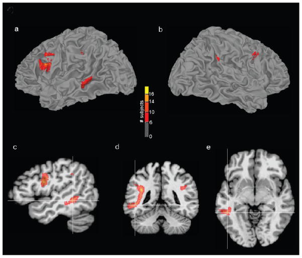
Figure 4. The Highest Anisotropy Arcuate Fasciculus Pathways Terminate Near Middle Temporal Gyrus.
For 19 left Wada patients, an image volume mask of the highest anisotropy (strongest 10%) AF pathways were transformed to standard space, and summed to create a single volume where each voxel value equaled the number of subjects sharing pathways at that location. This volume was thresholded (dark red=at least 6 subjects had common AF pathways, yellow=all subjects had common pathways) and displayed on a reconstruction of the left (a) and right (b) hemisphere white/gray matter border of the MNI N27 single-subject brain. The high anisotropy AF pathways were located disproportionately within the dominant hemisphere connecting Broca’s area (BA 44) to the middle temporal gyrus. Sagittal (c), coronal (d), and axial (e) orthogonal views are also shown with the crosshairs located near the posterior AF terminations at x=−46, y=−43, z=−7 mm (MNI brain coordinates).

