
Figure 2. Footprinting of a model GPCR – rhodopsin.
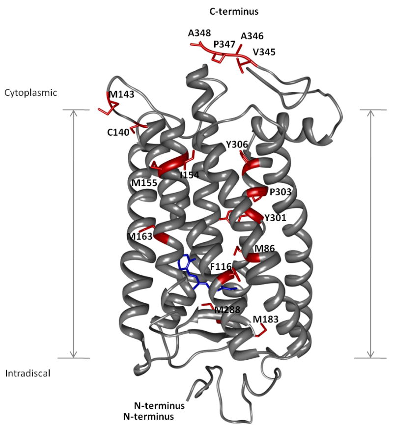
Rhodopsin is shown as gray ribbons whereas amino acid residues found to be modified following exposure to X-rays are shown as red sticks. The N- and C-termini are indicated together with the transmembrane region. The rhodopsin chromophore, 11-cis-retinal, is shown in blue near the intradiscal side of the membrane.