Abstract
Adenoviral vectors are highly efficient at transferring genes into cells and are broadly used in cancer gene therapy. However, many therapeutic genes are toxic to vector host cells and thus inhibit vector production. The truncated form of E2F-1 (E2Ftr), which lacks the transactivation domain, can significantly induce cancer cell apoptosis, but is also toxic to HEK-293 cells and inhibits adenovirus replication. To overcome this, we have developed binary- and single-vector systems with a modified tetracycline-off inducible promoter to control E2Ftr expression. We compared several vectors and found that the structure of expression cassettes in vectors significantly affects E2Ftr expression. One construct expresses high levels of inducible E2Ftr and efficiently causes apoptotic cancer cell death by activation of caspase-3. The approach developed in this study may be applied in other viral vectors for encoding therapeutic genes that are toxic to their host cells and/or inhibit vector propagation.
Keywords: Adenovirus, Gene, Expression, E2F-1, Truncated, Tet-Off, System, Apoptosis
Introduction
Adenovirus (Ad) is easy to manipulate in vitro, produces high titer stocks, and infects a broad range of mammalian cells. For these reasons, Ad vectors (Adv) are prefeered in basic and medical research. For cancer gene therapy, Adv are used to deliver genes or modified versions of genes into tumors. The products of therapeutic genes are generally toxic to cancer cells or cause apoptosis; thus, the gene products may be also toxic to human embryonic kidney HEK-293 cells used for Adv production and prevent vector construction and amplification.
The transcription factor E2F-1 plays important roles in the activation of expression of genes involved in cell cycle progression and growth. Recent studies by us and others have shown that Adv expressing E2F-1 efficiently induce apoptosis in cancer cells in vitro and in vivo (Dong et al., 1999; Dong et al., 2002; Elliott et al., 2001; Itoshima et al., 2000; Vorburger et al., 2005). However, the oncogenic property of promoting cell proliferation by wild type (wt) E2F-1, presumably by virtue of its ability to stimulate expression of cell cycle-promoting genes, limits its application in cancer gene therapy. To circumvent this barrier, the truncated forms of E2F-1 (E2Ftr) were generated, which lack the transactivation domain and cell cycle promoting effects (Bell, O’Prey, and Ryan, 2006; Fan and Bertino, 1997). Studies have showed that these mutants are potent inducers of apoptosis but are unable to induce cell cycle progression (Fan and Bertino, 1997; Hsieh et al., 1997; Melillo et al., 1994).
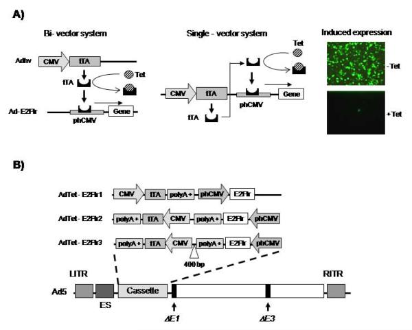
We previously attempted to create an Adv using the Cytomegalovirus (CMV) promoter to control E2Ftr (aa 1-375) without success despite multiple tries (Xiao-Mei Rao, unpublished results), likely because a high level of E2Ftr is toxic to HEK-293 cells and blocks Ad replication. To overcome this, we applied a tetracycline (Tet)-off inducible expression promoter (Gossen and Bujard, 1992) and developed inducible binary- and single-Adv systems to express E2Ftr (Fig. 1). Our binary-vector system consists of two vectors: an Ad helper virus (Adhv) and an Ad-E2Ftr vector. Adhv expresses Tet-regulated transactivator (tTA), which activates the E2Ftr expression cassette in Ad-E2Ftr. Without the helper vector, E2Ftr expression in Ad-E2Ftr is totally inhibited, so Ad-E2Ftr can be created and amplified in HEK-293 cells. High levels of the apoptotic E2Ftr protein are induced in cells only when Adhv is applied with Ad-E2Ftr. Although this system leads to high-level expression of E2Ftr and is useful in research, the absolute need for co-delivery of two separate vectors made the clinical use of this strategy untenable. Thus, we further developed a single-vector system in which both the tTA and E2Ftr expression cassettes are incorporated into one Adv. A helper vector is not required for E2Ftr expression in the single-vector system.
Figure 1. Schematic diagram of binary- and single Adv systems.
(A) CMV promoter drives the expression of the tTA protein. EGFP or E2Ftr is under the control of a synthetic minimal promoter composed of Tet-responsive element (TRE) and CMV mini-promoter (phcmv), which is silent unless activated by tTA. In the absence of Tet or Dox, tTA binds to phcmv and triggers the expression of EGFP or E2Ftr. When Tet is added to the medium, tTA is bound by Tet and unable to bind to phcmv and activate the expression of EGFP or E2Ftr. SK-MEL-2 cells in the absence or presence of Dox (1μg/ml) were infected with AdTet-EGFP at a MOI of 50. After 24 hours, cells were observed for EGFP expression under fluorescence microscopy and photographs were taken with Kodak MDS 290 software at × 20 magnification. (B) Schema of three single bicistronic Ads encoding E2Ftr, the left and right inverted terminal repeat sequences (LITR or RITR, respectively); encapsidation signal (ES) and E1/E3 deleted genes are shown in the Ad structure.
We compared the binary- and single-vector systems expressing inducible E2Ftr. We found that Ad-mediated high-level expression of E2Ftr is toxic to cancer cells as well as to vector host HEK-293 cells. E2Ftr efficiently induces cell death associated with poly (ADP-Ribose) polymerase (PARP) cleavage and procaspase-3 decrease, the hallmarks of apoptosis. Since many other genes have potential for cancer therapy may be also toxic to vector host cells and inhibit vector production, the approaches reported in this study may have broad applications in creation of viral vectors that will carry toxic cancer therapeutic genes.
Results
Binary- and single-vector systems
Figure 1A depicts the binary- and single-vector systems with Tet-off inducible transcription cassettes. In the bi-vector system, expression of E2Ftr from Ad-E2Ftr is controlled by the Tet-regulating promoter (phcmv), which is activated by tTA encoded in the helper vector Adhv. In the single-vector system, both transgene and tTA expression cassettes are inserted in the same vector. Expression of the enhanced green fluorescent protein (EGFP) reporter shows the efficient regulation of the Tet-off inducible promoter in the single-vector system (Fig. 1A). Figure 1B shows the schema of three different single bicistronic Adv encoding E2Ftr under the regulation of the Tet-off system. We did not insert a poly A signal in AdTet-E2Ftr1 because the Ad’s backbone can provide the viral E1 gene poly A signal. The tTA and E2Ftr expression cassettes in AdTet-E2Ftr2 are closely connected; while a 400-base pair (bp) fragment from the pcDNA3.1 (+/−) plasmid (Invitrogen, Carlsbad, CA) was inserted between the two cassettes in AdTet-E2Ftr3.
Regulation of E2Ftr expression in binary- and single Adv
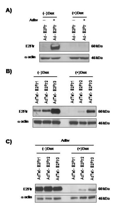
Human melanoma SK-MEL-2 cells were infected with Ad-E2Ftr alone (control) or co-infected with Adhv in the presence or absence of Doxycycline (Dox; 1 μg/ml), a derivative of Tet. At 48 hours after infection, E2Ftr expression was analyzed. E2Ftr expression from the binary system was only induced in the presence of Adhv and absence of Dox; adding Dox negatively regulated E2Ftr expression in cells (Fig. 2A).
Figure 2. Regulation of E2Ftr expression from binary- and single Adv systems.
(A) SK-MEL-2 cells in the absence or presence of Dox (1μg/ml) were infected with Ad-E2Ftr at a MOI of 50 or co-infected with Adhv at a MOI of 5. After 48 hours, cells were harvested and protein extracts were used for a western blot assay. (B) SK-MEL-2 cells in the absence or presence of Dox (1μg/ml) were infected with AdTet-E2Ftr1, AdTet-E2Ftr2, and AdTet-E2Ftr3 at a MOI of 50. After 48 hours, cells were harvested and protein extracts were used for a western blot assay. (C) SK-MEL-2 cells in the absence or presence of Dox (1μg/ml) were co-infected with AdTet-E2Ftr1, AdTet-E2Ftr2, and AdTet-E2Ftr3 at a MOI of 50 and Adhv at a MOI of 5. After 48 hours, cells were harvested, protein extracts were used for a western blot assay, and a mAb mouse-antihuman E2F-1 was used to detect E2Ftr. α-actin was used to demonstrate equal loading for each lane. Similar results were obtained in two additional experiments.
We further evaluated E2Ftr expression from the three constructs of the single-vector system. SK-MEL-2 cells were infected with AdTet-E2Ftr1, AdTet-E2Ftr2, or Ad-E2Ftr3 at a multiplicity of infection (MOI) of 50. Two days after infections, E2Ftr expression was analyzed. AdTet-E2Ftr1 and AdTet-E2Ftr2 expressed relatively lower levels of E2Ftr while AdTet-E2Ftr3 expressed a significantly high level of E2Ftr in the absence of Dox (Fig. 2B). Similar to the binary-vector system, adding 1 μg/ml of Dox in culture repressed E2Ftr expression in cells infected with AdTet-E2Ftr1, AdTet-E2Ftr2 or AdTet-E2Ftr3 (Fig. 2B). These data show that E2Ftr expression can be regulated with Dox in both single- and binary-vector systems.
Figure 2B shows that AdTet-E2Ftr1 and AdTet-E2Ftr2 expressed much lower levels of E2Ftr in cancer cells compared to AdTet-E2Ftr3. A possible cause that lowers E2Ftr expression may be attributed to the structures in AdTet-E2Ftr1 and AdTet-E2Ftr2 that restrict tTA production. To verify this, we investigated whether Adhv used in the bi-vector system may also increase E2Ftr expression in the single-vector AdTet-E2Ftr1 and AdTet-E2Ftr2 by providing tTA in trans. SK-MEL-2 cells were co-infected with Adhv plus AdTet-E2Ftr1, AdTet-E2Ftr2, or AdTet-E2Ftr3. In the absence of Dox, Adhv increased E2Ftr expression from AdTet-E2Ftr1 and AdTet-E2Ftr2 to levels similar to AdTet-E2Ftr3 (Fig. 2C). As expected, E2Ftr expression from all three vectors in Adhv co-infection was repressed in the presence of Dox (Fig. 2C). These results indicate that tTA levels are not optimized in AdTet-E2Ftr1 and AdTet-E2Ftr2 for maximal E2Ftr expression. However, AdTet-E2Ftr3 does not require Adhv to increase E2Ftr expression (Fig. 2B).
A truncated form of E2F-1 lacking the transactivation domain inhibits Ad replication
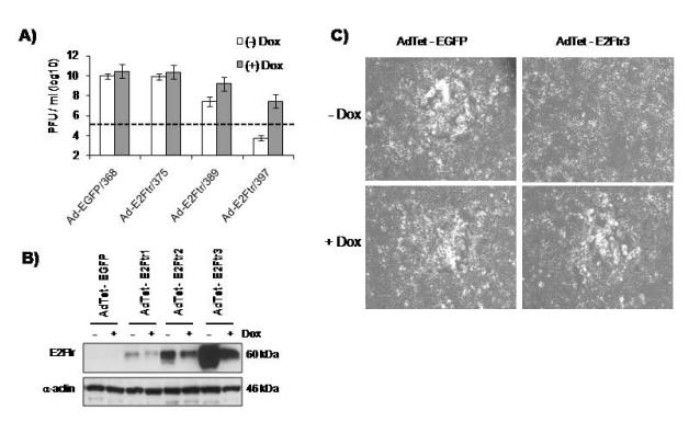
We previously attempted to apply the CMV promoter to control E2Ftr in Adv. However, such vector could not be rescued from plasmids with Ad backbone in HEK-293 cells. It is likely that the strong CMV-mediated high-level expression of E2Ftr may be toxic to HEK-293 cells and prevent viral vector generation. We investigated the effect of E2Ftr expression on Ad replication. HEK-293 cells were infected at a MOI of 5 with AdTet-EGFP, AdTet-E2Ftr1, AdTet-E2Ftr2, or AdTet-E2Ftr3 in the absence or presence of Dox (1μg/ml) in cell cultures, virus titers and E2Ftr expression were analyzed three days after infection. We found that there was no difference in the titers of AdTet-EGFP and AdTet-E2Ftr1 with or without Dox induction (Fig. 3A); AdTet-EGFP does not express E2Ftr and AdTet-E2Ftr1 expressed very low levels of E2Ftr (Fig. 3B). The addition of Dox had little effect on E2Ftr expression from AdTet-E2Ftr1, whereas Dox could repress E2Ftr expression from AdTet-E2Ftr2 or AdTet-E2Ftr3-infected cells (Fig. 3B). AdTet-E2Ftr2 and AdTet-E2Ftr3 produced higher levels of E2Ftr in the absence of Dox (Fig. 3B). Inhibition of E2Ftr by Dox resulted in increased AdTet-E2Ftr2 and AdTet-E2Ftr3 titers by 1000- and 10,000-fold, respectively, which correlates with repressed virus titers (Figs. 3A and B).
Figure 3. Effect of E2Ftr expresssion on Ad yield and lytic plaque formation.
HEK-293 cells in either the absence or presence of Dox (1μg/ml) were infected with AdTet-EGFP, AdTet-E2Ftr1, AdTet-E2Ftr2, or AdTet-E2Ftr3 at a MOI of 5. After 72 hours, cells were harvested and divided in two samples; one sample was used to conduct Western Blot analysis and the second sample was used to determine virus titers. (A) For Western blot, a mAb mouse-antihuman E2F-1 was used to detect E2Ftr. α-Actin was used to demonstrate equal loading for each lane. (B) For virus titering, cultures were collected at 72 hours after infection and went through three cycles of freezing and thawing to release viruses from the cells. Lysates were serially diluted to determine the titers in HEK-293 cells. The dashed line shows the level of viruses added in the cultures. Each point represents the mean of three independent experiments ± standard deviation (SD; bars). C) HEK-293 cells were cultured in presence or absence of Dox (1 μg/ml) and infected with AdTet-EGFP or AdTet-E2Ftr3 at a MOI of 5; a standard plaque assay was performed to assess formation of lytic plaques. A representative experiment is shown from three performed.
Because AdTet-E2Ftr3 produces the highest E2Ftr levels among the three vectors, further studies focused on this vector. We assessed virus plaque formation on HEK-293 plates transduced by AdTet-E2Ftr3. HEK-293 cells, in the absence or presence of Dox (1μg/ml), were infected with Ad-Tet-E2Ftr3 or AdTet-EGFP (control). One hour later, unadsorbed viruses were removed and 0.5% of an agarose layer with complete medium was added to the cells; plaque formation was monitored over time. AdTet-EGFP plaques appeared after 4 days on HEK-293 cell plates with or without Dox. No plaques were observed with AdTet-E2Ftr3-infected cells up to 7 days; adding Dox allowed AdTet-E2Ftr3 to form plaques (Fig. 3C).
Our results show that induced expression of E2Ftr strongly inhibits Ad replication. E2Ftr may act as repressor of the Ad E2 promoter by blocking wt E2F-1 access to the promoter. Because the Ad E2 gene is essential for viral DNA replication, overexpression of E2Ftr is not only toxic to HEK-293 cells, but may also directly inhibit Ad DNA replication.
Compare the effects of Dox and Tet on cell growth, E2Ftr expression, and virus yield
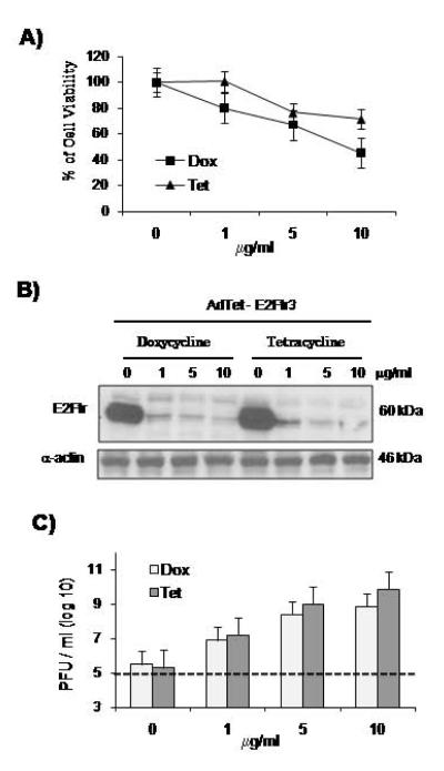
The application of a low concentration of Dox (1 μg/ml) could not strongly repress E2Ftr expression from the Tet-inducible vectors (Figs. 2 and 3). Therefore, 1 μg/ml Dox cannot completely release the E2Ftr-mediated inhibition of Ad replication; however, high doses of Dox were reported to be toxic to mammal cells (Ermak, Cancasci, and Davies, 2003; Fife and Sledge, 1998) and can decrease virus amplification as well. We previously used Tet to regulate a Tet-off expression system (Zhou and Beaudet, 2000). Tet may be less toxic to HEK-293 cells. We compared the toxicity of Dox versus Tet to HEK-293 cells using a 3-(4, 5-dimethylthiazol-2-)-2, 5-diphenyltetrazolium bromide (MTT) assay. Dox is more toxic to HEK-293 cells, as 10 μg/ml of Dox resulted in 50% of HEK-293 viable cells, while 80% of viable cells were observed at the same concentration of Tet (Fig. 4A). The high toxicity of Dox to HEK-293 cells could affect virus replication. We compared the effect of different doses of Dox and Tet on E2Ftr regulation. HEK-293 cells in presence of increasing doses of Dox or Tet (0, 1, 5, 10 μg/ml) were infected with AdTet-E2Ftr3 at a MOI of 5 and E2Ftr expression was analyzed 24 hours after infection. It was found that E2Ftr expression levels declined concomitantly with dose increase of Dox and Tet (Fig. 4B). Both drugs have similar regulatory effects on E2Ftr expression at their respective doses and at 10 μg/ml showed higher repression of E2Ftr expression. Comparing with Fig. 2B and Fig. 3B, more effective shutoff of E2Ftr with Dox and Tet is achieved in Fig. 4B, likely because of a lower MOI applied and earlier protein harvest.
Figure 4. Comparison between Dox and Tet in HEK-293 survival E2Ftr regulation, and Ad yield.
(A) Toxicity of Dox vs Tet at 0, 1, 5, and 10 μg/ml of concentration were compared in HEK-293 cells by a MTT assay at 72 hours after drug treatments. Each point represents the mean of three independent experiments ± (SD; bars). (B) HEK-293 cells in the presence of Dox or Tet at 0, 1, 5, and 10 μg/ml were infected with AdTet-E2Ftr3 at a MOI of 5. At 24 hours after infection, cell extracts were used for western blot assay and a mAb mouse-antihuman E2F-1 was used to detect E2Ftr. α-Actin was used to demonstrate equal loading for each lane. C) HEK-293 cells were infected with AdTet-E2Ftr3 at a MOI of 5. Ad yield was determined in the presence of Dox or Tet at 0, 1, 5, and 10 μg/ml of concentration. The cultures were collected three days after infection and went through three cycles of freezing and thawing to release viruses from the cells. Lysates were serially diluted to determine the titers in HEK-293 cells. The dashed line shows the level of viruses added in the cultures. Each point represents the mean of three independent experiments ± (SD; bars).
Because Tet is less toxic than Dox to HEK-293 cells and both Tet and Dox have similar regulatory effects on E2Ftr expression, we further anticipated that the application of Tet could result in a higher Ad yield. HEK-293 cells were infected with AdTet-E2Ftr3 at different doses of Dox or Tet. Although virus yield increased concomitantly with dose increases from both Dox and Tet, Tet 10 μg/ml resulted in nearly 10-fold higher virus titers than treatment with Dox at the same concentration (Fig. 4C). These results indicate that Tet induces higher AdTet-E2Ftr3 production as compared to Dox.
E2Ftr expression induces morphological changes in melanoma cells
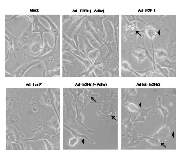
Changes on melanoma cell morphology were inspected after E2F-1 or E2Ftr expressions. AdTet-E2Ftr3 was compared with the binary vector Ad-E2Ftr and Ad-E2F-1 expressing wt E2F-1. SK-MEL-28 cells were uninfected (mock) or infected with Ad-LacZ, Ad-E2F-1, AdTet-E2Ftr3, or Ad-E2Ftr with or without Adhv. At 24 hours post-infection, melanoma cell morphology was inspected. Mock or cells infected with Ad-LacZ or Ad-E2Ftr without the Adhv vector showed no or little morphological changes. Light microscopic observations revealed morphological changes in SK-MEL-28 cells infected with Ad-E2F-1, Ad-E2Ftr plus Adhv, or AdTet-E2Ftr3 alone. Cells were characterized by rounding and loss of adherence to the flask. In addition to showing large cytoplasmic projections, some cells developed a vesiculated cytoplasm, associated with apoptotic cell death (Fig. 5). The results suggest that AdTet-E2Ftr3 alone can induce apoptotic cell death as did Ad-E2F-1 that expresses wt E2F-1, Ad-E2Ftr that requires an Adhv vector.
Figure 5. Effect of E2F-1 or E2Ftr expressions on melanoma cell morphology.
SKMEL-28 cells were uninfected (mock) or infected at a MOI of 100 with Ad-LacZ, Ad-E2F-1, AdTet-E2Ftr3, or Ad-E2Ftr alone or co-infected with Adhv at a MOI of 5. After 24 hours, cell morphology was analyzed by light microscopy. Apoptotic cells are indicated by arrowhead; vesiculated cells are indicated by arrow. Pictures were taken with bright-field optics at × 40 magnification.
AdTet-E2Ftr3-mediated cancer death via apoptosis
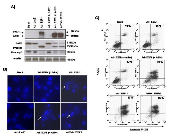
To confirm that AdTet-E2Ftr3 induces cell death by apoptosis, the vector was compared with Ad-E2F-1 or Ad-E2Ftr. SK-MEL-28 cells were uninfected (mock) or infected with Ad-LacZ, Ad-E2F-1, AdTet-E2Ftr3, Ad-E2Ftr, or Ad-E2Ftr plus Adhv. Induction of apoptosis was confirmed by detection of cleaved PARP, which is a substrate of caspase-3. Both PARP and caspase-3 are well known as apoptosis markers (Fernandes-Alnemri, Litwack, and Alnemri, 1994; Itoshima et al., 2000). Melanoma cells infected with Ad-E2F-1, AdTet-E2Ftr3, or Ad-E2Ftr with Adhv showed an apoptosis-specific PARP cleavage fragment (85-90 kDa) as well as a decrease in the proenzyme caspase-3/cysteine protease (CP)P32, whereas cleavage of PARP and decrease of caspase-3 were not present in mock or cells infected with Ad-LacZ or Ad-E2Ftr alone (Fig. 6A). In addition to activation of the caspases pathway, apoptosis was confirmed by Hoechst staining. No changes on nuclei morphology were observed in mock, Ad-LacZ, or Ad-E2Ftr (alone)-infected cells. In contrast, significant nuclei fragmentation was observed in cells infected with AdTet-E2Ftr3, Ad-E2F-1, or Ad-E2Ftr plus Adhv (Fig. 6B).
Figure 6. Evaluation of apoptosis induced by three different Adv expressing E2F-1 or E2Ftr.
SK-MEL-28 cells were uninfected (mock) or infected at a MOI of 100 with Ad-LacZ, Ad-E2F-1, AdTet-E2Ftr3, or Ad-E2Ftr alone or co-infection with Adhv at a MOI of 5. After 72 hours, cells were analyzed for activation of the caspases pathway and apoptosis. (A) Western blot was used to detect activation of the caspases pathway, a mAb mouse-antihuman E2F-1 was used to detect E2Ftr, a mAb mouse-antihuman PARP was used to detect PARP, and a mAb mouse-antihuman caspase-3/CPP3 was used to detect caspase-3. α-actin was used to demonstrate equal loading for each lane. (B) Hoechst staining was performed by adding Hoechst 33258 dye to a final concentration of 10μM to each well in the 24-well plate. Condensation and fragmentation of nuclei (examples indicated by white arrows) were observed under a fluorescence microscope (Olympus Microsystems) at × 20 magnification. (C) For annexin V staining, cells were stained with annexin V-PE and 7-Amino-actinomycin D (7-AAD); positive cells for annexin V-PE and 7-AAD staining were analyzed by FACScan flow cytometer with FlowJo software. Similar results were obtained in three independent experiments. A representative experiment is shown.
In apoptotic cells, the membrane phospholipid phosphatidylserine (PS) is translocated from the inner leaflet to the outer leaflet of the plasma membrane, thereby exposing PS to the external cellular environment. Annexin V is a dependent phospholipid-binding protein having a high affinity for PS and binds to cells with exposed PS. We used an annexin-V-phycoerythrin (PE) kit to determine the percentage of cells undergoing apoptosis. AdTet-E2Ftr3, Ad-E2F-1, and Ad-E2Ftr plus Adhv induced similar percentages of apoptosis at 87-, 70-, and 86%, respectively. In contrast, no significant apoptosis was detected in mock, Ad-LacZ, or Ad-E2Ftr (alone) infections (Fig. 6C). These results demonstrate that E2Ftr in the single-vector system can induce cancer apoptosis as efficiently as other vectors.
AdTet-E2Ftr3 strongly induces cancer cell death
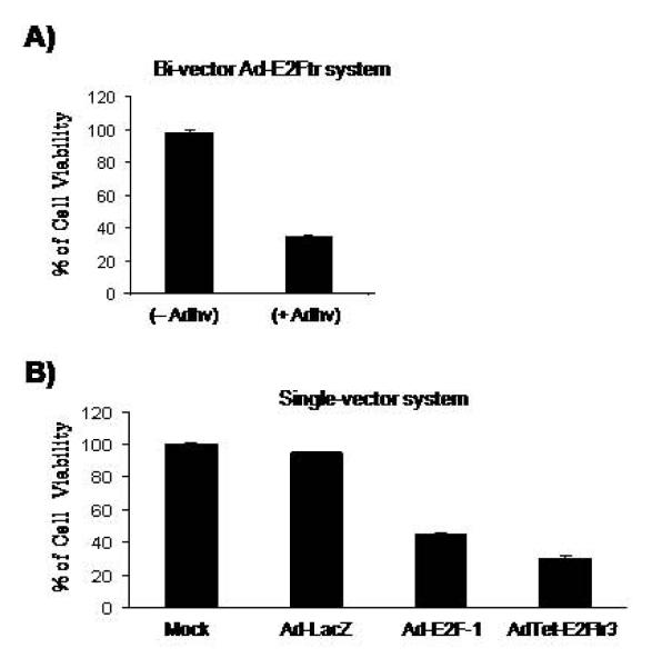
After observing that Ad-E2F-1 and AdTet-E2Ftr3 induce similar levels of apoptosis, we further compared the cytotoxic effect of these vectors on cancer cells. Cell death was analyzed by MTT assay at day 3 post-infection. As expected, Ad-E2Ftr only induces cancer cell killing in the presence of Adhv (Fig. 7A). AdTet-E2Ftr3 induced strong cancer death (70%), significantly better than wt E2F-1, which induced about 50% death (Fig. 7B). Mock and Ad-LacZ could not kill cancer cells in the same conditions. The results clearly show that AdTet-E2Ftr3 as a novel vector does not require a helper virus to induce cancer death. In addition, AdTet-E2Ftr3 can more efficiently destroy cancer cells than the Adv expressing wt E2F-1.
Figure 7. Assessment of melanoma cell killing activity from Adv encoding E2F-1 or E2Ftr.
SK-MEL-28 cells were infected with (A) Ad-E2Ftr alone (at a MOI of 100) or co-infected with Adhv at a MOI of 5 or (B) uninfected (mock) or infected at a MOI of 100 with Ad-LacZ, Ad-E2F-1, AdTet-E2Ftr3. At 72 hours after infection, cell viability was analyzed by MTT assay; each point represents the mean of three independent experiments ± (SD; bars).
Discussion
Despite wt E2F-1 efficiently inducing apoptosis in cancer cells in vitro and in vivo (Dong et al., 1999; Dong et al., 2002; Elliott et al., 2001; Itoshima et al., 2000; Vorburger et al., 2005), its oncogenic property of promoting cell proliferation by its ability to stimulate gene expression limits its application in cancer gene therapy. This concern could be avoided by using a E2Ftr that lacks the transactivation domain and cell cycle promoting effects (Bell, O’Prey, and Ryan, 2006; Fan and Bertino, 1997). Studies have shown that E2Ftr is a potent inducer of apoptosis but is unable to promote cell cycle progression (Fan and Bertino, 1997; Hsieh et al., 1997; Melillo et al., 1994).
We previously tried to construct an Adv encoding E2Ftr with the CMV promoter. However, such vector could not be generated, likely because E2Ftr is toxic to Ad host HEK-293 cells and/or inhibits vector amplification. For this reason, we developed binary- and single-inducible Adv. In the binary system, the helper vector Adhv expresses tTA to activate the E2Ftr gene carried in other vector. In a single-vector system, the tTA expressing cassette and E2Ftr gene are incorporated into one vector (Fig. 1). We constructed three single vectors, AdTet-E2Ftr1, AdTet-E2Ftr2, and AdTet-E2Ftr3, and compared them with the binary vector.
We observed that both AdTet-E2Ftr1 and AdTet-E2Ftr2 expressed low levels of E2Ftr and AdTet-E2Ftr3 expressed the highest E2Ftr level (Fig. 2B). The expression cassettes in AdTet-E2Ftr1 and AdTet-E2Ftr2 are closely connected, although they are in a different orientation and order. When Adhv, which can provide tTA in trans, was applied with AdTet-E2Ftr1 and AdTet-E2Ftr2 in infection, E2Ftr expression significantly increased to the levels observed with AdTet-E2Ftr3 (Fig. 2C). Adhv could not increase E2Ftr expression from AdTet-E2Ftr3 (Fig. 2B). The only difference between AdTet-E2Ftr2 and AdTet-E2Ftr3 is the 400-bp fragment inserted in the later vector. The results suggest that the structures of the E2Ftr cassette and the transcription activator tTA cassette in a single vector may affect each other when they are closely connected and cause inefficient E2Ftr induction. For maximal therapeutic gene expression, the optimum structure should be derived when an inducible single-vector system is applied to express toxic genes.
E2F-1 is a transcription factor that was first described as a nuclear activity that binds to the Ad E2 promoter (Kovesdi, Reichel, and Nevins, 1986a; Kovesdi, Reichel, and Nevins, 1986b). The Ad E2 early promoter contains two E2F binding sites; binding of E2F to the Ad promoter is important for expression of the virus E2 gene that is directly involved in viral DNA replication (Huang and Hearing, 1989). This binding is dependent on a region of E2F-1 between amino acids 284 and 358 (Helin and Harlow, 1994). The E2Ftr used in our study maintains this DNA binding region but lacks the transcription activation domain. We observed that E2Ftr significantly affects virus yield. High levels of E2Ftr of AdTet-E2Ftr3 correlate with significantly low virus production, especially when withdrawing Dox induces E2Ftr. In a viral plaque assay, we observed that plaques only formed in the presence of Dox in HEK-293 cells infected with AdTet-E2Ftr3, while the control AdTet-EGFP formed plaques with or without Dox. The results clearly show that E2Ftr strongly inhibits Ad replication that explains why we previously could not rescue an Ad-encoding E2Ftr controlled by the strong CMV promoter. E2Ftr protein may competitively bind to the viral E2a promoter and inhibits Ad vector amplification; the protein may also repress expression of cellular genes that are required for efficient Ad replication, such as cyclin E (Zheng et al., 2008). Furthermore, it is reasonable to believe that E2Ftr introduced into cancer cells can bind to and repress the promoters of genes that are normally activated by wt E2F for cell proliferation. Thus, we expect that using E2Ftr can inhibit tumor growth more efficiently than wt E2F-1. This hypothesis is supported by our animal studies of a melanoma xenograft model (J.G. Gomez-Gutierrez, et al. manuscript submitted).
The commercial distributor of the Tet-off system (Clontech Laboratories, Inc., Mountain View, CA) recommends using a 1μg/ml Dox concentration to inhibit the inducible promoter. However, to achieve efficient regulation of transgene expression, higher Dox concentrations are necessary (Figs. 2, 3, and 4). However, higher Dox concentrations are toxic to mammals cells (Ermak, Cancasci, and Davies, 2003) and induce cell death in vitro and in vivo (Fife and Sledge, 1998). We compared Dox and Tet and found them to have similar regulatory effects on E2Ftr expression, except that Dox was more toxic to HEK-293 cells (Fig. 4A). When Tet was used instead of Dox, approximately 10-fold higher titers were produced (Fig. 4C).
Our studies have shown that when Tet-off activator tTA is incorporated into the same Ad vector to induce E2Ftr, the vector structures significantly affect therapeutic gene expression. We have shown that separation of the tTA cassette from the therapeutic gene cassette with a 400-bp DNA fragment greatly increases the E2Ftr protein level. We further demonstrated that AdTet-E2Ftr3-mediated high-level expression of E2Ftr is toxic to cancer cells and efficiently induces apoptotic cancer cell death by increasing PARP cleavage and decreasing the proenzyme caspase-3/CPP32 (Figs. 6 and 7).
This is the first report of the creation of a single Adv system expressing the cancer-killing E2Ftr gene. This study demonstrates that we can optimize vector structures to create viral vectors expressing high levels of toxic genes. Because E2Ftr is so toxic to cancer cells, AdTet-E2Ftr3 may have an enormous advantage in cancer gene therapy. Many genes are selected for potential cancer gene therapy based on their properties of toxicity to destroy cancer cells. These genes are also likely to be toxic to viral vector host cells. Thereby, Adv, as well as other viral vectors intended to express powerful therapeutic genes, are generally difficult to generate. The approach reported in this study may be applied in constructions of Ad and other viral vectors for expression of other toxic genes.
Materials and Methods
Cell lines and culture conditions
Human melanoma cell lines SK-MEL-2 and SK-MEL-28 and human embryonic kidney cell line HEK-293 were purchased from the American Type Culture Collection (Rockville, MD). SK-MEL-2 and SK-MEL-28 cell lines were cultured in α-minimal essential medium (α-MEM). HEK-293 cells were cultured in minimal essential medium-α (MEM-α) supplemented with 10% heat-inactivated fetal bovine serum (FBS) and penicillin/streptomycin (100 U/mL). Cell culture reagents were obtained from Gibco BRL (Bethesda, MD). Cells were cultured in a 5% CO2 incubator at 37°C and the medium was changed every 2 or 3 days.
Recombinant Ad vectors
Eight replication-defective Adv were used. The Ad5CMV-E2F-1 (Ad-E2F-1) vector was deleted in the E1 subunit and contained the transgene E2F-1 under the control of the CMV promoter (Dong et al., 2002). Ad5CMV-LacZ (Ad-LacZ) vector was deleted in the E1 subunit and contained the transgene nuclear-localized β-galactosidase under the control of the CMV promoter and was used as a control as described (Dong et al., 2002). The Ad-E2Ftr expresses a truncated E2F-1 protein, which retains the E2F-1 DNA binding domain but lacks its transactivation domain. The expression of E2Ftr is under the regulation of Tet-responsive element (TRE) and the Tet-controlled transactivator virus (Adeno-X Tet-Off) was used as a helper virus (Adhv) to activate the Tet-responsive promoter (Dong et al., 2007; Hao et al., 2007). The other four Adv (AdTet-EGFP, AdTet-E2Ftr1, AdTet-E2Ftr2, and AdTet-E2Ftr3) were constructed by us based on the widely used Ad-Easy system (MP Biomedicals, Irvine, CA).
AdTet-EGFP was constructed as follows. The tTA protein and poly A signal (tTA/polyA) fragment was excised from pUHD15-1 with XbaI/HindIII and subcloned into pShuttle-tTA/polyA. The phcmv sequence was then excised from pBH72 with XhoI/HindIII and ligated into pΔE1sp1A, resulting in pΔE1sp1A-phcmv. The phcmv fragment was excised from pΔE1sp1A-phcmv with BamHI and subcloned into pShuttle-tTA in the BglII site, resulting in pShuttle-tTA/phcmv. Once this plasmid was obtained, a multiple cloning sequence (MCS) containing the sites ScaI, NheI, SpeI, PvuI, AgeI, and NotI were added upstream of tTA. The CMV promoter was amplified by polymerase chain reaction (PCR) from pShuttle-CMV with primers containing the restriction sites NheI and NotI, respectively. Primer sequences were:
5′- GCTAGCGCGTTACATAACTTACGGTA-3′ and
5′- GCGGCCGCGATCTGACGGTTCACTAAA-3′.
After PCR assay, a 530-bp product was obtained and ligated in the sites NheI/NotI from pShuttle-tTA/phcmv, resulting in the plasmid pShuttle-CMV/tTA/phcmv. EGFP was amplified by PCR from pIRES-EGFP using primers containing the restriction sites BglII and AscI, respectively. Primer sequences were:
5′- TATAGATCTACATGGTGAGCAAGGGCGAGG- 3′ and
5′- TGGCGCGCCGCCAGCTGGTTCTTTCCGCCT-3′.
After the PCR assay, a 1.0 kb fragment was obtained and ligated in the BglII/AscI sites from pShuttle-CMV/tTA/phcmv, resulting in the plasmid pShuttle-CMV/tTA/phcmv-EGFP.
AdTet-EGFP was a model for the construction of AdTet-E2Ftr1. E2Ftr was excised from plasmid pAdstE2Ftr with BamHI and cloned in the BglII restriction site from pShuttle-CMV/tTA/phcmv, resulting in the plasmid pShuttle-CMV/tTA/phcmv-E2Ftr.
AdTet-E2Ftr2 construction method
The phcmv/E2Ftr/polyA cassette was removed from the pAdstE2Ftr plasmid with the enzymes XhoI/HindIII and was ligated into a pShuttle vector in the SalI/HindIII sites, resulting in pShuttle-phcmv/E2Ftr/polyA. The CMV/tTA/polyA cassette was excised twice. It was first excised from pUHD15-1 with XhoI/HindIII and ligated in the XhoI/HindIII sites from pBluescript II KS +/− (Fermentas Inc., Glen Burnie, MD), resulting in pBSII-CMV/tTA/polyA. The CMV/tTA/polyA cassette was excised again from pBSII-CMV/tTA/polyA with XhoI/NotI and ligated in the XhoI/NotI sites from pShuttle-phcmv/E2Ftr/polyA, resulting in pShuttle-phcmv/E2Ftr/polyA-CMV/tTA/polyA.
AdTet-E2Ftr3 construction method
A 400-bp fragment containing a poly A signal of bovine growth hormone (BGHpA) and f1 origin was excised from the pcDNA3.1 (+/−) plasmid (Invitrogen) with XbaI/PsiI and ligated in the XbaI/EcoRV sites from pShuttle-phcmv/E2Ftr/polyA with the purpose of replacing the SV40 poly A sequence for BGHpA. The resulting plasmid was named pShuttle-phcmv/E2Ftr/BGHpA. The CMV/tTA/polyA fragment was then removed from pBSII-CMV/tTA/polyA with XhoI/NotI and ligated in the XhoI/NotI sites from pShuttle-phcmv/E2Ftr/BGHpA, resulting in pShuttle-phcmv/E2Ftr/BGHpA-CMV/tTA/polyA. These 4 shuttle vectors were used to generate recombinant Ads, according to the manufacture’s instructions accompanying the Ad-Easy system. Virus particles from AdTet-E2Ftr1, AdTet-E2Ftr2, and AdTet-E2Ftr3 were rescued from the host HEK-293 cells in the presence of 1μg/ml of Dox. Titers were determined using Adeno-X rapid titer kit (Clontech) in the presence of Tet (10 μg/ml).
For infections, 5×10⁵ SK-MEL-2 or SK-MEL-28 cells were plated in 60-mm tissue culture plates. The following day, the media was removed and the Adv were added in 0.5 ml of α-MEM at indicated concentrations of MOI plaque-forming units/cell. Mock infection was performed by treatment of cells with vehicle (media) only. After one hour of incubation at 37°C, the medium was removed and 3 ml of fresh α-MEM with 5% FBS was added. Cells were harvested at specific time points for analysis.
Western blot analysis
Cells were harvested and lysed in a radio immunoprecipitation assay (RIPA) buffer as described previously (Gomez-Gutierrez et al., 2006). Cell lysates were centrifuged and protein concentration was determined by Detergent Compatible (DC) protein assay (Bio-Rad, Hercules, CA). Equal amounts of cellular protein were electrophoresed in 10% sodium dodecyl sulfate-polyacrylamide gels (SDS-PAGEs) and transferred to Hybond-polyvinylidene difluoride (PVDP) membranes (Amersham, Arlington Heights, IL). The membranes were first incubated with the following primary antibodies (Abs): mouse-antihuman-E2F-1 monoclonal (m)Ab (Santa Cruz Biotech, Santa Cruz, CA) and rabbit-antihuman-α-actin polyclonal (p)Ab, mouse-antihuman-CPP32 mAb (Transduction Laboratories, Lexington, KY), and mouse-antihuman-PARP mAb (Calbiochem, San Diego, CA). Next, the membranes were incubated with antimouse immunoglobulin (Ig) or anti-rabbit Ig, peroxidase-linked, species-specific whole Abs (Amersham). Electrochemiluminescent (ECL) reagents were used to detect the signals according to the manufacturer’s instructions (Amersham).
Cellular proliferation
Cell proliferation was assessed at three days after drug treatment (Dox or Tet) or infection by measuring the conversion of the tetrazolium salt (MTT) to formazan according to the manufacturer’s instructions (Boehringer Mannheim, Indianapolis, IN) and as was reported previously (Gomez-Gutierrez et al., 2006). The supernatant from each plate was collected for measurement of absorbance at a wavelength of 570 nm. The results were expressed as the percentage of live cells.
Ad replication and titering
2×10⁵ HEK-293 cells were pleated in a 6-well plate in the absence or presence of Dox (1μg/ml). The next day, cells were infected with AdTet-EGFP, AdTet-E2Ftr1, AdTet-E2Ftr2, or AdTet-E2Ftr3 at a MOI of 5. After 1 hour of infection, unabsorbed virus particles were removed and fresh medium was added with or without Dox (1μg/ml). The cultures were collected after three days; cells went through three cycles of freezing and thawing to release viruses from the cells. Lysates were serially diluted to determine the titers in HEK-293 cells by standard infective unit (IFU) measurement.
Melanoma cell morphology
In a 12-well plate, 1 × 10⁵ SK-MEL-28 cells were uninfected (mock) or infected with Ad-LacZ, Ad-E2F-1, Ad-E2Ftr, Ad-E2Ftr plus Adhv (5 MOI), or AdTet-E2Ftr at a MOI of 100. After 24 hours, cell morphology was analyzed by light microscopy (Olympus Microsystems, Redwood City, CA). Pictures were taken in bright-field at ×40 magnification.
Detection of apoptosis
Hoechst staining was performed by adding Hoechst dye to a final concentration of 10μM to each well in the 24-well plate. Apoptotic cell death was determined by observation of apoptotic nuclei (i.e., fragmentation and condensation of nuclei) following Hoechst 33258 staining for 10 minutes at 37°C. Nuclei morphology was observed under a fluorescence microscope (Olympus Microsystems).
Annexin V staining
Annexin staining was performed as described previously (Gomez-Gutierrez et al., 2006) and according to the manufacturer’s instructions (Annexin V-PE Apoptosis Kit, Pharmingen, San Diego CA). Cells were analyzed by fluorescence-activated cell sorter scan (FACScan) flow cytometer (Becton Dickinson, Franklin Lakes, NJ) with FlowJo software (Tree Star Inc., Ashland, OR).
Acknowledgements
This work was supported by Award Numbers R01CA129975 (HSZ) and R01CA90784 (KMM) from the National Cancer Institute and G030983 (HSZ) and GMB081410 (KMM & HSZ) from the Kentucky Lung Cancer Research Program. AGG is recipient of a scholarship from National Council of Science and Technology (CONACYT) of Mexico. We thank Chuanlin Ding for annexin V analysis and Andrew Marsh for editing.
Footnotes
Publisher's Disclaimer: This is a PDF file of an unedited manuscript that has been accepted for publication. As a service to our customers we are providing this early version of the manuscript. The manuscript will undergo copyediting, typesetting, and review of the resulting proof before it is published in its final citable form. Please note that during the production process errors may be discovered which could affect the content, and all legal disclaimers that apply to the journal pertain.
References
- Bell LA, O’Prey J, Ryan KM. DNA-binding independent cell death from a minimal proapoptotic region of E2F-1. Oncogene. 2006;25(41):5656–63. doi: 10.1038/sj.onc.1209580. [DOI] [PubMed] [Google Scholar]
- Dong YB, Phelps AM, Yang HL, Jamshidi-Parsian A, Chen C, Hao H, Gomez-Gutierrez JG, Zhou HS, McMasters KM. Induction of apoptosis signal-regulating Kinase 1 by E2F-1 may not be essential for E2F-1-mediated apoptosis in melanoma cells. Tumour Biol. 2007;28(2):111–22. doi: 10.1159/000099370. [DOI] [PubMed] [Google Scholar]
- Dong YB, Yang HL, Elliott MJ, Liu TJ, Stilwell A, Atienza C, Jr., McMasters KM. Adenovirus-mediated E2F-1 gene transfer efficiently induces apoptosis in melanoma cells. Cancer. 1999;86(10):2021–33. doi: 10.1002/(sici)1097-0142(19991115)86:10<2021::aid-cncr20>3.0.co;2-1. [DOI] [PubMed] [Google Scholar]
- Dong YB, Yang HL, Elliott MJ, McMasters KM. Adenovirus-mediated E2F-1 gene transfer sensitizes melanoma cells to apoptosis induced by topoisomerase II inhibitors. Cancer Res. 2002;62(6):1776–83. [PubMed] [Google Scholar]
- Elliott MJ, Dong YB, Yang H, McMasters KM. E2F-1 up-regulates c-Myc and p14(ARF) and induces apoptosis in colon cancer cells. Clin Cancer Res. 2001;7(11):3590–7. [PubMed] [Google Scholar]
- Ermak G, Cancasci VJ, Davies KJ. Cytotoxic effect of doxycycline and its implications for tet-on gene expression systems. Anal Biochem. 2003;318(1):152–4. doi: 10.1016/s0003-2697(03)00166-0. [DOI] [PubMed] [Google Scholar]
- Fan J, Bertino JR. Functional roles of E2F in cell cycle regulation. Oncogene. 1997;14(10):1191–200. doi: 10.1038/sj.onc.1200940. [DOI] [PubMed] [Google Scholar]
- Fernandes-Alnemri T, Litwack G, Alnemri ES. CPP32, a novel human apoptotic protein with homology to Caenorhabditis elegans cell death protein Ced-3 and mammalian interleukin-1 beta-converting enzyme. J Biol Chem. 1994;269(49):30761–4. [PubMed] [Google Scholar]
- Fife RS, Sledge GW., Jr. Effects of doxycycline on cancer cells in vitro and in vivo. Adv Dent Res. 1998;12(2):94–6. doi: 10.1177/08959374980120012801. [DOI] [PubMed] [Google Scholar]
- Gomez-Gutierrez JG, Souza V, Hao HY, de Oca-Luna R. Montes, Dong YB, Zhou HS, McMasters KM. Adenovirus-mediated gene transfer of FKHRL1 triple mutant efficiently induces apoptosis in melanoma cells. Cancer Biol Ther. 2006;5(7):875–83. doi: 10.4161/cbt.5.7.2911. [DOI] [PubMed] [Google Scholar]
- Gossen M, Bujard H. Tight control of gene expression in mammalian cells by tetracycline-responsive promoters. Proc Natl Acad Sci U S A. 1992;89(12):5547–51. doi: 10.1073/pnas.89.12.5547. [DOI] [PMC free article] [PubMed] [Google Scholar]
- Hao H, Dong Y, Bowling MT, Gomez-Gutierrez JG, Zhou HS, McMasters KM. E2F-1 induces melanoma cell apoptosis via PUMA up-regulation and Bax translocation. BMC Cancer. 2007;7:24. doi: 10.1186/1471-2407-7-24. [DOI] [PMC free article] [PubMed] [Google Scholar]
- Helin K, Harlow E. Heterodimerization of the transcription factors E2F-1 and DP-1 is required for binding to the adenovirus E4 (ORF6/7) protein. J Virol. 1994;68(8):5027–35. doi: 10.1128/jvi.68.8.5027-5035.1994. [DOI] [PMC free article] [PubMed] [Google Scholar]
- Hsieh JK, Fredersdorf S, Kouzarides T, Martin K, Lu X. E2F1-induced apoptosis requires DNA binding but not transactivation and is inhibited by the retinoblastoma protein through direct interaction. Genes Dev. 1997;11(14):1840–52. doi: 10.1101/gad.11.14.1840. [DOI] [PubMed] [Google Scholar]
- Huang MM, Hearing P. The adenovirus early region 4 open reading frame 6/7 protein regulates the DNA binding activity of the cellular transcription factor, E2F, through a direct complex. Genes Dev. 1989;3(11):1699–710. doi: 10.1101/gad.3.11.1699. [DOI] [PubMed] [Google Scholar]
- Itoshima T, Fujiwara T, Waku T, Shao J, Kataoka M, Yarbrough WG, Liu TJ, Roth JA, Tanaka N, Kodama M. Induction of apoptosis in human esophageal cancer cells by sequential transfer of the wild-type p53 and E2F-1 genes: involvement of p53 accumulation via ARF-mediated MDM2 down-regulation. Clin Cancer Res. 2000;6(7):2851–9. [PubMed] [Google Scholar]
- Kovesdi I, Reichel R, Nevins JR. E1A transcription induction: enhanced binding of a factor to upstream promoter sequences. Science. 1986a;231(4739):719–22. doi: 10.1126/science.2935935. [DOI] [PubMed] [Google Scholar]
- Kovesdi I, Reichel R, Nevins JR. Identification of a cellular transcription factor involved in E1A trans-activation. Cell. 1986b;45(2):219–28. doi: 10.1016/0092-8674(86)90386-7. [DOI] [PubMed] [Google Scholar]
- Melillo RM, Helin K, Lowy DR, Schiller JT. Positive and negative regulation of cell proliferation by E2F-1: influence of protein level and human papillomavirus oncoproteins. Mol Cell Biol. 1994;14(12):8241–9. doi: 10.1128/mcb.14.12.8241. [DOI] [PMC free article] [PubMed] [Google Scholar]
- Vorburger SA, Hetrakul N, Xia W, Wilson-Heiner M, Mirza N, Pollock RE, Feig B, Swisher SG, Hunt KK. Gene therapy with E2F-1 up-regulates the protein kinase PKR and inhibits growth of leiomyosarcoma in vivo. Mol Cancer Ther. 2005;4(11):1710–6. doi: 10.1158/1535-7163.MCT-05-0036. [DOI] [PubMed] [Google Scholar]
- Zheng X, Rao XM, Gomez-Gutierrez JG, Hao H, McMasters KM, Zhou HS. Adenovirus E1B55K region is required to enhance cyclin E expression for efficient viral DNA replication. J Virol. 2008;82(7):3415–27. doi: 10.1128/JVI.01708-07. [DOI] [PMC free article] [PubMed] [Google Scholar]
- Zhou H, Beaudet AL. A new vector system with inducible E2a cell line for production of higher titer and safer adenoviral vectors. Virology. 2000;275(2):348–57. doi: 10.1006/viro.2000.0515. [DOI] [PubMed] [Google Scholar]


