Abstract
The objective of this study was to develop a pre-clinical large animal model for the in vivo hemodynamic testing of prosthetic valves in the aortic position without the need for cardiopulmonary bypass. Ten male pigs were used. A composite valved conduit was constructed in the operating room by implanting a prosthetic valve between two separate pieces of vascular conduits, which bypassed the ascending aorta to the descending aorta. Prior to applying a side-biting clamp to the ascending aorta for proximal grafting to the aortic anastomosis, an aorta to femoral artery shunt was placed just proximally to this clamp. The heart rate, cardiac output, Vmax, transvalvular pressure gradient, effective orifice area and incremental dobutamine stress response were assessed. A dose dependant increase with dobutamine was seen in terms of cardiac output, Vmax, and the peak transvalvular pressure gradient both in the native and in the prosthetic valve. However, the increment was much steeper in the prosthetic valve. No significant differences in cardiac output were noted between the native and the prosthetic valves. The described pre-clinical porcine model was found suitable for site-specific in-vivo hemodynamic assessment of aortic valvular prosthesis without cardiopulmonary bypass.
Keywords: Surgical instruments, Prosthetic Valve, Cardiopulmonary Bypass, Hemodynamic Assessment, Hemodynamic Processes
INTRODUCTION
The preclinical large animal study is one of the most important aspects of the assessment of a new valvular prosthesis (1). An appropriate animal model must be large enough to accommodate devices that can be implanted in humans, as both size and geometry influence the fluid mechanics (2). In the testing of aortic valvular prostheses in animals, the heterotopic mitral position has been the most frequently tested site, because direct aortic implantation is technically demanding. However, incongruities in the quality of hemodynamic stressors of the aortic and mitral positions exist, and the hemodynamic data derived from the mitral position may not necessarily represent the aortic position. Furthermore, for testing in the mitral position, not only is cardio-pulmonary bypass a prerequisite, but trained persons that can deal with the complexities and operation of these machines are required to be in attendance (4); all of which may add to excessive cost. To address these shortcomings, the current study was devised to develop a simple, safe, and cost efficient large animal model for performing site specific hemodynamic assessments in the aortic environment.
MATERIALS AND METHODS
Animals and preoperative preparation
Ten castrated male Hampshire swine, weighing 52.4±6 kg (40-60 kg) were used. The protocol used in this study was approved by the Animal Use and Care committee of the University of Ulsan. All animals received humane care in compliance with the principles stated in the Guide for Care and Use of Laboratory Animals (NIH Publication No. 85-23). From 12 hr before surgery, the animals were given free access to only water. The animals were sedated with an intramuscular injection of ketamine HCl (10-15 mg/kg) and placed in the supine position on the operating table. A blanket roll was used to aid thermoregulation. Electrocardiographic leads were positioned for continuous monitoring, and external defibrillation patches were placed on the back for emergent cardiac defibrillation. An additional intermittent infusion of ketamine was given intravenously via an ear vein as needed. Intubation for ventilator support was established by tracheostomy. General anesthesia was maintained with pancuronium bromide (2-4 mg/hr) and 1-3% enflurane. PaCO2 was maintained below 40 mmHg. Upon completing the experiment, the pigs were euthanized with a fatal intracardiac injection of saturated potassium chloride while under anesthesia.
Operative procedures
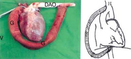
The pulmonary arterial pressure and cardiac output were monitored every 30 min with a Swan-Ganz catheter passed through the right internal jugular vein. Before opening the sternum, continuous arterial pressure monitoring was established through the right femoral artery, and through the left internal thoracic artery after sternotomy. The arterial pressure was continuously monitored and maintained at a mean level of 70 mmHg. To access the mediastinal structures, a standard median sternotomy was performed from the sternal notch to the xyphoid process level. For simultaneous exposure of the descending thoracic aorta, an L shaped lateral extension was made leftward from the distal tip of the sternotomy. Two individual vascular conduits of internal diameter 21 mm and length 30 cm were used to construct the composite valved conduit. The prosthetic valve, a 17 mm St. Jude HPJ 505 mechanical aortic heart valve (St. Jude Medical Europe, Brussels, Belgium), was sutured to the distal end of the conduit with continuous running suture of polypropylene #4-0. A 0.5 cm curtain of the vascular graft material was left distal to the prosthetic valve implantation site. The second graft conduit was sutured in an end to end fashion with #4-0 polypropylene continuous running suture, to the curtain of graft material distal to the prosthetic graft (Fig. 1). Basically, the vascular conduit was selected to closely match that of the prosthetic valve being tested in size. The conduit was sufficiently large to accommodate the slight variations in the external prosthetic valvular diameter that is seen among different manufacturers for any given valve size. Heparin 100 units/kg was administered intravenously for anticoagulation. A 21 Fr pediatric arterial perfusion cannula was connected to each end of a 3/8 inch diameter silicone tubing of 1 meter in length to construct an aorta to femoral artery bypass perfusion shunt. After separating the main pulmonary artery and the ascending aorta a braided nylon umbilical tape was passed under the ascending aorta with the aid of a right-angled Kelly. A single purse-string suture was created just distal to the non-coronary sinus with a braided multifilament #1-0 suture on the posterior aspect of the ascending aorta by pulling on the umbilical tape. The proximal femoral artery was then exposed for distal cannulation of the shunt. When the activated coagulation time exceeded 200 sec, the ascending aorta was shunted to the femoral artery with the silicone shunt, as described above. After ensuring good functioning of the aorta to femoral artery shunt, the ascending aorta was partially clamped with a side-biting Satinsky clamp just distal to the aorto-femoral shunt. The proximal end of the composite graft was anastomosed to the side clamped portion of the ascending aorta in an end- to-side fashion with a #4-0 polypropylene running suture. Due to the short length of the ascending aorta, clamping of the innominate artery together with the ascending aorta with the side-biting clamp was unavoidable. Consequently, the innominate artery was partially included in the proximal anastomosis between the ascending aorta and the composite valved conduit. The distal end of the composite valved conduit was anastomosed in and end-to side manner to the distal descending aorta at the diaphragmatic crux level with a continuous running suture of 4-0 polypropylene. Hemodynamic assessment of the native aortic valve was performed before cross clamping the proximal descending thoracic aorta, whereas assessment of the prosthetic aortic valve was performed after cross clamping the proximal descending thoracic aorta. Arterial pressure was monitored directly through the internal mammary and the femoral arteries as described above. The purpose of monitoring through the internal mammary artery was to check the arterial pressure during the cross clamping of the proximal descending thoracic aorta.
Fig. 1.
Photograph and schematic representation of the ascending aorta to desending thoracic aortic bypass with the composite valved conduit which was constructed intraoperatively.
DAO, Descending aorta; V, Position of valve; G, Graft.
Hemodynamic assessment
Echocardiography was performed using a 7.5-MHz transducer probe connected to a Vingmed CFM 725 ultrasound machine. An air free interface was created by interposing a thoroughly de-aired ultrasound gel-filled sterile latex glove between the probe and the vascular surface. Valvular function was assessed by color Doppler and by continuous or pulsedwave Doppler. Transvalvular pressure gradients were calculated from the maximal flow velocity across the valve as determined by continuous-wave Doppler. The conduit (native or graft) diameter, cardiac output (CO), ejection fraction (EF), time velocity integral of pulsed wave Doppler (PWTVI), time velocity integral of continuous wave Doppler (CWTVI), left ventricular end diastolic dimension (LVEDD), left ventricular end systolic dimension (LVESD), stroke volume (SV), and effective orifice area (EOA) were calculated. Preoperative baseline cardiac output prior to median sternotomy was obtained by using the thermodilution technique with the Swan Ganz Catheter. Subsequent cardiac output measurements through both the native aortic valve and the graft were calculated directly by echocardiography using the 2D Doppler technique. The time velocity integral of forward systolic flow was determined by pulsed Doppler at the left ventricular outflow tract. This value was then multiplied by the cross sectional area to obtain the stroke volume. PWTVI or CTVI, measures of flow velocity, were used to calculate the cardiac out-put according to the following formula;
Flow (Q)=Area (A)×velocity (V)
Cardiac output (CO)=Q×Heart rate/min
Echocardiographic data were collected preoperatively, postoperatively before drug infusion (control) and postoperatively after the infusion of dobutamine at 10 and 20 µg/kg/min.
Statistical analysis
All data are expressed as means±standard deviation (SD). The statistical package, SAS 6.12 (SAS Institute Inc., Cary North Carolina, U.S.A.) was used to perform the analyses. Echo data were compared using the Wilcoxon signed rank test between the native aortic valve and prosthetic valve. Mixed analysis was used to detect significant changes in dobutamine stress test results. A p value of less than 0.05 was considered statistically significant.
RESULTS
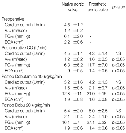
The results of the hemodynamic evaluation are summarized in Table 1. Measurements of the heart rate, cardiac output, transvalvular Vmax (m2/sec), peak gradient (PG)max (mmHg), and effective orifice area (EOA cm2) are shown. No significant differences were detected in, cardiac output, heart rate, and measured flow through the native aortic and prosthetic valves (p=ns). The transvalvular pressure gradient across the prosthetic valve was documented when it was noted that the difference in the calculated cardiac output between the native aortic annulus and the graft was negligible. The cardiac output and the heart rate increased equally in both the native and the prosthetic valves in a dobutamine dose dependent manner.
Table 1.
Postoperative echocardiographic data of aortic valve and new heart valve
Vmax, maximal velocity; PGmax, maximum pressure gradient; EOA, effective orifice area.
As the prosthetic valvular diameter was smaller than that of the native aortic valve, the mean EOA was also smaller (p<0.05). Accordingly, Vmax and PG in the resting state were significantly higher in the composite valved conduit than in the native aortic valve (p<0.05), and this difference was accentuated by incremental dobutamine infusion. Dobutamine in excess of 10 µg/kg/min significantly increased Vmax and PG versus the resting state in both native and prosthetic valves. Further increase in the dobutamine dosage to 20 µg/kg/min produced a further increase in Vmax and PG in the prosthetic valve, but not in the native aortic valve.
DISCUSSION
One of the prerequisites of developing a new cardiac valvular prosthesis is a successful pre-clinical large animal series. An appropriate animal model may provide information on valve performance, safety, and in vivo hemodynamics (1, 5). However, no suitable animal model is available that can yield such information for a prosthetic aortic valve. Many animal models have been used with variable results. These include sheep, goats, dogs, pigs, calves and primates (6). However, due to the unique limitations of each preclinical model, it is imperative to develop an appropriate animal model that best suits the purposes of the experiment at hand (7). The physical size and the rapid growth rate of calves present problems for postoperative husbandry and non-invasive valvular functional assessment (8, 9). Goats have been found to be a good model for valve testing, but their availability is often limited (10). The relative susceptibility of dogs to sepsis and thrombosis has marked these animals as less than optimal (11). Sheep (Ovis aris) are widely used for testing prosthetic cardiac valves. They have many favorable characteristics for prosthetic valve testing, as their blood pressures, heart rates, cardiac outputs and intra-cardiac pressures are similar to humans (12). Standard cardiopulmonary bypass techniques and anesthesia with minor modifications are possible in these animals without difficulty (7). However, the sheep is costly and not readily available in Korea. In addition, the short length of the ascending aorta, which is a common feature of cloven-footed animals (calves, sheep, and goats), gives way to the aortic bifurcation a short way downstream of the aortic valve. Consequently, prosthetic implantation directly into the aortic annulus is technically challenging (13, 14). The pig is more advantageous in terms of cost and availability, but it too shares the limitations of cloven footed animals of having a short ascending aortic trunk, a tendency to develop cardiopulmonary bypass complications, and presenting difficulties in terms of accessing the peripheral venous circulation, management and handling (7, 13, 15-17). Nevertheless, numerous reports on mechanical heart valves have been based on data derived from the pig (2, 13, 18, 19). After a careful deliberation of various animal studies, we selected the pig for the current study.
Thus we undertook the current study to develop a cost efficient, reliable animal model that allows a relatively simple site specific hemodynamic assessment of mechanical prosthetic devices in the arterial environment.
During the early part of the current experiment, several animals died of arrhythmias varying from atrial fibrillation with intractable rapid ventricular response to frank ventricular fibrillation. These animals were excluded from the current data and only data from those animals (n=10) with the final model were assessed. Ventricular fibrillation developed in three animals immediately after mechanical ventilator application. These animals did not receive any cardiac manipulation. In another animal, tension pneumothorax developed during tracheostomy after which ventricular fibrillation occurred. In yet another animal, ventricular fibrillation occurred after partial ascending aortic clamping. It is speculated that a sudden increase in ventricular afterload resulting from the cross clamp caused the ventricular fibrillation. The side-biting clamp may understandably produce supravalvular aortic stenosis in an unpredictable manner with variable acute after-load augmentation. Intolerable levels of this sudden unprotected afterload increase may have been the cause of death. Subsequently, to relieve the afterload build up, a temporary bypass shunting of the proximal ascending aorta, proximal to the cross clamp to the femoral artery, was performed. The proximal decompression afforded by this temporary shunt proved to be a key modification, and one that led to the success of the subsequent trials. The use of an artery to artery shunt as a means of partially bypassing the aorta with a heparin coated shunt is not a novel concept. Aorto-femoral shunting for thoracic aortic and great vessel procedures was described by Gott and colleagues (20), and the feasibility of implanting an aortic valvular device in the descending thoracic aorta to treat left ventricular outflow tract obstruction with an apicoaortic conduit was described by Norman et al. (21). The concepts underlying these ideas led to the final design of the current experimental model, which involved the use of a composite valved conduit to bypass the aorta from the proximal aorta to the distal aorta with the aid of an aorto-femoral bypass shunt.
With the current model, a site-specific functional evaluation of valvular prostheses without cardiopulmonary bypass in the high pressure arterial environment is possible Furthermore, the current model has the potential for successive testing of different valve unit types in a single animal in a cost efficient manner, i.e., one valve can simply be replaced by another between the two grafts. However, despite these purported advantages, the hemodynamic data must accurately reflect the actual hemodynamic performance of the valvular prosthesis being tested. The prosthetic valve that was used in this particular experiment had a small and fixed orifice, the latter of which is a common trait of mechanical prostheses. Consequently, a steeper Vmax and pressure gradient increase were observed in the prosthetic valve as compared to the native valve. This was especially evident with dobutamine increment (Table 1). With the native aortic valve serving as the control in each animal, direct hemodynamic assessments and comparisons with the native aortic valve were possible, as the cardiac output and heart rate remained constant when the respective hemodynamic data were being obtained from each site. Although some obligatory sharing or diversion of blood flow to the arch vessel during the hemodynamic evaluation of the composite valved conduit after proximal thoracic aortic cross clamping was inevitable, the differences in the measured cardiac output and flow between the native aortic valve and the valved conduit became insignificant once equilibrium had been reached between the arch vessel and the valved conduit. Therefore, it is our contention that the current model is suitable as a site-specific model for the hemodynamic assessment of valvular prostheses in the aortic position.
In terms of limitations, although some sharing of blood flow with the arch vessels during distal aortic clamping was unavoidable during the hemodynamic assessment of the prosthetic valve, the stated objectives of the study were fully satisfied as the differences in the cardiac output between the native valve as compared with the prosthetic valve were insignificant. Therefore, conclusions derived from the direct hemodynamic comparison of the prosthetic and native aortic valves in the current animal study are believed to be valid. The question of whether the current model should be regarded as being site specific is open to debate as the valvular device was actually implanted in a site other than the actual aortic annulus. However, it is our contention that functionally, this model is site specific, as the prosthetic aortic valve is continually subjected to the stressors of the high pressure systemic arterial system as is the annulus of the native aortic root. Issues regarding durability, thrombogenicity, calcification and other long term modes of valve failure are beyond the scope of the current model, as these parameters should be persued through a chronic animal series. Nevertheless, the current model proved useful at acquiring acute hemodynamic data relating to valve function.
In conclusion, the current model was found suitable for acquiring essential information on the hemodynamic performance of a valvular prosthesis, and cost and time efficient by obviating the cardiopulmonary bypass and by offering the potential for multiple valve testing in a single large animal.
Footnotes
This work was supported by The Korea Research Foundation Grant (KRF-99-042-F00127-F3300).
References
- 1.Schoen FJ, Levy RJ, Hilbert SL, Bianco RW. Antimineralization treatments for bioprosthetic heart valves: Assessment of efficacy and safety. J Thorac Cardiovasc Surg. 1992;104:1285–1288. [PubMed] [Google Scholar]
- 2.Gross DR, Dewanjee MK, Zhai P, Lanzo S, Wu SM. Successful prosthetic mitral valve implantation in pigs. ASAIO J. 1997;43:M382–M386. [PubMed] [Google Scholar]
- 3.Salerno CT, Pederson TS, Ouyang DW, Bolman RM, III, Bianco RW. Chronic evaluation of orthotopically implanted bileaflet mechanical aortic valves in adult domestic sheep. J Invest Surg. 1998;11:341–347. doi: 10.3109/08941939809032210. [DOI] [PubMed] [Google Scholar]
- 4.Chanda J, Kuribayashi R, Abe T. Valved conduit in the descending thoracic aorta in juvenile sheep: a useful, cost-effective model for accelerated calcification study in systemic circulation. Biomaterials. 1997;18:1317–1321. doi: 10.1016/s0142-9612(97)00065-3. [DOI] [PubMed] [Google Scholar]
- 5.Ouyang DW, Salerno CT, Pederson TS, Boldman RM, III, Bianco RW. Long-term evaluation of orthotopically implanted stentless bioprosthetic aortic valves in juvenile sheep. J Invest Surg. 1998;11:175–183. doi: 10.3109/08941939809098032. [DOI] [PubMed] [Google Scholar]
- 6.Rakow N, Nelson D, Boldt C, Pfenning K, McClay C, Waskiewicz J, Schecterle L, St Cyr J. Chronic orthotopic aortic valve implantation model. J Heart Valve Dis. 2000;9:822–827. [PubMed] [Google Scholar]
- 7.Salerno CT, Droel J, Bianco RW. Current state of in vivo preclinical heart valve evaluation. J Heart Valve Dis. 1998;7:158–162. [PubMed] [Google Scholar]
- 8.Braunwald NS, Bonchek LI. Prevention of thrombus formation on rigid prosthetic heart valves by the ingrowth of autogenous tissue. J Thorac Cardiovasc Surg. 1967;54:630–638. [PubMed] [Google Scholar]
- 9.Gallo I, Frater RWM. Experimental atrioventricular bioprosthetic valve insertion: A simple and successful technique. Thorac Cardiovasc Surg. 1983;31:288–290. doi: 10.1055/s-2007-1021998. [DOI] [PubMed] [Google Scholar]
- 10.Bjork VO, Sternlieb J. Artificial heart valve testing in goats. Scand J Thorac Cardiovasc Surg. 1986;20:97–102. doi: 10.3109/14017438609106483. [DOI] [PubMed] [Google Scholar]
- 11.Jones RD, Cross FS, Akao M. Bacteremia and thrombus on prosthetic heart valves in dogs. Circulation. 1969;39(Suppl I):1253–1256. doi: 10.1161/01.cir.39.5s1.i-253. [DOI] [PubMed] [Google Scholar]
- 12.Barnhart GR, Jones M, Ishihara T, Chavez AM, Rose DM, Ferrans VJ. Bioprosthetic valvular failure. Clinical and pathological observations in an experimental animal model. J Thorac Cardiovasc Surg. 1982;83:618–631. [PubMed] [Google Scholar]
- 13.Hasenkam JM, Ostergaard JH, Pedersen EM, Paulsen PK, Nygaard H, Schurizek BA, Johannsen G. A model for acute haemodynamic studies in the ascending aorta in pigs. Cardiovasc Res. 1988;22:464–471. doi: 10.1093/cvr/22.7.464. [DOI] [PubMed] [Google Scholar]
- 14.Hasenkam JM, Pedersen EM, Ostergaard JH, Nygaard H, Paulsen PK, Johannsen G, Schurizek BA. Velocity fields and turbulent stresses downstream of biological and mechanical aortic valve prostheses implanted in pigs. Cardiovasc Res. 1988;22:472–483. doi: 10.1093/cvr/22.7.472. [DOI] [PubMed] [Google Scholar]
- 15.Irwin E, Lang G, Clack R, St Cyr J, Runge W, Foker J, Bianco R. Long-term evaluation of prosthetic mitral valves in sheep. J Invest Surg. 1993;6:133–141. doi: 10.3109/08941939309141604. [DOI] [PubMed] [Google Scholar]
- 16.Swan H, Piermattei DL. Technical aspects of cardiac transplantation in the pig. J Thorac Cardiovasc Surg. 1971;61:710–723. [PubMed] [Google Scholar]
- 17.Jones RD, Dreher DM, Cross FS. The dog as a model for evaluating prosthetic heart valves. Ann Thorac Surg. 1989;48:S4–S5. doi: 10.1016/0003-4975(89)90616-4. [DOI] [PubMed] [Google Scholar]
- 18.Hasenkam JM, Nygaard H, Terp K, Riis C, Paulsen PK. Hemodynamic evaluation of a new bileaflet valve prosthesis: an acute animal experimental study. J Heart Valve Dis. 1996;5:574–580. [PubMed] [Google Scholar]
- 19.Grehan JF, Hilbert SL, Ferrans VJ, Droel JS, Salerno CT, Bianco RW. Development and evaluation of a swine model to assess the preclinical safety of mechanical heart valves. J Heart Valve Dis. 2000;9:710–720. [PubMed] [Google Scholar]
- 20.Donahoo JS, Brawley RK, Gott VL. The heparin-coated vascular shunt for thoracic aortic and great vessel procedures: a ten-year experience. Ann Thorac Surg. 1977;23:507–513. doi: 10.1016/s0003-4975(10)63692-2. [DOI] [PubMed] [Google Scholar]
- 21.Norman JC, Nihill MR, Cooley DA. Valved apico-aortic composite conduits for left ventricular outflow tract obstructions. A 4 year experience with 27 patients. Am J Cardiol. 1980;45:1265–1267. doi: 10.1016/0002-9149(80)90488-9. [DOI] [PubMed] [Google Scholar]


