Abstract
An approximately 9-month-old fox (Pseudalopex vetulus) was presented with malocclusion and deviation of the lower jaw to the right side. Orthodontic treatment was performed using the inclined plane technique. Virtual 3D models and prototypes of the head were based on computed tomography (CT) image data to assist in diagnosis and treatment.
Résumé
Prototypage rapide et technique de plan incliné dans le traitement des malformations maxillofaciales chez un renard. Un renard (Pseudalopex vetulus) âgé d’environ 9 mois a été présenté avec une malocclusion et une déviation de la mâchoire inférieure du côté droit. Un traitement d’orthodontie a été réalisé en utilisant la technique du plan incliné. Des modèles tridimensionnels virtuels et des prototypes de la tête se sont fondés sur des données de tomographie par reconstruction d’image afin de faciliter le diagnostic et le traitement.
(Traduit par Isabelle Vallières)
An approximately 9-month-old free-ranging 3.7-kg male fox (Pseudalopex vetulus) was presented to the Veterinary Hospital with malocclusion and deviation of the lower jaw to the right side. The animal was anesthetized and oral examination and computed tomography (CT) studies were performed.
Case description
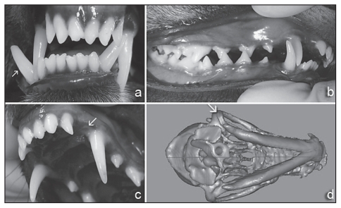
After premedication with acepromazine (Acepran 0.2%; Univet, São Paulo, Brazil) 0.05 mg/kg body weight (BW), IM, and meperidine (Dolosal; Cristália, Itapira, Brazil) 2 mg/kg BW, IM, anesthesia was induced with propofol (Propovan; Cristália, Itapira, Brazil) 2 mg/kg BW, IV, and maintained with isoflurane at 1.5% (Isoforine; Cristália, Itapira, Brazil) and oxygen at 1000 mL/min. Oral examination revealed a posterior crossbite involving the right mandibular 2nd premolar, right mandibular 3rd premolar, and right mandibular 4th premolar teeth with the right mandibular canine tooth immediately distal to the right maxillary canine tooth and tipped in a buccal direction (Figure 1a). The malocclusion caused the left mandibular canine tooth to impinge on the palate between the left maxillary 3rd incisor and canine teeth (Figures 1b and 1c).
Figure 1.
Photograph of the oral cavity of a 9-month-old fox showing distocclusion (Class II malocclusion) on the right side (posterior cross-bite) (a), palate lesion (arrow) caused by the left mandibular canine tooth (b), and right mandibular canine tooth tipped in a buccal direction (arrow) (c). Virtual 3D model (Magics X SP2) of the head based on CT image data showing malformation of the right condyle (arrow) (d).
Sequential transverse images of the head were acquired on a helical CT Scanner (Shimadzu SCT-7800CT) with the fox placed in dorsal recumbency. The scanning parameters were 120 kVp, 170 mA, 1.0 mm slice thickness, 1.0 mm interval, pitch of 2.0, and 1 s/rotation. A virtual 3D model (InVesalius software) of the head was generated based on CT image data. The InVesalius software was also used to edit and measure the original 3D virtual model obtained directly from the CT scan, and the Magics X SP2 was used to generate the final model to the prototype machine. The virtual 3D model of the head showed the right mandible to be shorter than the left mandible, malformation of the right condyle, and facial and mandibular asymmetry (Figure 1d). The physical prototype constructed by the 3DP (Z-Corp) technique enabled the preoperative planning.
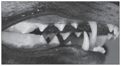
The orthodontic treatment goal was to correct the cross-bite and avoid palate injury, while preventing an increase in the maxillofacial discrepancy by applying the inclined plane technique. Acid etching was performed on the distal surface of the 3rd mandibular premolars and the teeth caudal to them. The etching gel (Acigel; SS White Artigos Dentários, Rio de Janeiro, Brazil) was rinsed off the teeth with water, followed by air drying of the tooth surfaces. Immediately after, glass ionomer (Vidrion; SS White Artigos Dentários, Rio de Janeiro, Brazil) and self-curing acrylic resin (Jet acrílico; Artigos Odontológicos Clássico, São Paulo, Brazil), were applied to the occlusal and vestibular faces of the maxillary and mandibular teeth, in the caudal region of the 3rd premolar tooth, forming a plane with a dorsomedial to ventrolateral inclination (Figure 2). The inclined plane was shaped with a diamond covered dental bur on a high-speed handpiece under water irrigation and then polished with a rubber cup and polishing paste on a low-speed handpiece.
Figure 2.
An inclined plane applied to the occlusal and vestibular faces of maxillary and mandibular teeth, in the caudal region of the 3rd premolar tooth, forming a plane with a dorsomedial to ventrolateral inclination.
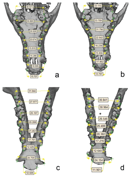
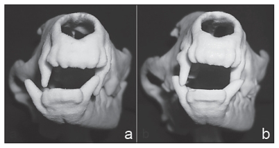
The fox adapted well to the treatment showing signs of discomfort only in the first 48 h. Evaluation of treatment was conducted on a monthly basis, with status changes registered by digital photography. The inclined plane was removed after 3 mo, and a new CT study was carried out as described previously. Stage I of periodontal disease was observed and was treated by complete dental cleaning. Visualization of 3D images in combination with rapid prototyping allowed execution of 3 sets of geometric measurements in the virtual model (Figures 3 and 4). The 3D models of both the maxilla and lower jaw were used to perform the measurements of distance between the same right and left tooth before inclined plane treatment and 3 mo after its application (Tables 1 and 2). The references were placed at the highest cuspid of each tooth (Figure 3). Comparison of the 3D models and prototypes confirmed improvement of facial and mandibular symmetry as well as occlusion after treatment (Figure 4). Furthermore, oral examination showed improvement of the occlusion, healing of the palate injury, and that the left mandibular canine tooth was not making contact with the palatal tissues while the inclined plane was in place. At this time, the fox was released into the wild.
Figure 3.
Maxilla 3D (a, b) and mandible 3D (c, d) models showing the measurements of distances between the same right and left tooth before inclined plane treatment (a, c) and 3 months after its application (b, d).
Figure 4.
Prototype models of the CT obtained before the inclined plane treatment (a) and 3 months after its application (b). Observe the improvement of the occlusion after treatment.
Table 1.
Measurementa (mm) between teeth of the maxilla before inclined plane treatment and 3 months after its application
| Teeth | Before treatment | After treatment | Variation (%) |
|---|---|---|---|
| Right maxillary 2nd molar to left maxillary 2nd molar | 30.572 | 33.115 | 8.32 |
| Right maxillary 1st molar to left maxillary 1st molar | 30.869 | 33.709 | 9.20 |
| Right maxillary 4th premolar to left maxillary 4th premolar | 29.928 | 31.709 | 5.95 |
| Right maxillary 3rd premolar to left maxillary 3rd premolar | 20.674 | 22.642 | 9.51 |
| Right maxillary 2nd premolar to left maxillary 2nd premolar | 15.810 | 16.238 | 2.70 |
| Right maxillary 1st premolar to left maxillary 1st premolar | 15.288 | 15.656 | 2.40 |
| Right maxillary canine to left maxillary canine | 18.595 | 17.862 | −3.94 |
| Right maxillary 3rd incisor to left maxillary 3rd incisor | 15.707 | 13.797 | −12.16 |
The distances are the means of 3 measurements.
Table 2.
Measurementsa (mm) between teeth of the mandible before inclined plane treatment and 3 months after its application
| Teeth | Before treatment | After treatment | Variation (%) |
|---|---|---|---|
| Right mandibular 3rd molar to left mandibular 3rd molar | 31.044 | 30.841 | −0.65 |
| Right mandibular 2nd molar to left mandibular 2nd molar | 27.977 | 30.964 | 10.67 |
| Right mandibular 1st molar to left mandibular 1st molar | 25.107 | 26.546 | 5.73 |
| Right mandibular 4th premolar to left mandibular 4th premolar | 25.208 | 26.488 | 5.08 |
| Right mandibular 3rd premolar to left mandibular 3rd premolar | 21.463 | 20.382 | −5.04 |
| Right mandibular 2nd premolar to left mandibular 2nd premolar | 17.761 | 17.216 | −3.07 |
| Right mandibular first premolar to left mandibular first premolar | 12.426 | 12.640 | 1.72 |
| Right mandibular canine to left mandibular canine | 24.759 | 24.154 | −2.44 |
| Right mandibular 3rd incisor to left mandibular 3rd incisor | 12.026 | 11.901 | −1.03 |
The distances are the means of 3 measurements.
Discussion
Abnormally positioned teeth may touch soft tissue structures, as observed in the present case, causing pain, discomfort, and trauma, while predisposing to periodontal disease (1). A malocclusion could be of genetic origin or caused by previous trauma (2). Environment, nutrition, dietary texture, stress, inflammation, injury, and infection may also contribute to the development of the abnormality (3). In the present case the cause of the malocclusion was unknown.
Angle’s classification system, based on tooth relationships used in human dentistry, has been adapted to categorize veterinary dental occlusion and malocclusion (4). There are 5 classes of occlusion or malocclusion: Class 0 (normal), Class I (neutroclusion), Class II (distoclusion), Class III (mesioclusion), and Class IV (mesiodistoclusion) (4,5). A Class II distoclusion exists when there is either a short mandible or an elongated maxilla as well as a possible wry bite (5). The present case was presented with a wry bite, meaning the right mandible was shorter than the left mandible. This suggested a unilateral distocclusion on the right side. Early treatment for class II malocclusion is frequently undertaken with the objective of correcting the skeletal disproportion by altering the growth pattern (6).
Among the alternatives to treatment of Class II malocclusion are selective tooth extraction, crown reduction with vital pulp therapy, and other types of active or passive orthodontic movement (5). Extraction was not considered in this case because the animal was to be released into the wild after treatment and would need to prey and defend itself. Crown reduction with vital pulp therapy may have been problematic as the vital pulp therapy could fail thus inducing pulpitis, pulp necrosis, and periapical diseases (4,5). Passive orthodontic movement with application of an inclined plane to the maxillary canines and incisors with appropriate inclinations is usually used in small animals (4). The aim in the present case, however, was to move the lower jaw using the Planas’ direct tracks to correct the distoclusion (7) and not exclusively the lower canine teeth.
The application of the Planas’ direct tracks concept represents an interesting tool for the correction of malocclusion when treatment is initiated at an early age (8). Planas’ direct tracks, named after Pedro Planas who conceived them, are called direct tracks because a resin is applied directly on the teeth (9). For a posterior crossbite, the inclined plane is contoured in a dorsomedial to ventrolateral direction on the lower deciduous molars of the crossbite side only (10). When perfectly applied, the “neuro-occlusal rehabilitation,” according to Planas, permits restoration of a functional balance for the masticatory apparatus at an early age, and subsequently reorients growth to a morphological normalization (11).
In the present case, the application of the inclined plane was performed on an animal that was young, but already had its permanent dentition. Computed tomography studies performed after the application of the orthopedic treatment, however, showed that Planas’ technique had achieved satisfactory results while modifying the skeletal structure and thus stimulating the development of the stomatognathic system. The friction process is physiological in most mammals, but it varies according to its intensity, velocity, and area of action, producing cutting edges (9). The high rate of skeletal changes, especially with growth of maxillary bone over a period of 3 mo, may have been related to the carnivorous diet, the growth stage of the fox, and the successful feeding with living prey.
The biophysical model (prototype), derived from the initial CT examination, aided treatment planning by allowing study of the region for application of the inclined plane. Additionally, comparison of the rapid prototyping biomodels before and after treatment showed a qualitative improvement of the occlusion. The CT images can be converted to 3D virtual objects with the aid of specially developed medical imaging reconstruction software programs. These programs are commercially available but can also be developed by research teams. The software used in the present case (InVesalius), developed by CenPRA, Brazil, can generate output directly for rapid prototyping. It is distributed free-of-charge and undergoes continuous development by the CenPRA research team (12). The software can reconstruct 3D virtual models from CT imaging modality based on the DICOM (Digital Imaging and Communication in Medicine) file format. This type of file is standardized to provide communication of patient information and medical imaging between physicians and it is commonly used in medical imaging examinations (13). The rapid prototyping output of the InVesalius is an industrial standard file format called STL (STereoLithography). The information contained in this file type is a surface representation of the 3D object through a mesh of triangles, described by the coordinates of their vertices (14). One of the difficulties faced while making the measurements with this type of file in the present case was in placing references in the images before and after treatment. This results from only being able to place the cursor onto the vertices of each very small triangle available from the STL mesh. To reduce this bias, the models were measured 3 times and the average value of each model was used.
In conclusion, the 3D models and the rapid prototyping opened up new possibilities for treatment planning. The inclined plane technique is a valuable alternative over selective tooth extraction or crown reduction with vital pulp therapy in the treatment of malocclusion. CVJ
Footnotes
Use of this article is limited to a single copy for personal study. Anyone interested in obtaining reprints should contact the CVMA office ( hbroughton@cvma-acmv.org) for additional copies or permission to use this material elsewhere.
References
- 1.Bellows J, editor. Small Animal Dental Equipment, Materials and Techniques. Ames, Iowa: Blackwell Publ; 2004. Orthodontic equipment, materials, and techniques; pp. 269–270. [Google Scholar]
- 2.Tholen M. Orthodontic. In: Bojrab MJ, editor. Small Animal Oral Medicine and Surgery. Philadelphia: Lea & Febiger; 1990. pp. 232–240. [Google Scholar]
- 3.Wiggs RB, Bloom BC. Exotic placental carnivore dentistry. Vet Clin North Am Exot Anim Pract. 2003;9:571–599. doi: 10.1016/s1094-9194(03)00038-0. [DOI] [PubMed] [Google Scholar]
- 4.Harvey CE, Emily PP, editors. Small Animal Dentistry. St. Louis, Missouri: Mosby; 1993. Occlusion, occlusive abnormalities, and orthodontic treatment; pp. 266–296. [Google Scholar]
- 5.Wiggs RB, Lobprise HB, editors. Veterinary Dentistry: Principles and Practice. Philadelphia: Lippincott-Raven; 1997. pp. 435–481. [Google Scholar]
- 6.Tulloch JF, Phillips C, Koch G, Proffit WR. The effect of early intervention on skeletal pattern in Class II malocclusion: A randomized clinical trial. Am J Orthod Dentofacial Orthop. 1997;111:391–400. doi: 10.1016/s0889-5406(97)80021-2. [DOI] [PubMed] [Google Scholar]
- 7.Moorrees CF. Thoughts on the early treatment of Class II malocclusion. Clin Orthod Res. 1998;1:97–101. doi: 10.1111/ocr.1998.1.2.97. [DOI] [PubMed] [Google Scholar]
- 8.Gribel MN, Gribel BF. Planas direct tracks in young patients with Class II malocclusion. World J Orthod. 2005;6:355–368. [PubMed] [Google Scholar]
- 9.Simões WA. Selective grinding and Planas’direct tracks as a source of prevention. J Pedod. 1981;5:298–314. [PubMed] [Google Scholar]
- 10.Ramirez-Yañes GO. Planas direct tracks for early cross-bitecorrection. J Clin Orthod. 2003;37:294–298. [PubMed] [Google Scholar]
- 11.Limme M. Interception in the primary dentition: Mastication and neuroocclusal rehabilitation. Orthod Fr. 2006;77:113–135. doi: 10.1051/orthodfr/200677113. [DOI] [PubMed] [Google Scholar]
- 12.Centro de Pesquisa Renato Archer (CenPRA) InVesalius Software for Medice Imaging Reconstruction [homepage on the Internet] Campinas: CenPRA; c2008. [Last accessed January 6, 2010]. Available from http://www.softwarepublico.gov.br/ [Google Scholar]
- 13.Caritá EC, Matos ALM, Azevedo-Marques PM. Image viewing tools used in a medical school. Radiol Bras. 2004;37:437–440. [Google Scholar]
- 14.Souza MA, Ricetti F, Centeno TM, Pedrini H, Erthal JL, Mehl A. Reconstrução de imagens tomográficas aplicada à fabricação de próteses por prototipagem rápida usando técnicas de triangulação. Proc Cong Latinoam Ingen Bioméd. 2001:1–5. [Google Scholar]


