to the editor: My attention has been drawn to an article titled “The looped heart does not save energy by maintaining the momentum of blood flowing in the ventricle” (4). The study questions previously postulated functional implications of cardiac looping (2).
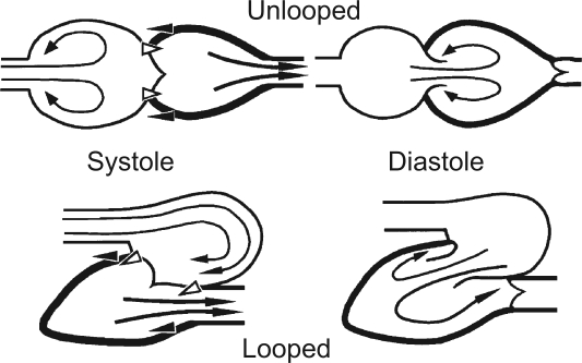
Watanabe et al. (4) used finite element numerical modeling to compare the hemodynamics of virtual left ventricles rather than whole hearts, with physiological and nonphysiological intraventricular flow paths. The differences of flow paths were induced by changes in the direction of inflow, tangential to the cavity in each case, but inclined either toward or away from the lateral wall. The geometries of the ventricles did not differ. Neither of them represented the ventricle of an unlooped heart if, as illustrated in Fig. 1, that were interpreted to mean a heart with an atrium, a ventricle, and an aorta lying in approximate alignment without a change of direction between ventricular inflow and outflow. In that sense, all hearts found in humans, and possibly all found in mature vertebrates, are looped, even in those with congenital malformations. A degree of direction change at the ventricular level and the inclusion of an atrial as well as a ventricular cavity within a pericardium seem to be fundamental features of the hearts of vertebrates.
Fig. 1.
Schematic illustration of the interactions between atrium, ventricle, and blood in a linearly arranged heart (top) contrasted with those of a looped heart (bottom), as described in the text (reprinted from Nature; see Ref. 2).
Watanabe et al. (4) reasonably refuted the concept of conservation of inflow momentum, which prompts me to reformulate the postulated functional advantages of cardiac looping that were put forward in relation to magnetic resonance studies of human intracardiac flow (2) (see Fig. 1).
Three factors are illustrated schematically in Fig. 1 [republished with permission from Nature (2)]. First, the tendency in a looped heart for inflows to be aligned tangential to a cavity predisposes to more stable intracavity flow. Second, the recirculation of inflowing streams in the atrial and ventricular cavities of a looped heart then tends to turn predominantly toward rather than away from the next cavity, which could facilitate an efficient onward passage (given the time relations at peak exercise, at least). Third, in a looped heart, the appropriate direction of the inertial recoil of a vigorously ejecting ventricle (black arrowheads pointing away from the direction of the acceleration of blood into the outflow tract) can enhance rather than suppress the structural coupling (indicated by the white arrowheads) of ventricular systole to atrial filling.
All three of these factors, even if trivial at rest, are likely to gain significance during strenuous exertion, a state likely to have been relevant to vertebrate survival through the course of evolution, but not necessarily so important to patients with cardiovascular disease. The third factor, concerning ventriculo-atrial coupling, is probably the most potent of the three. Of note, the velocities, volumes, and time courses of inflowing and outflowing streams, and hence the rates of change of momentum and the associated directional exchanges of force, change greatly from rest to strenuous exercise in humans. The heart rate of a physically fit young adult can increase more than threefold with exertion, and the cardiac output of an endurance-trained athlete, more than sixfold (3). The two peaks of early and late left ventricular filling of the resting heart become one during exertion, when a single peak of rapid ventricular filling is followed by vigorous ejection with little or no intervening isovolumetric period (1). In this state, the minimization of the dissipation of the kinetic energy of inflow would be functionally advantageous, allowing efficient transfers of energy between inflowing blood, myocardium, outflowing blood, and arterial walls. Even on exercise, only part of the momentum of blood flowing into a chamber is likely to be conserved and redirected. There will also be an initial deceleration and decline of momentum as streamlines diverge, with an associated force field that may contribute to the expansion of the cavity, priming the myocardium for its subsequent contraction.
GRANT
P. J. Kilner is supported by the British Heart Foundation.
DISCLOSURES
No conflicts of interest are declared by the author.
REFERENCES
- 1.Kilner PJ, Henein MY, Gibson DG. Our tortuous heart in dynamic mode—an echocardiographic study of mitral flow and movement in exercising subjects. Heart Vessels 12: 103–110, 1997 [DOI] [PubMed] [Google Scholar]
- 2.Kilner PJ, Yang GZ, Wilkes AJ, Mohiaddin RH, Firmin DN, Yacoub MH. Asymmetric redirection of flow through the heart. Nature 404: 759–761, 2000 [DOI] [PubMed] [Google Scholar]
- 3.Warburton DE, Gledhill N, Jamnik VK. Reproducibility of the acetylene rebreathe technique for determining cardiac output. Med Sci Sports Exerc 30: 952–957, 1998 [DOI] [PubMed] [Google Scholar]
- 4.Watanabe H, Sugiura S, Hisada T. The looped heart does not save energy by maintaining the momentum of blood flowing in the ventricle. Am J Physiol Heart Circ Physiol 294: H2191–H2196, 2008 [DOI] [PubMed] [Google Scholar]


