Abstract
Esophageal leiomyoma derived from the muscularis mucosae (MM) is a rare condition, and the optimal modality for diagnosis and treatment is controversial. Endoscopic ultrasonography can provide an accurate image of esophageal layer structure, providing information on lesion suitability for potential endoscopic therapy. We attempted to investigate the diagnostic value of a transendoscopic balloon-tipped miniature ultrasonic endoprobe for small esophageal leiomyomas derived from MM. We resected 7 small esophageal leiomyomas derived from MM by endoscopic mucosal resection (EMR), all of which were diagnosed by a balloon-tipped endoprobe. The endosonographic and pathologic features of 7 cases of small esophageal leiomyomas derived from MM were compared. The balloon-tipped endoprobe clearly showed all 7 small esophageal leiomyomas derived from MM, even those under 5 mm in size (smallest lesion, 3.0 mm). The endosonographic characteristics of small esophageal leiomyomas derived from MM were a hypoechoic mass with smooth, regular, and a well-defined outer margin and homogenous inner echogram arising from the second hypoechoic layer. Complete resections were possible in all 7 cases by EMR without any complications. Tumor size was 3.0 - 13.5 mm (mean 7.8 mm) in maximum diameter. In all cases, endosonographic findings by endoprobe were exactly concordant with pathologic finding in determining the tumors depth in the esophageal wall, tissue origin and characteristics, growth pattern, and size. We detail the balloon-tipped endoprobe is a simple, convenient, and very useful in making accurate diagnosis of small esophageal leiomyomas derived from the MM and the appropriate applications of EMR.
Keywords: Endoscopic ultrasonography, leiomyoma, esophagus
INTRODUCTION
The leiomyoma is the most common type of submucosal tumor of the esophagus.1 The diagnosis of esophageal leiomyoma is increasing now due to increased physician concern and the greater availability of endoscopic ultrasonography (EUS). Esophageal leiomyomas may arise from either the muscularis mucosae (MM) or muscularis propria (PM) layer of the esophagus, but the differentiation of origin may be difficult in small tumors by conventional EUS. As the high frequency ultrasonic endoprobe has developed, the detection of origin of small tumor seems to be easier.2 Small esophageal leiomyomas derived from the MM seem to be less rare but debate persists about the best modality for diagnosis and treatment. Removal of esophageal leiomyomas from the MM may seem to be excessive in light of their benign nature, but patients can be relieved from anxiety, and the presence of a potential precancerous lesion can be abolished. When the tumor is confirmed to have arisen from the MM, it can be removed far more safely by endoscopy. It is often difficult to acquire accurate images of small-sized esophageal submucosal tumors due to luminal compression artifact and crude images by low frequency ultrasound of conventional EUS. Endoprobe provides accurate imaging of the layer structure of the esophagus and potential suitability for endoscopic therapy or not as it produces high frequency ultrasonic beams, and is easier to access the lesion than conventional EUS. Endoprobe can also help confirm the separation of layers after injection of saline into the submucosa.
Over two years, we have resected seven esophageal leiomyomas derived from the MM using the technique of endoscopic mucosal resection (EMR). In this study, we attempted to assess the efficacy of an endoprobe for small esophageal leiomyomas originating from the MM that were often difficult to assess with conventional EUS.
MATERIALS AND METHODS
Over the past two years, EMR was performed on seven patients with esophageal leiomyomas derived from MM confirmed with endoprobe. We defined the indications for EMR as a hypoechoic tumor originating from the second hypoechoic layer of the esophagus with a size less than 15 mm in diameter.
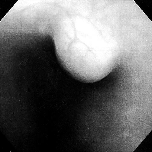
The patients consisted of five males and two females, age ranged from 24 - 58 years (mean, 46). A routine endoscopy was performed to reveal the mucosal aspect of the tumor and to check for additional lesions in the upper gastrointestinal tract. In all patients, there were no mucosal abnormalities on the tumor surface (Fig. 1).
Fig. 1.
Endoscopic finding of case No. 4 demonstrates a submucosal tumor with intact overlying mucosa.
After routine endoscopy, endoprobe technique was performed using an UM-2R (12 MHz) or UM-3R (20 MHz) ultrasonic probe with balloontipped catheter (MH-246R) through the forceps channel. The display unit was EU-M30 (Olympus Optical Co. Ltd., Tokyo, Japan).
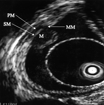
By scanning the normal esophagus with a 20 MHz endoprobe, a seven layer image can be obtained: the first echogenic layer corresponding to the mucosa, the second an echolucent MM layer, the third an echogenic submucosa, the fourth an hypoechoic inner circular PM layer, the fifth a thin, echogenic intermuscular septum, the sixth a hypoechoic outer longitudinal PM layer, and the seventh a broad hyperechoic adventitia and periesophageal fat layer because there is no covering serosa in the esophagus (Fig. 2).
Fig. 2.
High frequency ultrasonic endosonographic finding of case No. 4 shows a clear homogenous hypoechoic mass derived from the second layer of the esophagus (M, mass; MM, lamina muscularis mucosae; SM, submucosa; PM, proper muscle).
The principles of the EMR and its potential risks were explained to the patients and family, and informed consent was formally obtained. Standard routine EMR or EMR-using transparent cap (EMR-C) was performed.
RESULTS
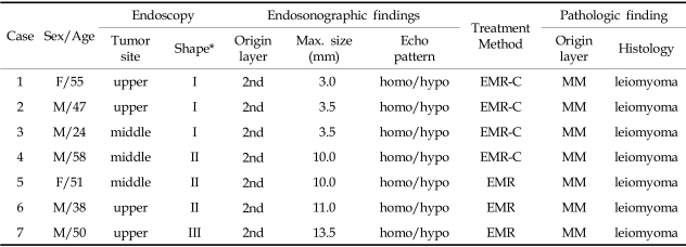
Table 1 summarizes the clinical and histologic features found in seven cases. All the patients had no symptoms related to the leiomyoma, and the lesions were found during checkup incidentally. The locations of the tumors in the esophagus were four in the upper third of the esophagus and three in the middle third.
Table 1.
Characteristics of Patients with Esophageal Leiomyomas Derived from MM
*by Yamada's Classification.
homo/hypo, homogenous/hypoechoic; EMR, endoscopic mucosal resection; EMR-C, EMR using transparent cap; MM, lamina muscularis mucosae.
Endoscopy revealed hemispherical bulging lesions with intact overlying mucosa measuring 4 to 15 mm in diameter (Fig. 1). Balloon-tipped endoprobe clearly showed all 7 small esophageal leiomyomas derived from MM, even for tumors less than 5 mm in size (smallest lesion, 3.0 mm). All tumors were depicted by endoprobe as homogenous inner hypoechoic echograms, well-demarcated masses with smooth and regular outer margin derived from the second hypoechoic layer of the esophagus, and with an intact muscularis propria (Fig. 2).
Complete resection was possible in all 7 cases by EMR or EMR-C without any severe complications. The size of the completely removed tumors ranged from 3.0 to 13.5 mm (mean 7.8 mm) in maximum diameter.
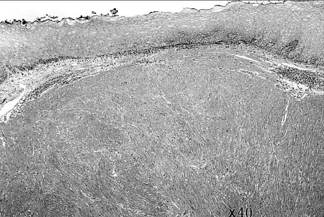
The resected tumors were of firm consistency, smoothly rounded, grayish-white or yellowish on section, without any degenerative changes. They were confirmed as the leiomyomas derived MM layer of the esophagus, as shown by contiguous lamina muscularis mucosae at their peripheral edges and the usual bundles of smooth muscle fibers, microscopically. There were very few mitotic figures (Fig. 3).
Fig. 3.
Microscopic finding of resected specimens demonstrates the leiomyoma derived from the muscularis mucosae of the esophagus. Uniform, interlacing bundles of spindle-like cells without any mitotic bodies are seen (H&E stain, × 40).
After a follow-up of 3 months with repeated endoscopy, no recurrence was seen in all cases and the resected mucosal lesions were completely healed with only linear scarring contracted change remaining.
In all cases, endosonographic findings by endoprobe were exactly concordant with pathologic findings in determining the tumor's depth in the esophageal wall, tissues origin and characteristics, growth pattern, and size of esophageal leiomyoma derived from MM.
DISCUSSION
Leiomyoma is the most common type of esophageal benign tumor and may occur in the MM or PM layer of the esophagus.1-3 Typically MM origin leiomyoma of the esophagus are intramural, single, round to oval protruding masses like a polyp less than 2 cm in diameter, covered with intact normal mucosa, and usually are found incidentally.2,3 Tumor growth seems to be slow, however, the rate of growth, the period needed to observe a change, and size of tumor at risk of malignant transformation are uncertain. The leiomyomas derived from the MM layer of the esophagus usually protrude into the lumen and are likely to become polypoid or pedunculated due to the effects of the constant downward drag of food and peristalsis.2,3 Such MM originating polypoid leiomyomas are ideal candidates for endoscopic resection and can be removed safely with current modernized endoscopic resection methods. In our study, seven leiomyomas originating from the MM were all polypoid or pedunculated in shape suitable for endoscopic removal.
In recent years, EUS has improved the detection rate and differentiation of leiomyomas in the upper gastrointestinal tract.4 EUS can determine the layer of origin, direction of growth, and histologic consistency of the tumor as it can demonstrate echogenic differences between the various submucosal tumors, reflecting their etiologic differences. Criteria for the identification of a leiomyoma derived from the MM of the esophagus are: 1) presence of hypoechoic mass of the second layer of the esophageal wall with well-defined boundaries, 2) homogenous internal echo pattern, and 3) normal overlying mucosa. EUS with high frequency ultrasonic probe is superior to other imaging modalities for detecting the origins of leiomyomas and estimating histologic characteristics of tumors,5,6 as a conventional EUS has difficulty capturing the precise images of small leiomyomas in the esophagus because of difficulty in direct visualization of the tumors and its poor imaging resolution with low frequency ultrasound. High frequency ultrasonic endoprobe can provide direct endoscopic visualization of a small tumor during ultrasonic scanning and it is easier to access a small mural esophageal lesion. A precise diagnosis of small esophageal leiomyoma derived from MM can be achieved by demonstrating fine layer structure and high endosonograhic - histologic concordance with high frequency ultrasonic endoprobe. It is valuable in assessing the origin and size of the tumor and for determining whether or not endoscopic resection is required.3,7 In our study, a 3 mm sized small esophageal leiomyoma derived from MM, which was too small to be demonstrated with conventional EUS, was clearly diagnosed with balloon-tipped high frequency ultrasonic endoprobe. In all of our cases, endosonographic findings by endoprobe were exactly concordant with pathologic findings of resected tumors. Leiomyomas have a tendency toward continuous endoluminal growth and the possibility of malignant transformation.8 Preoperative pathological diagnosis of a smooth muscle cell tumor is impossible because the differentiation of benign and malignant smooth muscle tumors is mainly based on the mitotic figures of several fields of entire resected specimen. Esophageal leiomyomas derived from the MM in our study, showed a tendency to become more pedunculated as the size increased. The endoscopic resection of a small tumor, less than 2 cm in size, and limited to the mucosa or submucosa could be performed with negligible risk of perforation.7,9,10 Endoprobe is an important diagnostic method for smooth muscle tumors of the esophagus and the assessment of potential candidates for endoscopic resection. It is believed that endoprobe can have a great impact in the therapeutic decision-making in small esophageal leiomyomas because of its capacity to clearly define the size and layer of tumor origin.11,12
In conclusion, balloon-tipped endoprobe is an easy and very convenient technique to scan the small MM-derived leiomyomas of the esophagus, even for tumors under 5 mm, useful in making an accurate diagnosis of esophageal leiomyoma derived from MM, and in decision-making for appropriate application of EMR.
References
- 1.Attah EB, Hajdu SI. Benign and malignant tumors of the esophagus at autopsy. J Thorac Cardiovasc Surg. 1968;55:396–404. [PubMed] [Google Scholar]
- 2.Massari M, Lattuada E, Zappa MA, Pieri G, Cioffi U, Simone MD, et al. Evaluation of leiomyoma of the esophagus with endoscopic ultrasonography. HepatoGastroenterology. 1997;44:727–731. [PubMed] [Google Scholar]
- 3.Kajiyama T, Sakai M, Torii A, Kishimoto H, Kin G, Uose S, et al. Endoscopic aspiration lumpectomy of esophageal leiomyomas derived from the muscularis mucosae. Am J Gastroenterol. 1995;90(3):417, 422. [PubMed] [Google Scholar]
- 4.Tio TL, Tytgat GNJ, den Hartog Jager FCA. Endoscopic ultrasonography for the evaluation of smooth muscle tumors in the upper gastrointestinal tract: an experience with 42 cases. Gastrointest Endosc. 1990;36:342–350. doi: 10.1016/s0016-5107(90)71061-9. [DOI] [PubMed] [Google Scholar]
- 5.Faivre J, Bory R, Moulinier B. Benign tumors of oesophagus: Value of endoscopy. Endoscopy. 1978;10:264–268. doi: 10.1055/s-0028-1098306. [DOI] [PubMed] [Google Scholar]
- 6.Takada N, Higashino M, Osugi H, Tokuhara T, Kinoshita H. Utility of endoscopic ultrasonography in assessing the indications for endoscopic surgery of submucosal esophageal submucosal tumors. Surg Endosc. 1999;13:228–230. doi: 10.1007/s004649900950. [DOI] [PubMed] [Google Scholar]
- 7.Kojima T, Takahashi H, Parra-Blanco A, Kohsen K, Fujita R. Diagnosis of submucosal tumor of the upper GI tract by endoscopic resection. Gastrointest Endosc. 1999;50:516–522. doi: 10.1016/s0016-5107(99)70075-1. [DOI] [PubMed] [Google Scholar]
- 8.Solomon MP, Rosenblum H, Rosato FE. Leiomyoma of the esophagus. Ann Surg. 1984;199:246–248. doi: 10.1097/00000658-198402000-00020. [DOI] [PMC free article] [PubMed] [Google Scholar]
- 9.Jacobs WH, Bruns D. Endoscopic electrosurgical polypectomies of the upper gastrointestinal tract. Am J Gastroenterol. 1977;68:241–248. [PubMed] [Google Scholar]
- 10.Yu JP, Luo HS, Wang XZ. Endoscopic treatment of submucosal lesions of the gastrointestinal tract. Endoscopy. 1992;24:190–193. doi: 10.1055/s-2007-1010460. [DOI] [PubMed] [Google Scholar]
- 11.Xu GQ, Zhang BL, Li YM, Chen LH, Ji F, Chen WX, et al. Diagnostic value of endoscopic ultrasonography for gastrointestinal leiomyoma. World J Gastroenterol. 2003;9:2088–2091. doi: 10.3748/wjg.v9.i9.2088. [DOI] [PMC free article] [PubMed] [Google Scholar]
- 12.Fleischer DE. Endoscopic resection of gastrointestinal tumors. Endoscopy. 1993;25:479–481. doi: 10.1055/s-2007-1010371. [DOI] [PubMed] [Google Scholar]


