Abstract
Background
The photopigment melanopsin has been suggested to act as a dominant photoreceptor in nonvisual photoreception including resetting of the circadian clock (entrainment), direct tuning or masking of vital status (activity, sleep/wake cycles, etc.), and the pupillary light reflex (PLR). Pituitary adenylate cyclase-activating polypeptide (PACAP) is exclusively coexpressed with melanopsin in a small subset of retinal ganglion cells and is predicted to be involved extensively in these responses; however, there were inconsistencies in the previous reports, and its functional role has not been well understood.
Methodology/Principal Findings
Here we show that PACAP-deficient mice exhibited severe dysfunctions of entrainment in a time-dependent manner. The abnormalities in the mutant mice were intensity-dependent in phase delay and duration-dependent in phase advance. The knockout mice also displayed blunted masking, which was dependent on lighting conditions, but not completely lost. The dysfunctions of masking in the mutant mice were recovered by infusion of PACAP-38. By contrast, these mutant mice show a normal PLR. We examined the retinal morphology and innervations in the mutant mice, and no apparent changes were observed in melanopsin-immunoreactive cells. These data suggest that the dysfunctions of entrainment and masking were caused by the loss of PACAP, not by the loss of light input itself. Moreover, PACAP-deficient mice express an unusually early onset of activities, from approximately four hours before the dark period, without influencing the phase of the endogenous circadian clock.
Conclusions/Significance
Although some groups including us reported the abnormalities in photic entrainments in PACAP- and PAC1-knockout mice, there were inconsistencies in their results [1], [2], [3], [4]. The time-dependent dysfunctions of photic entrainment in the PACAP-knockout mice described in this paper can integrate the incompatible data in previous reports. The recovery of impaired masking by infusion of PACAP-38 in the mutant mice is the first direct evidence of the relationship between PACAP and masking. These results indicate that PACAP regulates particular nonvisual light responses by conveying parametric light information—that is, intensity and duration. The “early-bird” phenotype in the mutant mice originally reported in this paper supposed that PACAP also has a critical role in daily behavioral patterns, especially during the light-to-dark transition period.
Introduction
Behavioral and physiological adaptations to external day/night cycles are regulated by a variety of environmental cues. Among these cues, light is considered to be the most universal and efficient [5]. Light induces phase-shifting of the circadian clock (entrainment), direct tuning or masking of vital status (activity, sleep/wake cycles, hormone secretion), and pupil constriction, so that the adaptations can be achieved [6], [7], [8], [9]. These ‘nonvisual’ light responses have been suggested to be predominantly mediated by the novel photopigment melanopsin [10], [11].
Pituitary adenylate cyclase-activating polypeptide (PACAP) belongs to the vasoactive intestinal polypeptide (VIP)/glucagons/secretin family and plays pleiotropic roles as a neurotransmitter, neuromodulator and neurotrophic factor [12]. In the retina, PACAP is exclusively expressed with melanopsin [13] in a small subset of retinal ganglion cells [14], [15]. PACAP-containing RGCs innervate widespread brain areas including the suprachiasmatic nucleus (SCN: the master circadian clock in mammals), paraventricular zone (the area involved in masking) and olivary pretectal tract (OPT: the crucial node in the pupillary reflex circuit) [16]. Previous studies using transgenic mice [1], [2], [3], [4] or SCN slice cultures [17], [18] suggested the involvement of PACAP signaling in light- and/or glutamate (a major light transmitter)–induced phase shifts. However, varieties and inconsistencies among these results have complicated our understanding of the role of PACAP: slice cultures analyses suggest that PACAP is both an inducer and a modulator of phase shifts, because PACAP at nanomolar concentrations is alone sufficient to induce phase shifts [17], yet higher concentrations of PACAP enhance or repress glutamate-induced phase delay or advance, respectively [18]. Mice lacking PACAP (Adcyap1 −/−) or the PACAP–specific receptor PAC1 (PAC1 −/−) show a common impairment of phase advance, but intricate differences in phase delay [1], [2], [3], [4]. In addition, relationships between PACAP and other nonvisual light responses were reported [2], [4], [19], but discrepancies in their results were also found.
To clarify the functional roles of intrinsic PACAP in nonvisual photoreception, we examined entrainment, masking and the pupillary light reflex (PLR) in Adcyap1 −/− mice. The present study indicates that PACAP controls particular nonvisual light responses; that is, entrainment and masking. In mediating these two responses, we suggest that PACAP transmits light information such as irradiances and durations. Additionally, PACAP has been shown to affect daily activity patterns under a light/dark (LD) cycle through one or more non-entrainment mechanisms.
Results
Entrainment
As described previously [1], Adcyap1 −/− mice could synchronize with a 12 hr light:12 hr dark LD cycle (12L∶12D) and retained behavioral periodicity under constant dark (DD) conditions (Fig. S1A). We tested the entrainment function of Adcyap1 −/− mice by exposing them to a short light pulse at the indicated circadian time (CT). A dim light pulse at early subjective night (CT15) (10 or 20lx, 5 min) induced phase delay of locomotor activity rhythms in Adcyap1 −/− mice, similar in magnitude to that in Adcyap1 +/+ mice, but a brighter pulse (100lx, 5 min) failed to produce any further shifting in the mutants ( Fig. 1A and Fig. S1A). This impaired phase delay was not restored, even when the duration of light stimulation was prolonged to 30 min ( Fig. 1A and Fig. S1A). Consistent with the behavioral deficits, light–induced phosphorylation of extracellular signal-regulated kinase 1/2 (ERK) in the SCN, which is suggested to be a highly sensitive light detector and a determinant of the magnitude of phase delay [20], reached a ceiling at 20lx preventing further phosphorylation in Adcyap1 −/− mice at 100lx ( Fig. 1B and Fig. S1B). Unlike phase delay, phase advance elicited by a light pulse at late subjective night (5 min, CT21) was not altered in Adcyap1 −/− mice, either at 20 or 100lx ( Fig. 1C ). However, Adcyap1 −/− mice showed a significant smaller phase advance when the light duration was prolonged to 30 min ( Fig. 1C ).
Figure 1. Impaired light-induced behavioral phase shift in Adcyap1 −/− mice.
(A) Quantification of light-induced phase delay at CT15 (n = 5–7 for 5 min, n = 4–5 for 30 min). (B) Numbers of phosphorylated ERK-immunoreactive (pERK-Ir.) cells in the SCN (n = 4–7). (C) Quantification of light-induced phase advance at CT21 (n = 4–5). Values denote means ± SEM. * p<0.05 versus Adcyap1 +/+ mice at 100lx, ## p<0.01 versus each genotype at 0lx, & p<0.05 versus Adcyap1 +/+ mice at 20lx; Mann-Whitney U test followed by Kruskal-Wallis Test.
Masking Responses
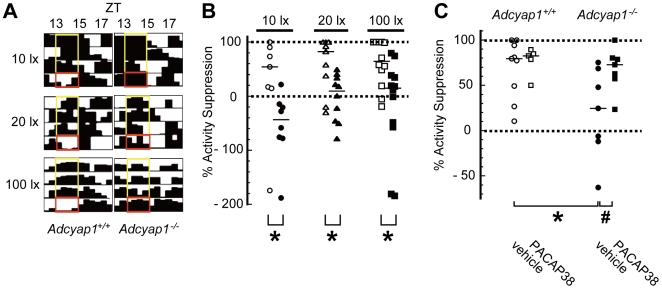
Masking modulates vital status with time by detecting ambient luminance [8], [9]; for example, nocturnal animals are active when the surroundings are dark (<∼0.2lx; positive masking) and passive in a brighter milieu (>∼10lx; negative masking) [21]. Such reactions occur without influencing the underlying circadian clock. We assessed masking by quantifying the amount of activity induced by a two-hour light pulse during early night, a stimulus that is specifically designed for the estimation of masking. Adcyap1 +/+ mice exhibited clear light-induced locomotor suppression (negative masking) at each light intensity ( Fig. 2A ). Because this negative masking in Adcyap1 +/+ mice was predominantly occurred in the first an hour during a two-hour-light exposure, we compared suppression rates during the first hour only between two genotypes. Adcyap1 +/+ mice showed more than 50% suppression of activity rate at all of the light intensities investigated, but this negative masking was significantly impaired in Adcyap1 −/− mice, at all intensities tested, and several mutants paradoxically became active (paradoxical positive masking), especially at the lowest light intensity ( Fig. 2A, B ). These abnormalities were ameliorated by intracerebroventricular administration of PACAP38 (20 pmol) 30 min before exposure to 100lx light, whereas this concentration of PACAP38 had no effect on the amount of activity in Adcyap1 +/+ mice ( Fig. 2C ).
Figure 2. Impaired activity masking in Adcyap1 −/− mice.
(A) Wheel-running records for ZT12–18. Actograms are magnified to show direct responses to a two-hour light pulse at ZT13–15 on test days (red boxes) and no pulse during a comparable time on the three previous nights (yellow boxes). (B) Quantification of % activity suppression during the first hour (ZT13–14). The solid bar in each group denotes the median (n = 7–12). Open symbols: Adcyap1 +/+, closed symbols: Adcyap1 −/−. (C) The effect of PACAP supplementation (20 pmol) on masking responses 30 min before photic stimulation at ZT13 (100lx). The solid bar in each group denotes the median (n = 7–8). * p<0.05 versus Adcyap1 +/+ in each intensity of light or vehicle-treated Adcyap1 +/+, # P<0.05 versus vehicle-treated Adcyap1 −/−; Mann-Whitney U test.
PLR
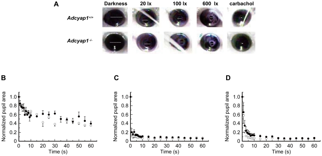
There were no differences in pupil sizes under scotopic conditions between genotypes (Adcyap1 −/−, 1.06±0.18 mm2; Adcyap1 +/+, 0.98±0.10 mm2). A PLR was detected in Adcyap1 −/−mice in response to 460–490 nm blue light, and this was similar in magnitude and time course to that detected in Adcyap1 +/+ mice at all light intensities (two–way repeated-measures ANOVA, Fig. 3B–D ). Additionally, the minimal pupil areas attained by Adcyap1 −/− mice in 1 min of steady light were comparable to those of Adcyap1 +/+ mice ( Fig. 3A ).
Figure 3. PLR in Adcyap1 −/− mice.
(A) Photographs of pupillary constriction 1 min post-irradiation. Darkness: scotopic conditions; carlbachol: 1 min after topical instillation of carlbachol under dark conditions. White broken lines indicate pupillary diameters. (B–D) Time courses of pupillary miosis during 1 min post-irradiation [(B) 20lx; (C) 100lx; (D) 600lx]. The graphs indicate normalized pupil area relative to time 0. Values denote means ± SEM (n = 4–7). No statistical differences were seen in all light intensities (two–way repeated-measures ANOVA).
Retinal Morphology, Innervation and PACAP-Related Gene Expression
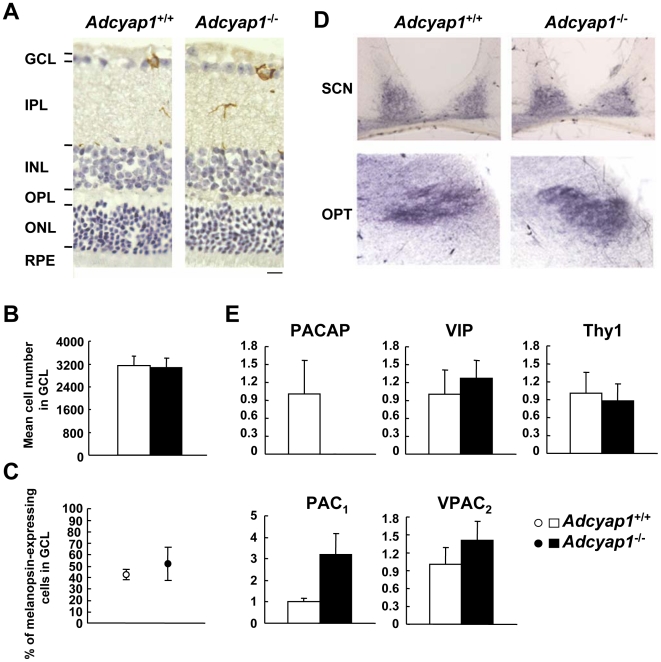
PACAP exerts neurotrophic and neuroprotective effects and modulates cellular differentiation and survival [12]. In the retina, PACAP promotes retinal development through its specific receptor, PAC1 [22], [23]. Thus, we examined whether there was any remodeling in terms of morphology, neuronal projections and PACAP-related gene expression, in the retinas of Adcyap1 −/− mice. Adcyap1 −/− mice showed retention of somatic and dendritic/axonal layers, and melanopsin expression in the ganglion cell layer ( Fig. 4A–C ). Anterograde tracing with the use of horseradish peroxidase–labeled wheat germ agglutinin disclosed normal projections from RGCs to the SCN or OPT in the mutants ( Fig. 4D ). Gene expression analysis ( Fig. 4E ) indicated that there were no changes in the expression of VIP (a peptide that has approximately 70% amino acid sequence identity with PACAP), VPAC2 (VIP receptor subtype 2: a receptor for both PACAP and VIP), and Thy1 (thymus cell antigen 1, theta: a marker of the retinal ganglion cell layer) [24]. PAC1 expression showed a tendency to increase in Adcyap1 −/− mice, but this difference did not reach statistical significance (P = 0.10). We confirmed the complete loss of PACAP mRNA in Adcyap1 −/− eye.
Figure 4. Retinal morphology, innervation and expression of PACAP-related genes in the eye.
(A) Immunohistochemical analysis of melanopsin localization (brown) in the retina. Slices were counterstained with hematoxylin. GCL: ganglion cell layer; IPL: inner plexiform layer; INL: inner nuclear layer; OPL: outer plexiform layer; ONL: outer nuclear layer; RPE: retinal pigment epithelium. Scale bar = 10 µm. (B) Mean cell number in the GCL (n = 4). (C) Percentage of melanopsin-expressing cells in the GCL (n = 4). (D) Bright-field photomicrographs of retinal innervation to the SCN and the OPT, stained purple. (E) Expression levels of PACAP-related genes in the whole eye (n = 3–4). Means ± SEM are presented.
Daily Activity Rhythms
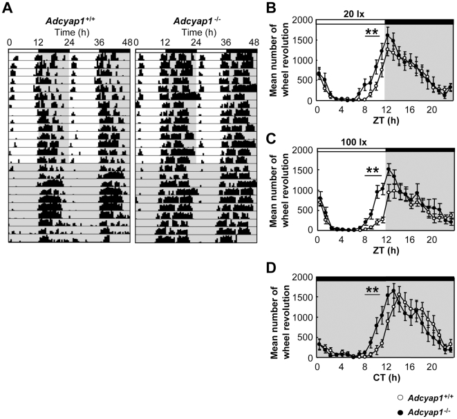
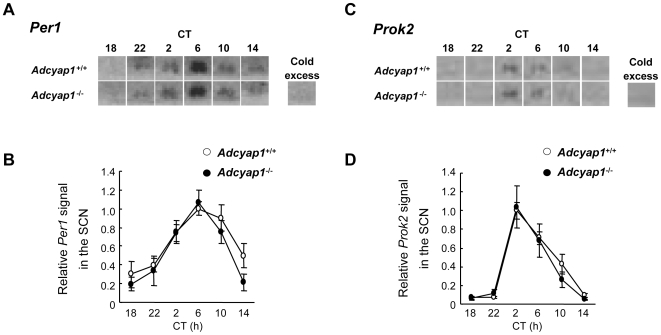
Adaptation of daily activity rhythms to environmental LD cycles is reported to be attained by harmonization with entrainment and masking [7]. We tested the effects of the deficits in entrainment and masking found in Adcyap1 −/− mice on their behavioral adaptation. Under LD cycles with a 20 or 100lx light phase, Adcyap1 −/− mice showed robust behavioral rhythms nearly identical to those in Adcyap1 +/+ mice ( Fig. 5A, B ); however the mutants unexpectedly exhibited an earlier onset of activities, about four hours before the dark phase, but showed a normal offset latencies after the dark phase ( Fig. 5A, B ). These aberrant activities were still observed in subsequent DD conditions ( Fig. 5A, C ). We hypothesized that this early onset of activity was ascribable to molecular clock disturbances with a positive phase angle; therefore, we examined the circadian fluctuation of Period1 (Per1: a major clock gene that determines the free-running period) [25] and Prokineticin2 (Prok2: a clock-controlled gene that suppresses day-time activity) [26] transcripts in the SCN. Adcyap1 +/+ mice showed remarkable circadian fluctuations in both genes with the same peak times ( Fig. 6 ), as reported previously [25], [26]. Inconsistent with our hypothesis, Adcyap1 −/− mice showed an almost all identical phase angle and waveform to those of Adcyap1 +/+ mice. Additionally, the free-running period in DD conditions was not altered in Adcyap1 −/− mice (Adcyap1 −/−, 23.7±0.09 hours, n = 12; Adcyap1 +/+, 23.8±0.08 hours, n = 12).
Figure 5. Early onset of activities in Adcyap1 −/− mice.
(A) Representative double-plotted actogram in LD cycles (light phase 20lx) and subsequent DD conditions. Dark phases are shaded. (B–C) Quantification of wheel-running activities in the LD cycles with a 20lx (B) or 100lx (C) light phase (n = 12). (D) Quantification of wheel-running activities in subsequent DD conditions (after 20lx LD cycle; n = 12). Values denote means ± SEM ** p<0.01 versus Adcyap1 +/+ during ZT/CT 8–11, two–way repeated-measures ANOVA.
Figure 6. Circadian fluctuations of Per1 and Prok2 in the SCN under constant dark conditions.
Photomicrographs (A, C) represent mRNA expression in the SCN by in situ hybridization and line charts (B, D) show the expression levels of Per1 (A, B; n = 3–6) and Prok2 (C, D; n = 4–6). As a control, in situ hybridization was performed in the presence of a 100–fold cold excess of unlabeled probes.
Discussion
The present study shows that PACAP regulates particular nonvisual light responses in time–, irradiance– and duration–dependent manners, and that a PACAP deficit leads to an unusually early onset of activities without changing the phase of the molecular clock. Our findings suggest that PACAP transmits parametric light information, such as fluctuation of light intensities or durations that occur continuously under natural conditions, and that PACAP contributes to formation of daily activity patterns, notably at dusk, by a non–clock entrainment system.
Previous anatomical studies of PACAP/melanopsin-containing retinal innervation suggested roles of PACAP/melanopsin in a broad variety of nonvisual photoreception [16], [27]. In particular, the SCN and OPT are major target areas of PACAP/melanopsin–positive RGCs. Unlike the anatomy-based prediction that PACAP is especially important for functions of these major target areas, Adcyap1 −/− mice exhibited abnormalities in entrainment and masking, but normal PLRs. This could be attributed to differences in the number of melanopsin-expressing RGCs projecting to the area involved in each of these functions. Melanopsin-expressing RGCs represent a large majority of the retinal projections to the entrainment center, the SCN, and the area underlying masking, the ventral subparaventricular zone (approximately 70% of the total retinal input), but only a minority of the retinal projection to the critical node of PLR circuit OPT (about 10%) [27].
The circadian clock is reset daily by environmental timing cues such as dusk and dawn; these stimuli induce phase delay or advance of circadian rhythms, respectively. In this study, we found that intrinsic PACAP exquisitely controls both phase delay and advance in time-, irradiance-, and duration-dependent manners, not merely inducing phase shifts. At early subjective night, PACAP appears to transmit information regarding light of a high intensity, because Adcyap1 −/− mice lost the ability to exhibit further phase delay shifting and ERK phosphorylation in the presence of bright light (100lx). This suggestion is supported by previous in vitro studies showing that PACAP potentiates glutamatergic signaling at early subjective night [28] and glutamate-induced phase delay of the firing rhythms in the SCN [18]. At brighter irradiances, light duration seems unlikely to be a critical factor for phase delay, since Adcyap1 +/+ mice show maximal amplitudes of phase delay after a short (5 min) light pulse, consistent with a previous report [29]. This irradiance-dependent profile of PACAP could explain previous discrepancies in Adcyap1 −/− mice, such as a normal phase delay at 20lx (30 min) [1], but an impaired phase delay at 50 or 500lx (15 min) [2]. Also our results were consistent with the results in the PAC1-dificient (PAC1 −/−) mice showed blunted phase delay at 300lx (30 min) [4]. Unlike phase delay, PACAP seems to be involved in light-induced phase advance at late subjective night, in a duration–dependent manner, because Adcyap1 −/− mice exhibited a significantly impaired phase advance in response to a 30 min light pulse (with 100lx in this study, and 20lx in our previous one [1]), but not to a 5 min light pulse, regardless of the irradiances. This finding is consistent with previous reports of deficits in the phase advances of mice lacking PACAP signaling [2], [3], [4]. These researchers used a light duration longer than 5 min (PAC1 −/− mice: 30 min, ≥300lx; Adcyap1 −/− mice: 15 min, 500lx). Therefore, intrinsic PACAP signaling is supposed to participate in phase advance events, upon and after 5 min of light exposure. By contrast, no in vitro studies agree with the present finding, because PACAP inhibits glutamate-induced phase advances of firing rhythms at higher concentrations [18], but produces a phase advance at lower concentrations [17]. This might be attributed to differences between the physiological and pharmacological concentrations of PACAP. The varied effects of PACAP in early- and late-subjective night are supposed to be caused by the circadian regulations in signaling cascade of glutamate and PACAP in the SCN, but further analyses are necessary.
Although Adcyap1 −/− mice exhibited severe deficits in masking, they incompletely detected luminance, as their suppression rates only slightly increased in increasing light intensities. These results are consistent with the recent reports using PAC1 −/− mice [4]. These incomplete responses were observed even when Adcyap1 −/− mice were exposed to bright light (100lx; an intensity sufficient to induce maximal negative masking) [21], but were completely rescued by supplementation with PACAP38. Importantly, PACAP38 administration did not exert any effects on the basal activity levels of Adcyap1 +/+ mice. Therefore, these data indicate that the loss of PACAP is the primary cause of disturbed negative masking and that PACAP is essential to maintain the photic sensitivity of this response. These results, taken together with the results on entrainment, suggest that PACAP-mediated transmission of parametric light information is prerequisite for both entrainment and masking, but that PACAP regulates each function in a distinct manner. Masking disturbances in Adcyap1−/− mice could be also related to melanopsin-mediated phototransduction, because melanopsin is a photoreceptor that is exclusively colocalized in PACAP-expressing ganglion cells in the retina [13], and because melanopsin is suggested to dominantly mediate the transduction of photic information for negative masking [21], [30]. Unexpectedly, several Adcyap1−/− mice showed a locomotor increase in response to all three intensities of light. This ‘paradoxical’ positive masking might occur due to diminished negative masking as a result of disturbances in melanopsin/PACAP signaling, leaving positive masking, which is driven by rod/cone cells [31], [32]. Since there were no remarkable changes in retinal structures or PLR in Adcyap1−/− mice, their dysfunctions of entrainment and masking are suggested to arise not from light input per se, but from loss of PACAP in a neuronal circuit after the retina.
Adcyap1−/− mice showed early onset of activities under LD cycles and in DD conditions, without changing the phase of the circadian clock, indicating that there is no relation between predark activities and molecular clock phase. There remains the possibility that impaired entrainment in mutants is causal for predark activities, but this idea seems unlikely to be supported because phase of the molecular clocks are almost identical between the two genotypes, and the daily behavioral patterns were also identical with the predark periods being the only exception. Another possibility is aberrant masking. Masking, especially positive masking, has been suggested to have a circadian phase dependency. Several lines of evidence indicate that maximal increases in positive masking occur around the onset of the night phase, even in constant conditions [33]. Other lines of evidence indicate a strong correlation of paradoxical positive masking with light/dark transition periods [7]. In addition, several transgenic mice with masking disturbances often exhibited unusual activities during the transitions between light and dark [21], [34], [35]. Among these mutant mice, the phenotypes of melanopsin−/− mice strongly resemble those of Adcyap1−/− mice in terms of diminished negative masking and predark activities in both LD and DD conditions [21], [36]. Thus, these previous reports seem to agree with the idea that deficits in negative masking contribute to early onset of activities in Adcyap1−/− mice. Further analyses are warranted to investigate the underlying mechanism.
Materials and Methods
Ethics Statement
All animal care and handling procedures were approved by the Institutional Animal Care and Use Committee of Osaka University, the Guiding Principles for the Care and follow the United States National Institutes of Health Guide for the Care and Use of Laboratory Animals.
Mice
The generation of Adcyap1 −/− mice by gene-targeting has been reported previously [37]. The null mutation was backcrossed ten times onto a CD1 (ICR) genetic background. We should note that each mouse was kept in the insulated box. The boxes were equipped with an electric bulb for illumination and a measuring device (a far-infrared apparatus or a running wheel) (Bio-Medica, Japan) to monitor the behavioral activities of the mouse. Thus all experiments described below were performed without irritating mice by moving the cages and without affected by the neighbor mice.
Entrainment
Light-induced phase shifts of locomotor activity rhythms were examined as described [1]. Briefly, mice were transferred to constant dark (DD) conditions after being entrained to a 24-hour LD (12L∶12D) cycle, with monitoring of their locomotor activity by far-infrared apparatuses. After more than eight days in DD conditions, animals were exposed to a white light pulse of the indicated intensities at circadian time (CT)15 or CT21, and their behavioral rhythms were further recorded. The phase shifts were calculated based on the distance between two regression lines drawn from the onset of activity before and after the light pulse as described in [38], supported by the custom-made period-detection software based on autocorrelation [39].
Masking Responses
Mice were entrained to a 24-hour LD (12L∶12D) cycle, with monitoring of their locomotor activity by running wheels. A light pulse was given to the animals for 2 hours from Zeitgeber time (ZT)13. The percentage of activity suppression during the first hour after the stimulus was calculated using the following formula [30]: % activity suppression = (B–A)/B * 100, where A is the amount of activity during the first hour after ZT13 on the day on which light stimulations were given, and B is the average activity level during a comparable time period during the previous three days.
Intracerebroventricular Injection
Central administration of PACAP was performed as described [40]. Briefly, mice were anesthetized and a guide cannula was implanted into the lateral ventricle using a stereotaxic apparatus (Narishige, Japan). After at least eight days for recovery, mice were injected with 20 pmol of PACAP38 (Peptide Institute, Japan; 1 µl min−1, total volume 2 µl), under dim red light, 30 min before photic stimulation (100lx) at ZT13; this amount of PACAP38 seems to be within the physiological range [41] and could completely ameliorate the affective disturbance in Adcyap1 −/− mice [40]. Control mice were administered with Ringer's solution. At the end of the experiments, successful administration was verified by the infusion of Evance Blue.
Wheel-Running Activity Rhythms
Adult male mice were individually housed under a 24-hour LD [12 h L (20 or 100lx)∶12 h D] cycle and their activity rhythms were measured for at least eight days using wheel-running apparatuses. Subsequently, mice were transferred to DD conditions and the free running period (τ) was calculated as described [1]. Wheel revolution was quantified using data from three consecutive days, from the sixth day before the start of DD conditions to the fourth day before the start of DD conditions, inclusive, or from the sixth day to the eighth day in DD conditions.
Slice Preparation, Immunohistochemistry and In Situ Hybridization
LD-entrained mice were transferred to DD conditions and their brains were removed on the 2nd day of DD conditions. To assess ERK phosphorylation, animals were exposed to a light pulse (20 or 100lx) or no pulse (0lx) at CT15, and then perfused with 4% paraformaldehyde in PBS including 1 mM sodium orthovanadate and 0.2 mM phenylmethyl sulfonyl fluoride to inhibit phosphatase activity, at an appropriate time after light stimulation (7.5 min for the 20lx stimulus and 15 min for the 100lx stimulus). We previously confirmed that the level of ERK activation elicited by these stimuli reached a peak at these times. For melanopsin immunostaining, mice were perfused with 4% paraformaldehyde in PBS as described previously [1]. Twenty µm brain sections including the SCN and 5 µm ocular sections were prepared using a cryomicrotome (CM1900, Leica, Germany). Immunohistochemistry and subsequent quantification were undertaken as previously described [1]. Briefly, we used a 1∶1000 dilution of a rabbit anti-phospho-p44/42 Map Kinase (Thr202/Tyr204) antibody (Cell Signaling) for phosphorylated ERK (pERK) or a 1∶1000 dilution of an anti-melanopsin antibody (a gift from Dr. King–Wai Yau), and quantified the number of pERK- or melanopsin-immunoreactive cells in a series of sections (five sections of the SCN and ten sections of the retina per animal). The percentage of melanopsin-expressing cells was calculated as (melanopsin-expressing RGCs/total RGCs)×100.
Pupillometry
Adult male mice, dark–adapted for 1–3 h, were exposed to 1 min of blue light (wavelength: 460–490 nm; light intensities: 20, 100 or 600lx; MVX10, Olympus) without anesthesia. Temporal changes in pupillary responses were archived through an infrared video recorder (Sony, Japan). All tests were conducted between ZT3 and ZT7. Pupil constriction was quantified as described previously [42]. In order to confirm the intrinsic function of the pupillary sphincter, parasympathetic activation by topical administration of carbachol (1 M, Sigma) was also assessed.
Anterograde Tracing
Mice were anaesthetized with ketamine (60 mg/kg, ip) prior to unilateral intravitreal injection of 2 µl of horseradish peroxidase–labeled wheat germ agglutinin. Subsequently, mice were individually housed for two days and then perfused with 1.25% glutaraldehyde and 0.5% paraformaldehyde. Fifty µm sections were immunostained as described above.
Reverse Transcription and Real-Time PCR
Adult male mice were killed during ZT14–16 and their left eyes were immediately enucleated under dim red light. Total RNA was extracted using an RNeasy MinElute Cleanup Kit (Qiagen), including DNase I treatment following homogenization in Buffer RLT. One-hundred ng of total RNA was reverse-transcribed using random primers and SuperScript III reverse transcriptase (Invitrogen). Real-time PCR was performed as described previously [43] with some modifications. Briefly, amplification of cDNAs was executed using a Dynamo SYBR Green qPCR kit (Finnzymes) for the PACAP gene and a SYBR Green Real-time PCR Master Mix (Toyobo, Japan) for the other genes. Beta-actin was amplified as an internal control using both PCR kits. The PCR reaction for Thy1 was performed as follows: forward primer 5′–gtcgctctcctgctctcagtcttg–3′, reverse primer 5′ –tcatccttggtggtgaagttggc–3′, 40 cycles of denaturation at 95°C for 5 sec, annealing at 58°C for 5 sec and elongation at 72°C for 20 sec.
In Situ Hybridization
Brains of mice, kept under DD conditions, were removed at indicated circadian times, then immediately frozen by isopentane, and stored at −80°C until use. Twenty µm brain sections including the SCN and 5 µm ocular sections were prepared using a cryomicrotome (CM1900, Leica, Germany). In situ hybridization was performed as described previously [37] using antisense probes containing the coding regions of mouse Per1 (GenBank accession number AF022992; nucleotides 763–1977), and the mouse Bv8 variant 3 precursor (i.e., Prok2; GenBank accession number AF182066; nucleotides 166–414; from a vector gifted by Takeda Pharmaceutical Co. Ltd.).
Supporting Information
(A) Representative double-plotted actogram during constant dark conditions. Red arrows indicate photic stimulation for 5 or 30 minutes at CT15. Paired red dashed lines represent onset and ending of activity. (B) Photomicrographs showing light-induced ERK phosphorylation, 7.5 minutes (20 lx) or 15 minutes (100 lx) after light stimulation at CT15. Scale bar = 100 µm.
(1.51 MB PDF)
Acknowledgments
We thank Dr. King–Wai Yau (Johns Hopkins University) for providing the melanopsin antibody, Takeda Pharmaceutical Co. Ltd. for gifting the plasmid vector carrying the Prok2 gene and Dr. Hajime Sawai (Osaka University) for teaching the anterograde tracing method.
Footnotes
Competing Interests: The authors have declared that no competing interests exist.
Funding: This research was partly supported by Grants-in-Aid for Scientific Research from Japan Society for the Promotion of Science, the Sankyo Foundation of Life Science, and the Uehara Memorial Foundation. The funders had no role in study design, data collection and analysis, decision to publish, or preparation of the manuscript.
References
- 1.Kawaguchi C, Tanaka K, Isojima Y, Shintani N, Hashimoto H, et al. Changes in light-induced phase shift of circadian rhythm in mice lacking PACAP. Biochem BiophysRes Commun. 2003;310:169–175. doi: 10.1016/j.bbrc.2003.09.004. [DOI] [PubMed] [Google Scholar]
- 2.Colwell CS, Michel S, Itri J, Rodriguez W, Tam J, et al. Selective deficits in the circadian light response in mice lacking PACAP. Am J Physiol Regul Integr Comp Physiol. 2004;287:R1194–1201. doi: 10.1152/ajpregu.00268.2004. [DOI] [PubMed] [Google Scholar]
- 3.Hannibal J, Jamen F, Nielsen HS, Journot L, Brabet P, et al. Dissociation between light-induced phase shift of the circadian rhythm and clock gene expression in mice lacking the pituitary adenylate cyclase activating polypeptide type 1 receptor. J Neurosci. 2001;21:4883–4890. doi: 10.1523/JNEUROSCI.21-13-04883.2001. [DOI] [PMC free article] [PubMed] [Google Scholar]
- 4.Hannibal J, Brabet P, Fahrenkrug J. Mice lacking the PACAP type I receptor have impaired photic entrainment and negative masking. Am J Physiol Regul Integr Comp Physiol. 2008;295:R2050–2058. doi: 10.1152/ajpregu.90563.2008. [DOI] [PubMed] [Google Scholar]
- 5.Aschoff J. Masking and parametric effects of high-frequency light-dark cycles. Jpn J Physiol. 1999;49:11–18. doi: 10.2170/jjphysiol.49.11. [DOI] [PubMed] [Google Scholar]
- 6.Berson DM. Strange vision: ganglion cells as circadian photoreceptors. Trends Neurosc. 2003;26:314–320. doi: 10.1016/S0166-2236(03)00130-9. [DOI] [PubMed] [Google Scholar]
- 7.Mrosovsky N. Masking: history, definitions, and measurement. Chronobiol Int. 1999;16:415–429. doi: 10.3109/07420529908998717. [DOI] [PubMed] [Google Scholar]
- 8.Redlin U. Neural basis and biological function of masking by light in mammals: suppression of melatonin and locomotor activity. Chronobiol Int. 2001;18:737–758. doi: 10.1081/cbi-100107511. [DOI] [PubMed] [Google Scholar]
- 9.Smale L, Lee T, Nunez AA. Mammalian diurnality: some facts and gaps. J Biol Rhythms. 2003;18:356–366. doi: 10.1177/0748730403256651. [DOI] [PubMed] [Google Scholar]
- 10.Doyle S, Menaker M. Circadian photoreception in vertebrates. Cold Spring Harb Symp Quant Biol. 2007;72:499–508. doi: 10.1101/sqb.2007.72.003. [DOI] [PubMed] [Google Scholar]
- 11.Guler AD, Altimus CM, Ecker JL, Hattar S. Multiple photoreceptors contribute to nonimage-forming visual functions predominantly through melanopsin-containing retinal ganglion cells. Cold Spring Harb Symp Quant Biol. 2007;72:509–515. doi: 10.1101/sqb.2007.72.074. [DOI] [PubMed] [Google Scholar]
- 12.Vaudry D, Falluel-Morel A, Bourgault S, Basille M, Burel D, et al. Pituitary adenylate cyclase-activating polypeptide and its receptors: 20 years after the discovery. Pharmacol Rev. 2009;61:283–357. doi: 10.1124/pr.109.001370. [DOI] [PubMed] [Google Scholar]
- 13.Hannibal J, Hindersson P, Knudsen SM, Georg B, Fahrenkrug J. The photopigment melanopsin is exclusively present in pituitary adenylate cyclase-activating polypeptide-containing retinal ganglion cells of the retinohypothalamic tract. J Neurosci. 2002;22:RC191. doi: 10.1523/JNEUROSCI.22-01-j0002.2002. [DOI] [PMC free article] [PubMed] [Google Scholar]
- 14.Hattar S, Liao HW, Takao M, Berson DM, Yau KW. Melanopsin-containing retinal ganglion cells: architecture, projections, and intrinsic photosensitivity. Science. 2002;295:1065–1070. doi: 10.1126/science.1069609. [DOI] [PMC free article] [PubMed] [Google Scholar]
- 15.Dacey DM, Liao HW, Peterson BB, Robinson FR, Smith VC, et al. Melanopsin-expressing ganglion cells in primate retina signal colour and irradiance and project to the LGN. Nature. 2005;433:749–754. doi: 10.1038/nature03387. [DOI] [PubMed] [Google Scholar]
- 16.Hannibal J, Fahrenkrug J. Target areas innervated by PACAP-immunoreactive retinal ganglion cells. Cell Tissue Res. 2004;316:99–113. doi: 10.1007/s00441-004-0858-x. [DOI] [PubMed] [Google Scholar]
- 17.Harrington ME, Hoque S, Hall A, Golombek D, Biello S. Pituitary adenylate cyclase activating peptide phase shifts circadian rhythms in a manner similar to light. J Neurosci. 1999;19:6637–6642. doi: 10.1523/JNEUROSCI.19-15-06637.1999. [DOI] [PMC free article] [PubMed] [Google Scholar]
- 18.Chen D, Buchanan GF, Ding JM, Hannibal J, Gillette MU. Pituitary adenylyl cyclase-activating peptide: a pivotal modulator of glutamatergic regulation of the suprachiasmatic circadian clock. Proc Natl Acad Sci U S A. 1999;96:13468–13473. doi: 10.1073/pnas.96.23.13468. [DOI] [PMC free article] [PubMed] [Google Scholar]
- 19.Hatanaka M, Tanida M, Shintani N, Isojima Y, Kawaguchi C, et al. Lack of light-induced elevation of renal sympathetic nerve activity and plasma corticosterone levels in PACAP-deficient mice. Neurosci Lett. 2008;444:153–156. doi: 10.1016/j.neulet.2008.08.030. [DOI] [PubMed] [Google Scholar]
- 20.Butcher GQ, Dziema H, Collamore M, Burgoon PW, Obrietan K. The p42/44 mitogen-activated protein kinase pathway couples photic input to circadian clock entrainment. Journal of Biological Chemistry. 2002;277:29519–29525. doi: 10.1074/jbc.M203301200. [DOI] [PubMed] [Google Scholar]
- 21.Mrosovsky N, Hattar S. Impaired masking responses to light in melanopsin-knockout mice. Chronobiol Int. 2003;20:989–999. doi: 10.1081/cbi-120026043. [DOI] [PubMed] [Google Scholar]
- 22.Silveira MS, Costa MR, Bozza M, Linden R. Pituitary adenylyl cyclase-activating polypeptide prevents induced cell death in retinal tissue through activation of cyclic AMP-dependent protein kinase. J Biol Chem. 2002;277:16075–16080. doi: 10.1074/jbc.M110106200. [DOI] [PubMed] [Google Scholar]
- 23.Borba JC, Henze IP, Silveira MS, Kubrusly RC, Gardino PF, et al. Pituitary adenylate cyclase-activating polypeptide (PACAP) can act as determinant of the tyrosine hydroxylase phenotype of dopaminergic cells during retina development. Brain Res Dev Brain Res. 2005;156:193–201. doi: 10.1016/j.devbrainres.2005.02.016. [DOI] [PubMed] [Google Scholar]
- 24.Barnstable CJ, Drager UC. Thy-1 antigen: a ganglion cell specific marker in rodent retina. Neuroscience. 1984;11:847–855. doi: 10.1016/0306-4522(84)90195-7. [DOI] [PubMed] [Google Scholar]
- 25.Tei H, Okamura H, Shigeyoshi Y, Fukuhara C, Ozawa R, et al. Circadian oscillation of a mammalian homologue of the Drosophila period gene. Nature. 1997;389:512–516. doi: 10.1038/39086. [DOI] [PubMed] [Google Scholar]
- 26.Cheng MY, Bullock CM, Li C, Lee AG, Bermak JC, et al. Prokineticin 2 transmits the behavioural circadian rhythm of the suprachiasmatic nucleus. Nature. 2002;417:405–410. doi: 10.1038/417405a. [DOI] [PubMed] [Google Scholar]
- 27.Gooley JJ, Lu J, Fischer D, Saper CB. A broad role for melanopsin in nonvisual photoreception. J Neurosci. 2003;23:7093–7106. doi: 10.1523/JNEUROSCI.23-18-07093.2003. [DOI] [PMC free article] [PubMed] [Google Scholar]
- 28.Michel S, Itri J, Han JH, Gniotczynski K, Colwell CS. Regulation of glutamatergic signalling by PACAP in the mammalian suprachiasmatic nucleus. BMC Neurosci. 2006;7:15. doi: 10.1186/1471-2202-7-15. [DOI] [PMC free article] [PubMed] [Google Scholar]
- 29.Bauer MS. Irradiance responsivity and unequivocal type-1 phase responsivity of rat circadian activity rhythms. Am J Physiol. 1992;263:R1110–1114. doi: 10.1152/ajpregu.1992.263.5.R1110. [DOI] [PubMed] [Google Scholar]
- 30.Panda S, Provencio I, Tu DC, Pires SS, Rollag MD, et al. Melanopsin is required for non-image-forming photic responses in blind mice. Science. 2003;301:525–527. doi: 10.1126/science.1086179. [DOI] [PubMed] [Google Scholar]
- 31.Mrosovsky N, Foster RG, Salmon PA. Thresholds for masking responses to light in three strains of retinally degenerate mice. J Comp Physiol A. 1999;184:423–428. doi: 10.1007/s003590050341. [DOI] [PubMed] [Google Scholar]
- 32.Mrosovsky N, Salmon PA, Foster RG, McCall MA. Responses to light after retinal degeneration. Vision Res. 2000;40:575–578. doi: 10.1016/s0042-6989(99)00207-2. [DOI] [PubMed] [Google Scholar]
- 33.Mrosovsky N. In praise of masking: behavioural responses of retinally degenerate mice to dim light. Chronobiol Int. 1994;11:343–348. doi: 10.3109/07420529409057251. [DOI] [PubMed] [Google Scholar]
- 34.Doyle SE, Castrucci AM, McCall M, Provencio I, Menaker M. Nonvisual light responses in the Rpe65 knockout mouse: rod loss restores sensitivity to the melanopsin system. Proc Natl Acad Sci U S A. 2006;103:10432–10437. doi: 10.1073/pnas.0600934103. [DOI] [PMC free article] [PubMed] [Google Scholar]
- 35.Debruyne JP, Noton E, Lambert CM, Maywood ES, Weaver DR, et al. A clock shock: mouse CLOCK is not required for circadian oscillator function. Neuron. 2006;50:465–477. doi: 10.1016/j.neuron.2006.03.041. [DOI] [PubMed] [Google Scholar]
- 36.Mrosovsky N, Hattar S. Diurnal mice (Mus musculus) and other examples of temporal niche switching. J Comp Physiol A. 2005;191:1011–1024. doi: 10.1007/s00359-005-0017-1. [DOI] [PubMed] [Google Scholar]
- 37.Hashimoto H, Shintani N, Tanaka K, Mori W, Hirose M, et al. Altered psychomotor behaviors in mice lacking pituitary adenylate cyclase-activating polypeptide (PACAP). Proc Natl Acad Sci U S A. 2001;98:13355–13360. doi: 10.1073/pnas.231094498. [DOI] [PMC free article] [PubMed] [Google Scholar]
- 38.Daan S, Pittendrigh CS. A functional analysis of circadian pacemakers in nocturnal rodents. II. The variability of phase responce curves. J Comp Physiol A. 1976;106:253–266. [Google Scholar]
- 39.Asahara H, Yokoi S, Isojima Y, Yamanaka C. The auto-correlation periodgram and the phasegram: for automated detection of unusual animal behavior. In: Ulusoy U, Kundu HK, editors. Future Systems for Earhquake Early Warning. New York: Nova Science Publishers, Inc; 2008. pp. 67–82. [Google Scholar]
- 40.Hashimoto H, Hashimoto R, Shintani N, Tanaka K, Yamamoto A, et al. Depression-like behavior in the forced swimming test in PACAP-deficient mice: amelioration by the atypical antipsychotic risperidone. J Neurochem. 2009;110:595–602. doi: 10.1111/j.1471-4159.2009.06168.x. [DOI] [PubMed] [Google Scholar]
- 41.Fang J, Payne L, Krueger JM. Pituitary adenylate cyclase activating polypeptide enhances rapid eye movement sleep in rats. Brain Res. 1995;686:23–28. doi: 10.1016/0006-8993(95)00443-t. [DOI] [PubMed] [Google Scholar]
- 42.Lucas RJ, Douglas RH, Foster RG. Characterization of an ocular photopigment capable of driving pupillary constriction in mice. Nat Neurosci. 2001;4:621–626. doi: 10.1038/88443. [DOI] [PubMed] [Google Scholar]
- 43.Mabuchi T, Shintani N, Matsumura S, Okuda-Ashitaka E, Hashimoto H, et al. Pituitary adenylate cyclase-activating polypeptide is required for the development of spinal sensitization and induction of neuropathic pain. J Neurosci. 2004;24:7283–7291. doi: 10.1523/JNEUROSCI.0983-04.2004. [DOI] [PMC free article] [PubMed] [Google Scholar]
Associated Data
This section collects any data citations, data availability statements, or supplementary materials included in this article.
Supplementary Materials
(A) Representative double-plotted actogram during constant dark conditions. Red arrows indicate photic stimulation for 5 or 30 minutes at CT15. Paired red dashed lines represent onset and ending of activity. (B) Photomicrographs showing light-induced ERK phosphorylation, 7.5 minutes (20 lx) or 15 minutes (100 lx) after light stimulation at CT15. Scale bar = 100 µm.
(1.51 MB PDF)


