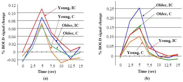
Fig. 2.
The within-group whole-brain differential activation during successful flanker trials (“Incongruent – Congruent”) (voxel-based p value ≤ 0.005 and whole-brain corrected p value ≤ 0.021) revealed a location shift of the IFG/MFG cluster with aging. The mean impulse response functions for older and younger group in conditions IC (Incongruent) and C (Congruent) are plotted at (a) the right IFG/MFG cluster found from the younger group, and (b) the right IFG/MFG/PCG cluster found from the older group.

