Abstract
Background
Both HIV and TB cause a state of heightened immune activation. Immune activation in HIV is associated with progression to AIDS. Prior studies, focusing on persons with advanced HIV, have shown no decline in markers of cellular activation in response to TB therapy alone.
Methodology
This prospective cohort study, composed of participants within a larger phase 3 open-label randomized controlled clinical trial, measured the impact of TB treatment on immune activation in persons with non-advanced HIV infection (CD4>350 cells/mm3) and pulmonary TB. HIV load, CD4 count, and markers of immune activation (CD38 and HLA-DR on CD4 and CD8 T cells) were measured prior to starting, during, and for 6 months after completion of standard 6 month anti-tuberculosis (TB) therapy in 38 HIV infected Ugandans with smear and culture confirmed pulmonary TB.
Results
Expression of CD38, and co-expression of CD38 and HLA-DR, on CD8 cells declined significantly within 3 months of starting standard TB therapy in the absence of anti-retroviral therapy, and remained suppressed for 6 months after completion of therapy. In contrast, HIV load and CD4 count remained unchanged throughout the study period.
Conclusion
TB therapy leads to measurable decreases in immune activation in persons with HIV/TB co-infection and CD4 counts >350 cells/mm3.
Introduction
Cellular markers of immune activation, CD38 and HLA-DR on CD4 and CD8 T lymphocytes, are elevated in HIV infected individuals. Opportunistic infections such as tuberculosis (TB) lead to further increases in immune activation and stimulate HIV viral replication [1]. Sulkowski et al. showed that successful treatment of opportunistic infections, excluding TB, led to declines in HIV load and decreased immune activation [2]. One would predict that treatment of TB would also lead to declines in HIV load and increases in CD4 count, but studies addressing this question have been contradictory. Studies within the United States and Europe have shown a decrease in HIV load in response to TB therapy [3], [4]. In contrast, studies from sub-Saharan Africa have failed to see declines in HIV load after several months of TB therapy [5]–[7]. It is a similar story for CD4 counts. In HIV negative individuals active TB disease induces CD4 lymphopenia which is responsive to TB therapy [8]. However, in persons with HIV/TB co-infection, treatment of TB has not impacted CD4 counts [5], [6].
Immune activation has been implicated as a critical driver of HIV disease progression. Expression of CD38 on CD8 T cells has been shown to be a strong predictor of HIV progression and death [9]. Despite multiple studies demonstrating increased CD38 and/or HLA-DR expression on CD8 T cells in persons with or without HIV co-infection during active TB there are few studies showing declines in these cellular markers of immune activation in response to TB treatment. Our earlier studies have shown that TB exerts its greatest effect on survival in persons with early stage HIV [10]. We therefore hypothesized that TB therapy alone in TB patients with less advanced HIV would lead to measurable declines in immune activation and corresponding declines in HIV load. To test this hypothesis we prospectively followed a cohort of 38 HIV positive persons with baseline CD4 counts >350 cells/mm3 in Uganda who had smear and culture confirmed pulmonary TB and was placed on standard anti-TB therapy.
Materials and Methods
We present a cohort study of participants in a phase 3 open-labeled randomized clinical trial measuring the impact of early HAART combined with standard TB therapy versus TB therapy alone in persons with pulmonary TB and HIV infection with high CD4 counts. Subjects, between the ages of 18 and 60, were recruited from the Uganda-Case Western Reserve University Research Collaboration within the Mulago Hospital Complex in Kampala, Uganda. All subjects gave written informed consent. This sub-study focused on the anti-retroviral therapy naïve HIV positive persons with smear positive pulmonary TB and CD4 counts >350 cells/mm3 who were started on a standard regimen of directly observed TB therapy (two months of isoniazid, rifampin, pyrazinamide, and ethambutol, followed by four months of isoniazid and rifampin). Mid-way through study enrollment daily cotrimoxazole was added in response to new international guidelines. At the 3, 6, 9, and 12 month study visits 84%, 92%, 84%, and 87% of the participants were taking daily cotrimoxazole. The study protocol was approved by the institutional review boards of Case Western Reserve University (CWRU), University of California at San Francisco, the Joint Clinical Research Center in Kampala, Uganda and the Ugandan National Council for Science and Technology (Figures S1–S3).
Enrollment for the larger clinical trial began in October 2004. The protocol for this trial, supporting CONSORT checklist and flow diagram of enrollment are available as supporting information (Checklist S1, Protocol S1, Figure S4). In this substudy, data was accrued between March 2006 and February 2009. Participants gave informed consent and had blood obtained for immune analysis at baseline, 3, 6, 9, and 12 months after enrollment. All persons with flow data at baseline, 3 and/or 6 months, and 9 and/or 12 months were included in the analysis (n = 38). All patients completed TB treatment by month 6. Immunologic studies were performed at the Joint Clinical Research Center in Kampala, Uganda.
Plasma HIV-RNA copy levels (HIV load) were measured using Amplicor quantitative restriction transcriptase-polymerase chain reaction assay (Roche Amplicor 1.5) according to manufacturer instructions. The lower limit of detection of the assay was 400 copies/mm3.
Whole blood was collected in sodium heparin tubes and 200 ul was aliquotted to each of the 12×75 mm test tubes. For flow cytometry the following antibodies were used: anti-CD4 allophycocyananin (APC), anti-CD8 APC, anti-HLA-DR phycoerythrin (PE), and anti-CD38 phycoerythrin Cy5 (PE Cy5). Mouse monoclonal isotypic controls conjugated with PE, PE Cy5, and APC were used to determine non-specific binding and to set gating boundaries. All antibodies were obtained from BD Pharmingen (San Diego, California).
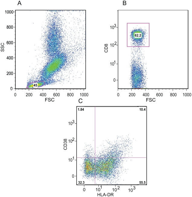
Using 4 color flow cytometry (Becton Dickinson FACSCalibur) 25–50,000 cells were analyzed for each condition. Initial gating was on the lymphocytes based on forward and side scatter. CD4 or CD8 T cell populations were then determined. T cells were then evaluated for the percentage of cells expressing HLA-DR and CD38 (Figure 1).
Figure 1. Representative Flow cytometric approach to define immune activation of CD8 T cells.
A. Gating on lymphocytes based on forward and side scatter properties. B. Gating on CD8 T cells. C. Expression of CD38 and HLA-DR on CD8 T cells.
Non-parametric rank tests were performed to compare median changes in immune activation from baseline to months 3, 6, 9, and 12; and to compare levels at 6 months with 9 and 12 months (the period after TB therapy was completed). Data were stratified by cotrimoxazole use to determine its effect on immune activation at each time point. All analyses were performed using Statistical Analysis Software (SAS) Version 9.1 (SAS Institute, Cary, NC).
Results
Of the 38 HIV infected persons with sputum smear and culture confirmed pulmonary TB the mean age was 32 years (range 19–54) and 24 were males (63%) and 14 female. The median baseline HIV load was 4.4 log10 copies/ml (range 2.0–5.9 log10 copies/mL) and the median CD4 count was 610 cells/mm3 (range 374–1368). 36 of 38 persons were sputum culture negative at 2 months, and all 38 were culture negative by 4 months while on TB treatment. Among these 38 persons there was only one death which occurred 10 months after completion of TB therapy.
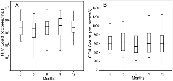
There was no significant change in median HIV log10 load as compared to baseline at 3 (4.3 log10 copies/mL), 6 (4.5 log10 copies/mL), 9 months (4.6 log10 copies/mL), or 12 months (4.4 log10 copies/mL) (Figure 2A). Additionally there was no significant change in median CD4 count from a baseline value of 610 cells/mm3, to 637 cells/mm3 at 3 months, 540 cells/mm3 at 6 months, 602 cells/mm3 at 9 months, and 608 cells/mm3 at 12 months. (Figure 2B).
Figure 2. Change in HIV Load and CD4 Counts in response to TB therapy.
A. HIV log10 RNA viral loads on standard TB therapy at baseline, 3, 6, 9, and 12 months. B. CD4 counts at baseline, 3, 6, 9, and 12 months (n = 38 for all time points). The boxes indicate the interquartile ranges, the horizontal lines transecting the boxes indicate the medians, and the whiskers indicate the highest and lowest values. All values with P>.05.
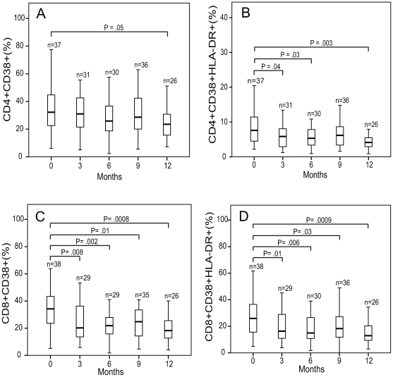
The change in median percentage of CD4 cells expressing CD38 was unchanged from baseline (32%), to 3 (31%), 6 (26%), and 9 months (29%), but measured a significant decline at 12 months (24%) (Figure 3A). The percentage of CD4 cells that were double positive, expressing both HLA-DR and CD38, declined from a baseline value of 8% to 6% (P<.05) at 3 months, 5% (P<.05) at 6 months, at 9 months the change from baseline was no longer significant (6%) (P>.05), but regained significance at 12 months (4%) (P<.01) (Figure 3B).
Figure 3. Change in immune activation in response to TB therapy.
Percentage expression of CD38 (A,C), and CD38/HLA-DR (B,D) on CD4 and CD8 T cells at baseline, 3, 6, 9, and 12 months. The boxes indicate the interquartile ranges, the horizontal lines transecting the boxes indicate the medians, and the whiskers indicate the highest and lowest values. Significant P values (<.05) are indicated.
The median percentage of CD8 cells expressing CD38 declined from 34% at baseline to 20% (P<.01) at 3 months, 22% (P<.01) at 6 months, to 25% (P<.05) at 9 months, to 18% at 12 months (P<.001) (Figure 3C). The percentage of CD8 cells that were double positive, expressing both HLA-DR and CD38, declined from 26% at baseline to 16% (P<.01) at 3 months, 15% (P<.01) at 6 months, to 18% (P<.05) at 9 months, and to 13% at 12 months (P<.001) (Figure 3D). HLA-DR expression alone on CD4 and CD8 cells was not significantly changed from baseline. Thus, the expression of CD38 and the co-expression of CD38/HLA-DR on CD8 cells were strongly affected by TB therapy while decreased expression of these markers on CD4 cells was less pronounced.
The primary analysis used for figures 2 and 3 was Wilcoxon rank test for non-parametric values. Data sets were also analyzed by Sign test which confirmed all values of statistical significance. Additionally, stratified analysis showed no significant impact of cotrimoxazole use on immune activation of CD8 T cells. When accounting for cotrimoxazole use the combined expression of CD38/HLA-DR on CD4 T cells was no longer significant at 6 months but remained so at 12 months.
Discussion
In this prospective cohort study of persons with HIV (CD4 counts>350 cells/mm3) and pulmonary TB we measured a significant decline in CD8 T cell immune activation that persisted for at least 6 months after completion of TB therapy. We did not observe decreased HIV load or increased CD4 counts in response to TB therapy.
Both HIV and MTB infection cause immune activation. How HIV infection leads to chronic immune activation is poorly understood. Depletion of gut mucosal lymphocytes and transudation of LPS into the systemic circulation has been postulated by some. Alternatively, HIV and its viral gene products can directly activate the immune system [11]. In active TB, macrophages release pro-inflammatory cytokines (IL-1, IL-6, TNF-α) in response to mycobacterial proteins and glycolipids, resulting in CD4 and CD8 T lymphocyte recruitment and activation at the site of infection and systemically [12]. CD38 and HLA-DR expression on CD8 cells, markers of immune activation, are higher in HIV/TB co-infection than in persons with either HIV infection or TB alone [13].
In HIV/TB co-infection, TB leads to increased HIV viral replication both in the lungs and systemically. TNF-α produced in response to mycobacterial proteins induces HIV viral replication through nuclear factor kappa β (NFK-β) binding to the 5′ end of the long terminal repeat of HIV [12]. Immune activation and inflammatory cytokines induced by opportunistic infections such as MTB can drive HIV viral replication. Thus one would predict that TB treatment should lead to a decrease in immune activation and a corresponding decline in HIV load.
Our finding that reduced immune activation did not result in a reduction in HIV load suggests that there may not be a linear relationship between immune activation and viral replication in HIV/TB. The anti-inflammatory/-infective properties of cotrimoxazole and rifampin could contribute to decreasing immune activation. However, when data was stratified for cotrimoxazole use there was no impact on CD38 expression. The lack of a rebound six months after completing TB therapy argues against a role for rifampin. The possible disconnect between immune activation and HIV load is further supported by our observation that up to 24% of HIV/TB co-infected Ugandans have low baseline HIV viral loads that are independent of CD4 counts and severity of TB [14]. While studies performed in the United States and Great Britain show that TB treatment decreases HIV load [3], [4], our study is consistent with similar work from sub-Saharan Africa that found no change [6], [7].
Our most striking finding was a clear decline in immune activation seen as early as 3 months into treatment. Percentage expression of both CD38 alone and co-expression of CD38/HLA-DR on CD8 T cells was significantly decreased after 3 and 6 months of standard TB therapy and remained decreased for at least 6 months after completion of therapy. Additionally, there was a trend towards decreased expression of CD38 and HLA-DR on CD4 T cells. These findings are at odds with Morris et al. who failed to see a decline in immune activation in their sub-Saharan HIV/TB co-infected cohort [6]. Although both cohorts were in sub-Saharan Africa, our cohort had much higher CD4 counts at baseline (mean of 642 cells/mm3) compared to the Morris cohort (mean CD4 count of 184 cells/mm3). It is quite possible that normal regulatory patterns that induce macrophages and T cells to produce pro-inflammatory cytokines are disrupted in advanced HIV disease and are less likely to normalize in response to TB therapy [15]. In contrast, in less advanced HIV infection TB therapy may be more likely to re-establish normal cytokine responses leading to a corresponding decline in immune activation. Epidemiologic studies have shown that treatment of TB has a greater impact on survival in HIV infected persons with higher CD4 counts (>200 cells/mm3), and little impact on those with more advanced HIV disease [10].
The failure of CD4 counts to increase in response to TB therapy is also intriguing. TB in non-HIV infected persons induces lymphopenia that returns to normal within one month of treatment [8]. This response may be offset by the increased T cell apoptosis in HIV/TB [12]. Our findings are in-line with others who have shown no increase in CD4 counts with TB therapy in HIV infected individuals [5], [6]. Conversely one might argue that the relatively stable CD4 counts over 1 year, as opposed to a steady decline, are indirect evidence for further immunologic benefit.
Limitations of our study include the lack of control group and limited number of patients. The ideal control group would be similar persons with HIV/TB co-infection that are followed and do not receive TB treatment. For obvious reasons this study will not be performed. Alternatively, one might compare our group to those with HIV infection alone, but these persons even if having similar CD4 and HIV loads at baseline are unlikely to have similar baseline levels of immune activation [16]. Despite following only 38 persons longitudinally we were able to find a measurable decline in immune activation. One cannot exclude the possibility that our study lacked the power to detect more subtle, but still significant changes in HIV load and CD4 counts.
In conclusion, we found that TB therapy in HIV/TB co-infected persons with CD4 counts >350 cells/mm3 leads to significant declines in immune activation of CD8 T cells without a measurable impact on HIV load or CD4 counts. Our findings suggest that declines in immune activation rather than changes in HIV load or CD4 count may explain prior epidemiologic observations that TB treatment leads to survival benefits in HIV/TB co-infected patients with less advanced HIV disease.
Supporting Information
Trial Protocol for the parent study.
(2.10 MB DOC)
CONSORT Checklist
(0.19 MB DOC)
IRB approval from the Ugandan Council of Science and Technology.
(0.03 MB PDF)
IRB Approval from the Joint Clinical Research Center in Kampala, Uganda.
(0.04 MB PDF)
IRB approval from University Hospitals-Case Medical Center.
(0.22 MB PDF)
Enrollment Flow diagram.
(0.03 MB DOC)
Acknowledgments
This study was completed only through the dedicated work of many individuals. We would like to thank the staff of the Uganda-Case Western Reserve University Research Collaboration TB Project Clinic and the Joint Clinical Research Center in Kampala, Uganda. In particular, the authors would like to thank Pierre Peters, Joy Baseke, Phineas Gitta, Jalia Birabwa, Abdunulu Mbabaali, Caroline Kumukama, Michael Odie, Michael George Mujwiga, Harriet Kose-Kayanja and Dennis Dobbs for their parts in the conduct of the study. We are most grateful to the patients who participated in the study.
Trial: Anti-HIV Drugs for Ugandan Patients With HIV and Tuberculosis.
Registry #:NCT0007847
Footnotes
Competing Interests: The authors have declared that no competing interests exist.
Funding: Financial support for study provided by: NIH grants RO1-AI 51219, T32-HL07889, Center for AIDS Research Developmental Pilot Grant Award, Case/UHC Center for AIDS Research: AI36219, Tuberculosis Research Unit NIH Contract No. HHSN266200700022C/NO1-AI70022, and AI95383. The funders had no role in the study design, data collection and analysis, decision to publish, or preparation of the manuscript.
References
- 1.Lawn SD, Butera ST, Folks TM. Contribution of immune activation to the pathogenesis and transmission of human immunodeficiency virus type 1 infection. Clin Microbiol Rev. 2001;14:753–777. doi: 10.1128/CMR.14.4.753-777.2001. [DOI] [PMC free article] [PubMed] [Google Scholar]
- 2.Sulkowski MS, Chaisson RE, Karp CL, Moore RD, Margolick JB, et al. The effect of acute infectious illnesses on plasma human immunodeficiency virus (HIV) type 1 load and the expression of serologic markers of immune activation among HIV-infected adults. J Infect Dis. 1998;178:1642–1648. doi: 10.1086/314491. [DOI] [PubMed] [Google Scholar]
- 3.Goletti D, Weissman D, Jackson RW, Graham NM, Vlahov D, et al. Effect of Mycobacterium tuberculosis on HIV replication. Role of immune activation. J Immunol. 1996;157:1271–1278. [PubMed] [Google Scholar]
- 4.Dean GL, Edwards SG, Ives NJ, Matthews G, Fox EF, et al. Treatment of tuberculosis in HIV-infected persons in the era of highly active antiretroviral therapy. Aids. 2002;16:75–83. doi: 10.1097/00002030-200201040-00010. [DOI] [PubMed] [Google Scholar]
- 5.Kalou M, Sassan-Morokro M, Abouya L, Bile C, Maurice C, et al. Changes in HIV RNA viral load, CD4+ T-cell counts, and levels of immune activation markers associated with anti-tuberculosis therapy and cotrimoxazole prophylaxis among HIV-infected tuberculosis patients in Abidjan, Cote d'Ivoire. J Med Virol. 2005;75:202–208. doi: 10.1002/jmv.20257. [DOI] [PubMed] [Google Scholar]
- 6.Morris L, Martin DJ, Bredell H, Nyoka SN, Sacks L, et al. Human immunodeficiency virus-1 RNA levels and CD4 lymphocyte counts, during treatment for active tuberculosis, in South African patients. J Infect Dis. 2003;187:1967–1971. doi: 10.1086/375346. [DOI] [PubMed] [Google Scholar]
- 7.Lawn SD, Shattock RJ, Acheampong JW, Lal RB, Folks TM, et al. Sustained plasma TNF-alpha and HIV-1 load despite resolution of other parameters of immune activation during treatment of tuberculosis in Africans. Aids. 1999;13:2231–2237. doi: 10.1097/00002030-199911120-00005. [DOI] [PubMed] [Google Scholar]
- 8.Jones BE, Oo MM, Taikwel EK, Qian D, Kumar A, et al. CD4 cell counts in human immunodeficiency virus-negative patients with tuberculosis. Clin Infect Dis. 1997;24:988–91. doi: 10.1093/clinids/24.5.988. [DOI] [PubMed] [Google Scholar]
- 9.Fahey JL, Taylor JM, Detels R, Hofmann B, Melmed R, et al. The prognostic value of cellular and serologic markers in infection with human immunodeficiency virus type 1. N Engl J Med. 1990;322:166–172. doi: 10.1056/NEJM199001183220305. [DOI] [PubMed] [Google Scholar]
- 10.Whalen CC, Nsubuga P, Okwera A, Johnson JL, Hom DL, et al. Impact of pulmonary tuberculosis on survival of HIV-infected adults: a prospective epidemiologic study in Uganda. Aids. 2000;14:1219–1228. doi: 10.1097/00002030-200006160-00020. [DOI] [PMC free article] [PubMed] [Google Scholar]
- 11.Grossman Z, Meier-Schellersheim M, Paul WE, Picker LJ. Pathogenesis of HIV infection: what the virus spares is as important as what it destroys. Nat Med. 2006;12:289–295. doi: 10.1038/nm1380. [DOI] [PubMed] [Google Scholar]
- 12.Toossi Z. Virological and immunological impact of tuberculosis on human immunodeficiency virus type 1 disease. J Infect Dis. 2003;188:1146–1155. doi: 10.1086/378676. [DOI] [PubMed] [Google Scholar]
- 13.Hertoghe T, Wajja A, Ntambi L, Okwera A, Aziz MA, et al. T cell activation, apoptosis and cytokine dysregulation in the (co)pathogenesis of HIV and pulmonary tuberculosis (TB). Clin Exp Immunol. 2000;122:350–357. doi: 10.1046/j.1365-2249.2000.01385.x. [DOI] [PMC free article] [PubMed] [Google Scholar]
- 14.Srikantiah P, Wong JK, Liegler T, Walusimbi M, Mayanja-Kizza H, et al. Unexpected low-level viremia among HIV-infected Ugandan adults with untreated active tuberculosis. J Acquir Immune Defic Syndr. 2008;49:458–460. doi: 10.1097/QAI.0b013e31817e9fb4. [DOI] [PMC free article] [PubMed] [Google Scholar]
- 15.Goletti D, Carrara S, Vincenti D, Giacomini E, Fattorinia L, et al. Inhibition of HIV-1 replication in monocyte-derived macrophages by Mycobacterium tuberculosis. J Infect Dis. 2004;189:624–633. doi: 10.1086/381554. [DOI] [PubMed] [Google Scholar]
- 16.Villacian J, Tan GB, Teo LF, Paton N. The effect of infection with Mycobacterium tuberculosis on T-cell activation and proliferation in patients with and without HIV co-infection. J of Infect. 2005;51:408–412. doi: 10.1016/j.jinf.2004.11.011. [DOI] [PubMed] [Google Scholar]
Associated Data
This section collects any data citations, data availability statements, or supplementary materials included in this article.
Supplementary Materials
Trial Protocol for the parent study.
(2.10 MB DOC)
CONSORT Checklist
(0.19 MB DOC)
IRB approval from the Ugandan Council of Science and Technology.
(0.03 MB PDF)
IRB Approval from the Joint Clinical Research Center in Kampala, Uganda.
(0.04 MB PDF)
IRB approval from University Hospitals-Case Medical Center.
(0.22 MB PDF)
Enrollment Flow diagram.
(0.03 MB DOC)


