Abstract
Our recent studies indicate that endoplasmic reticulum (ER) stress causes INS-1 cell apoptosis by a Ca2+-independent phospholipase A2 (iPLA2β)-mediated mechanism that promotes ceramide generation via sphingomyelin hydrolysis and subsequent activation of the intrinsic pathway. To elucidate the association between iPLA2β and ER stress, we compared β-cell lines generated from wild type (WT) and Akita mice. The Akita mouse is a spontaneous model of ER stress that develops hyperglycemia/diabetes due to ER stress-induced β-cell apoptosis. Consistent with a predisposition to developing ER stress, basal phosphorylated PERK and activated caspase-3 are higher in the Akita cells than WT cells. Interestingly, basal iPLA2β, mature SREBP-1 (mSREBP-1), phosphorylated Akt, and neutral sphingomyelinase (NSMase) are higher, relative abundances of sphingomyelins are lower, and mitochondrial membrane potential (ΔΨ) is compromised in Akita cells, in comparison with WT cells. Exposure to thapsigargin accelerates ΔΨ loss and apoptosis of Akita cells and is associated with increases in iPLA2β, mSREBP-1, and NSMase in both WT and Akita cells. Transfection of Akita cells with iPLA2β small interfering RNA, however, suppresses NSMase message, ΔΨ loss, and apoptosis. The iPLA2β gene contains a sterol-regulatory element, and transfection with a dominant negative SREBP-1 reduces basal mSREBP-1 and iPLA2β in the Akita cells and suppresses increases in mSREBP-1 and iPLA2β due to thapsigargin. These findings suggest that ER stress leads to generation of mSREBP-1, which can bind to the sterol-regulatory element in the iPLA2β gene to promote its transcription. Consistent with this, SREBP-1, iPLA2β, and NSMase messages in Akita mouse islets are higher than in WT islets.
Keywords: Apoptosis, Diabetes, ER Stress, Mitochondria, Phospholipase, Signal Transduction
Introduction
β-Cell loss due to apoptosis contributes to the progression and development of Type 1 or Type 2 diabetes mellitus (T1DM2 or T2DM, respectively). This is supported by autopsy studies that reveal reduced β-cell mass in obese T2DM subjects in comparison with obese non-diabetic subjects (1, 2) and reveal that the loss in β-cell function in non-obese T2DM is associated with decreases in β-cell mass (3, 4). Other evidence suggests that cytokines cause β-cell apoptosis during the development of autoimmune T1DM (5–8). It is therefore important to understand the mechanisms underlying β-cell apoptosis if this process is to be prevented or delayed.
β-Cell mass is regulated by a balance between β-cell replication/neogenesis and β-cell death resulting from apoptosis (9, 10). Findings in rodent models of T2DM (10, 11) and in human T2DM (3, 4) indicate that the decrease in β-cell mass in T2DM is not attributable to reduced β-cell proliferation or neogenesis but to increased β-cell apoptosis (12). In addition to the intrinsic and extrinsic apoptotic pathways, apoptosis due to prolonged endoplasmic reticulum (ER) stress (12, 13) has been reported in various diseases, including Alzheimer and Parkinson diseases (14).
Evidence from the Akita (15, 16) and NOD.k iHEL (17) mouse models suggests that ER stress can also lead to the development of diabetes mellitus as a consequence of β-cell apoptosis. Further, mutations in genes encoding the ER stress-transducing enzyme pancreatic ER kinase (PERK) (18) and the ER-resident protein involved in degradation of malfolded ER proteins have been clinically linked to diminished β-cell health (19, 20). Other reports suggest that ER stress may also play a prominent role in the autoimmune destruction of β-cells during the development of T1DM (6, 21, 22). Because the secretory function of β-cells endows them with a highly developed ER and the β-cell is one of the cells most sensitive to nitric oxide (23), it is not unexpected that β-cells exhibit a heightened susceptibility to autoimmune-mediated ER stress (24, 25). In support of this, Wolfram syndrome, which is associated with juvenile onset diabetes mellitus, is recognized to be a consequence of chronic ER stress in pancreatic β-cells (21, 26).
In view of the evidence suggesting that ER stress-induced β-cell apoptosis may be a factor in the development of diabetes mellitus, it was of interest to elucidate the mechanisms involved. Recent work from our laboratory led to the identification of a Ca2+-independent phospholipase A2 (iPLA2β) as a key participant in ER stress-mediated apoptosis of INS-1 insulinoma cells. The iPLA2β, classified as a Group VIA isoform of iPLA2, is a member of a large family of PLA2s (27) that is cytosolic and does not require Ca2+ for activity (28–30). It is activated by ATP, is inhibited by the bromoenol lactone suicide substrate (BEL) inhibitor of iPLA2β (31), and is recognized to have a role in signal transduction (28, 31–39). We found that induction of ER stress with the sarcoendoplasmic reticulum Ca2+-ATPase inhibitor thapsigargin causes INS-1 cell apoptosis that is amplified by overexpression of iPLA2β and is inhibited by BEL (40).
Further work indicated that ER stress activates iPLA2β in INS-1 cells, leading to neutral sphingomyelinase (NSMase) induction and generation of ceramides via hydrolysis of sphingomyelins (41). Subsequently, the ceramides trigger mitochondrial apoptotic processes and cell death ensues. Consistent with this proposed mechanism, inhibition of iPLA2β or NSMase suppresses sphingomyelin hydrolysis and ceramide generation, mitochondrial abnormalities, and apoptosis (42).
Although a link between iPLA2β and ER stress-induced apoptotic pathway was gleaned from the above observations, it was derived from studies in which a chemical agent was used to induce ER stress and iPLA2β expression levels were genetically manipulated in an insulinoma cell line. Of importance was to determine whether a similar link existed under conditions where ER stress developed in the β-cell in the absence of chemical intervention. Here, we present studies in a β-cell line established from Akita mice (43), which contain a spontaneous mutation of the insulin 2 gene (Ins2) (C96Y) that results in misfolding insulin in the ER leading to development of hyperglycemia/diabetes due to ER stress-induced β-cell apoptosis (44, 45). Our findings reveal for the first time that predisposition to ER stress is associated with increased expression of iPLA2β and that its expression is modulated by activation of SREBP-1 (sterol-regulatory element-binding protein-1). We also present the first evidence in Akita mouse islet β-cells that substantiates these findings.
EXPERIMENTAL PROCEDURES
Materials
The sources for the material used were as follows: 16:0/[14C]18:2-GPC (55 mCi/mmol), rainbow molecular mass standards, and ECL reagent (Amersham Biosciences); low melting agarose (Applied Biosystems Inc., Foster City, CA); Coomassie reagent, SDS-PAGE supplies, and Triton X-100 (Bio-Rad); mitochondrial membrane potential detection kit (Cell Tech. Inc., Mountain View, CA); paraformaldehyde (Electron Microscopy Sciences, Ft. Washington, PA); xylene (Fisher); secondary antibody goat anti-rabbit IgG Alexa Fluor 594 (Invitrogen); secondary antibody Cy3-affinipure bovine anti-goat IgG(H + L) (Jackson ImmunoResearch Laboratories Inc., West Grove, PA); DiOC6(3) and Hoechst dye and Slow Fade® light antifade kit (Molecular Probes, Inc., Eugene, OR); peroxidase-conjugated goat anti-rabbit IgG antibody, TUNEL kit (Roche Applied Science); Lipofectamine 2000 (Invitrogen); siRNA kit (Qiagen, Valencia, CA); primary antibodies (Santa Cruz Biotechnology, Inc., Santa Cruz, CA); agarose, collagenase, protease inhibitor mixture, thapsigargin, common reagents, and salts (Sigma); tissue marking dye (Triangle Biomedical Sciences Inc. (Durham, NC) and Vector Laboratories, Inc. (Burlingame, CA).
Cell Culture and Treatment
The Akita and wild type (WT) cells were generated as follows. A transgenic mouse line that harbored the modified human insulin promoter connected to the SV40 T antigen coding region on C57BL/6 mice (IT3 line) was kindly provided by Dr. Jun-Ichi Miyazaki (46). Male offspring were mated with female Akita mice (Ins2+/AK) (45) on a C57BL/6 background. Insulinomas that developed in the pancreas of the progeny were removed and cultured in Dulbecco's modified Eagle's medium containing high glucose and 15% fetal bovine serum (43). Pancreatic β-cell lines were established from the cells grown as monolayers on culture dishes. Several cell lines were obtained for Ins2+/AK and for wild type Ins2+/+.
The cells were cultured in Dulbecco's modified Eagle's medium (containing 10 μl of β-mercaptoethanol/200 ml) as described (43). The medium was exchanged every 2 days, and the cells were split, as required, once a week. Cells (∼25–30 passages) were grown to 80% confluence in cell culture dishes and harvested following treatment with vehicle (DMSO, 0.50 μl/ml) or thapsigargin (0.5 μm). The cells were harvested at various times (2–16 h) and prepared for various analyses described below. All incubations were done at 37 °C under an atmosphere of 95% air, 5% CO2.
In Situ Detection of DNA Cleavage by TUNEL and DAPI Staining
Cells were harvested and washed twice with ice-cold PBS and then immobilized on slides by cytospin (40) and fixed with 4% paraformaldehyde (in PBS, pH 7.4, 1 h, room temperature). The cells were then processed for TUNEL staining analyses as described (42). Incidence of apoptosis was assessed under a fluorescence microscope (Nikon Eclipse TE300) using a fluorescein isothiocyanate filter. Cells with TUNEL-positive nuclei were considered apoptotic. DAPI staining was used to determine the total number of cells in a field. A minimum of three fields per slide was used to calculate the percentage of apoptotic cells.
Assessment of Mitochondrial Membrane Potential (ΔΨ)
Loss of ΔΨ is an important step in the induction of cellular apoptosis (47). Akita and WT cell ΔΨ was initially measured using a commercial kit by flow cytometry (BD Biosciences) as described (42). Fluorescence in cells was monitored at an excitation wavelength of 488 nm. Although these analyses yield quantitative results, they were hampered by the tendency of these cells to clump together, causing the fluorescence peaks to be broad. To address this issue, a second method to monitor ΔΨ was established. Cells were plated in a 6-well plate with the coverslips and cultured up to semiconfluence. After washing with PBS twice at room temperature, the coverslips were incubated with DiOC6(3) solution (175 nm) for 15 min under an atmosphere of 5% CO2, 95% air (37 °C). The Hoechst reagent (5 μg/ml) was then added to stain the nucleus. After 20 min, the coverslips were rinsed with PBS, mounted on slides, and the cells were immediately examined using a confocal laser-scanning microscope (Zeiss) with a 488-nm argon laser and a 405-nm diode laser.
Immunoblotting Analyses
Cells were harvested and sonicated, and an aliquot (30 μg) of lysate protein was analyzed by SDS-PAGE (8 or 15%), transferred onto Immobilon-P polyvinylidene difluoride membranes, and processed for immunoblotting analyses as described (40). The targeted factors and the primary antibody concentrations were as follows: phosphorylated PERK (pPERK) (1:1000), iPLA2β (T-14) (1:500), caspase-3 (1:1000), SREBP-1 (1:1000), pAkt (1:1000), and tubulin (1:2000). The secondary antibody concentration was 1:10,000. Immunoreactive bands were visualized by ECL.
Assay for iPLA2β Activity
Cytosol fraction was prepared from Akita and WT cells and harvested, and protein concentration was determined using Coomassie reagent. Ca2+-independent PLA2 catalytic activity in an aliquot of cytosolic protein (30 μg) was assayed under zero Ca2+ conditions (no added Ca2+ plus 10 mm EGTA) in the presence of 16:0/[14C]18:2-GPC (5 μm) as the substrate, and specific enzymatic activity was quantitated as described (40). To verify that the phospholipase activity is manifested by iPLA2β, activity was also assayed in the presence of ATP (10 mm) or BEL (1 μm).
Sphingomyelin Analyses by ESI/MS/MS
Sphingomyelins are formed by reaction of a ceramide with CDP-choline, and similar to GPC lipids, they contain a phosphocholine as the polar headgroup. This feature of sphingomyelins facilitates identification of sphingomyelin molecular species by constant neutral loss scanning of trimethylamine ([M + H]+ − N(CH3)3) or constant neutral loss of 59 as described (41). The prominent ions in the total ion current spectrum are those of the even mass PC molecular species, and these mask the odd mass sphingomyelin signals (41). Constant neutral loss of 59, however, facilitates emergence of signals for sphingomyelin species at odd m/z values, reflecting loss of nitrogen. Lipid extracts were prepared as above in the presence of a 14:0/14:0-GPC (m/z 684, 8 μg) internal standard, which is not an endogenous component of β-cell lipids, and analyzed by ESI/MS/MS. Sphingomyelins content in the samples was determined based on standard curves generated using commercially available brain and egg sphingomyelins with a known percentage of each fatty acid constituent and 14:0/14:0-GPC (m/z 684, 8 μg) as an internal standard as described (41). Total (pmol) sphingomyelin species in each fraction was determined and normalized to total phosphate (μmol of PO4).
Transient Transfection of Dominant Negative (DN) SREBP-1
A DN SREBP-1 vector (Addgene plasmid 8885) was obtained from Addgene (Cambridge, MA). The plasmid developed by Kim and Spiegelman (49) expresses truncated ADD1 with a tyrosine to alanine mutation at amino acid 320. WT and Akita cells were cultured as described above and transfected 1 day before 90–95% confluence using Lipofectamine 2000 (Invitrogen). Briefly, DN SREBP-1 was diluted in 50 μl of Opti-MEM I reduced serum medium and mixed gently. After a 5-min incubation, DN SREBP-1 was mixed with diluted Lipofectamine 2000 (total volume, 100 μl). The mixture was incubated for 20 min at room temperature and used to transfect WT and Akita cells cultured in a 6-well plate. The cells were incubated at 37 °C under an atmosphere of 95% air, 5% CO2 for 48 h prior to experimentation.
Suppression of iPLA2β Expression
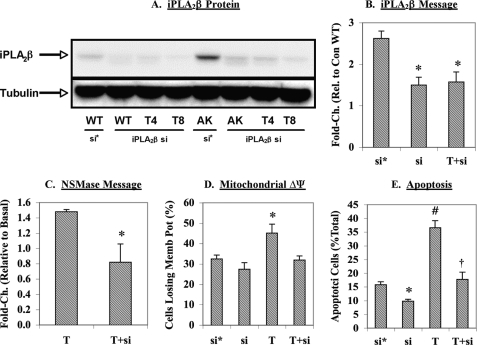
To knock down iPLA2β, wild type and Akita cells were transfected with iPLA2β siRNA according to the manufacturer's instructions (Qiagen, Valencia, CA). Cells were seeded in wells with culture medium containing serum and antibiotics and allowed to reach 50–80% confluence. The cells were then transfected with either control siRNA (si* in Fig. 7) or iPLA2β siRNA (si in Fig. 7). The siRNAs were prepared in dilution buffer and mixed gently with RNAiFect transfection reagent. The mixture was then incubated at room temperature for 10–15 min to allow formation of transfection complexes. The complexes were then gently added to the cells. The cells were cultured with the transfection complexes at 37 °C under an atmosphere of 95% air, 5% CO2 for 72 h prior to experimentation.
FIGURE 7.
Effects of iPLA2β knockdown on NSMase induction, mitochondrial membrane potential loss, and apoptosis in Akita cells. Wild type and Akita cells were transfected with either control siRNA (si*) or iPLA2β siRNA (si) and subsequently treated with DMSO vehicle or thapsigargin (T) (1 μm). The cells were harvested at 8 h for various experimental protocols, each done 3–5 times. A, iPLA2β protein. Cell lysates were prepared, and 30-μg protein aliquots were processed for immunoblotting. B, iPLA2β message. Total RNA was prepared, and cDNA was generated for qRT-PCR of iPLA2β message. *, significantly different from the control siRNA group, p < 0.01. C, NSMase message. Total RNA was prepared, and cDNA was generated for qRT-PCR of NSMase message. *, significantly different from the thapsigargin group, p < 0.05. D, mitochondrial membrane potential. The percentage of cells losing ΔΨ was determined using the flow cytometry protocol as described for Fig. 3. *, significantly different from all other groups, p < 0.05. E, apoptosis. The incidence of cell death was assessed using the TUNEL assay. *, iPLA2β siRNA group significantly different from the control siRNA group, p < 0.001. #, thapsigargin group significantly different from the control siRNA group, p < 0.0001. †, thapsigargin plus iPLA2β siRNA group significantly different from the thapsigargin group, p < 0.005. The means ± S.E. for each measurement are presented in B–E.
Islet Isolation
Male mice (4–5 weeks of age) were anesthetized with ketamine/xlyazine mixture (0.75 μl/g body weight) prior to euthanization by cervical dislocation. The abdomen was isolated, and pancreata were isolated as described (50). The common bile duct was clamped at the duodenum-bile duct junction, and collagenase/Krebs-Ringer buffer (5 ml) was injected into the pancreas via the duct. Once the pancreas was completely distended, it was removed and placed in a scintillation vial with collagenase/Krebs-Ringer buffer (2.5 ml) and incubated in a 37 °C water bath for 13 min. The vial was then vigorously shaken for 90 s, followed by washing (three times) of the pancreas with Krebs-Ringer buffer containing 1 mm CaCl2 (50 ml). The pancreas was then resuspended in incomplete RPMI 1640 (without FBS or penicillin/streptomycin, 25 ml) and poured through a 70-μm cell strainer into a Petri dish. The cells were washed further with incomplete RPMI 1640 (75 ml). The cell strainer was then inverted and rinsed with complete RPMI 1640 (10% fetal bovine serum, 2× penicillin/streptomycin, 25 ml) to collect the remaining islets in solution. The islets were then hand-picked under a microscope, counted, and incubated overnight in media under an atmosphere of 5% CO2, 95% and 37 °C. Typical islet yields ranged from 25 to 50 from the Akita mice and 150 from WT mice. The islets were used to prepare total RNA for message and for immunostaining analyses, as described below.
Quantitative Reverse Transcription-PCR (qRT-PCR)
Total RNA was prepared from cells and islets cells using the RNeasy kit (Qiagen Inc.) as described (41, 42). cDNA was then generated using the SuperScriptII kit (Invitrogen) and heat-inactivated (70 °C for 15 min). PCR amplifications were performed using the SYBR Green PCR kit in an ABI 7000 detection system (Applied Biosystems). The primers were designed based on known mouse sequences for iPLA2β, SREBP-1, NSMase, and control 18 S provided in the GenBankTM data base with identification numbers 53357, 20787, 20598, and 19791, respectively. The sense/antisense primer sets were as follows: iPLA2β, gccctggccattctacacagta/cacctcatccttcatacggaagt; SREBP-1, ccagagggtgagcctgacaa/agcctctgcaatttccagatct; NSMase, ctgagtacagccgacagaagga/gggccagttcccaagctt; 18 S, gccgctagaggtgaaattcttg/cattcttggcaaatgctttcg.
Immunostaining
Islets were fixed in 10% formalin and 5 μl of tissue marking dye, followed by the addition of 150 μl of low melting agarose. The mixture was spun down quickly to settle the islets at one surface of the agarose, which was then allowed to solidify. The islet-containing blocks were then processed, and paraffin sections (8–10 μm) were prepared for staining for iPLA2β and SREBP-1. The sections were incubated overnight with primary antibodies (1:25), washed with PBS (4 × 30 min), incubated for 2–3 h with secondary antibodies (1:100 of Cy3 for iPLA2β and Alexa Fluor 594 for SREBP-1), and washed with PBS (three times for 10 min each). DAPI stain (20–30 μl) was then added, and the sections were sealed with a coverslip using nail polish.
Statistical Analyses
Data were converted to mean ± S.E. values, and Student's t test was applied to determine significant differences between two samples (p < 0.05). The n value for each analysis is indicted in the figure legends.
RESULTS
Expression of ER Stress Factors in Akita β-Cells
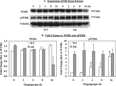
As part of the unfolded protein response, various ER factors are induced to alleviate ER stress. One such marker of unfolded protein response activation is pPERK (51). Because the Akita β-cells are reported to be a spontaneous model of ER stress, we compared pPERK levels in these cells with those in WT β-cells. As shown in Fig. 1, basal pPERK is greater in the Akita relative to wild type cells. Although exposure to thapsigargin induced pPERK expression in both the WT and Akita cells, they peaked earlier and remained higher in the Akita cells (Fig. 1B). These observations are consistent with the original findings of increased ER molecular chaperones and ER stress response element-binding transcription factors in the Akita cells (43) and confirm that the Akita cells are predisposed to developing ER stress and that they are a good model for the study of underlying mechanisms that contribute to β-cell apoptosis due to ER stress.
FIGURE 1.
Basal expression of ER stress factors and apoptotic factors. WT and AK β-cells were harvested at various times after exposure to thapsigargin (0.50 μm), and 30 μg of lysate protein was processed for immunoblotting analyses for PERK and pPERK. Tubulin was used as a loading control. These analyses were done three separate times. A, immunoblot. A representative blot for PERK and pPERK is presented. B, densitometry. The ratios of PERK- and pPERK-immunoreactive bands to the corresponding tubulin band were determined, and means ± S.E. of -fold change relative to WT at 0 h are presented. * and †, significantly different from corresponding WT, p < 0.001 and p < 0.05, respectively. #, significantly different from WT at 0 h, p < 0.05.
Reduced Sphingomyelins in Akita β-Cells
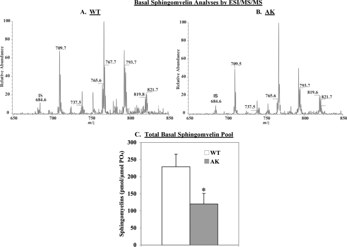
We reported that ER stress-induced ceramide generation via NSMase-catalyzed sphingomyelin hydrolysis was a critical contributor to INS-1 cell apoptosis (41, 42). If this pathway is activated by ER stress, this should be reflected by reduced abundances of sphingomyelin molecular species in the Akita cells. To examine this possibility, WT and Akita cells were harvested under basal conditions and analyzed by ESI/MS/MS. The spectra in Fig. 2 display tracings of Li+ adducts of sphingomyelin species in cell lysates after the addition of the 14:0/14:0-GPC internal standard, which is represented in the spectrum by its (M + Li+)+ ion (m/z 684). Similar to the INS-1 cells, the major sphingomyelin species endogenous to WT and Akita β-cells are 16:0 (m/z 709), 18:0 (m/z 737), 22:0 (m/z 693), 24:1 (m/z 819), and 24:0 (m/z 821). The spectra were acquired by monitoring constant neutral loss of 59, as described (52), in WT (Fig. 2A) and Akita (Fig. 2B) cells. As reflected by the decreases in the intensity of ions representing them, the relative abundances of the sphingomyelin molecular species are decreased in the Akita cells in comparison with WT cells. Normalization of individual sphingomyelin molecular species to lipid phosphorus revealed the 16:0, 18:0, 22:0, 24:0, and 24:1 species in the Akita cells to be 47 ± 9, 22 ± 2, 75 ± 4, 95 ± 4, and 78 ± 10% of WT cells, respectively. The total pool of sphingomyelins (pmol/μmol of PO4) revealed a 48% decrease in sphingomyelin abundance in the Akita cells, relative to the WT cells (Fig. 2C). These findings indicate that hydrolysis of sphingomyelins is ramped up in the Akita cells and suggest the possibility of consequential triggering of mitochondrial abnormalities.
FIGURE 2.
Sphingomyelin analyses by ESI/MS/MS. Phospholipids were extracted from WT and AK β-cells, and the abundances of sphingomyelin molecular species were analyzed by ESI/MS/MS in the presence of 14:0/14:0-GPC (m/z 684) internal standard. The WT (A) and AK (B) spectra were obtained by monitoring constant neutral loss of 59. C, total sphingomyelin pool. Content of each sphingomyelin molecular species (pmol) was determined in each group and normalized to total phosphate (μmol of PO4). The means ± S.E. (n = 3) of the total (pmol/μmol of PO4) sphingomyelin pools are presented. *, AK group significantly different from WT, p < 0.05.
Compromised ΔΨ in Akita β-Cells
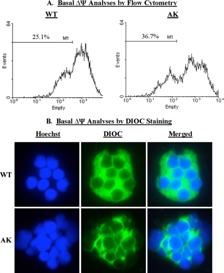
Loss of ΔΨ is an important step in the induction of cellular apoptosis (47). Because thapsigargin-induced ER stress caused a loss in INS-1 cell ΔΨ (42) in an NSMase-dependent manner, we examined whether there was a similar compromising of ΔΨ in the Akita cells. As in the earlier study, we used a flow cytometry protocol to monitor ΔΨ in a suspension of cells to which a fluorescent Mito Flow reagent was added. This reagent concentrates in the mitochondria of healthy cells, but the mitochondria of cells undergoing apoptosis become compromised and accumulate less of the reagent, and this is reflected by a decrease in the fluorescence signal and the appearance of a second peak that is left of the original. The spectra presented in Fig. 3A reflect fluorescence measurement in 10,000 cells, and the percentage of cells (mean of four separate experiments) losing ΔΨ was analyzed by the application software and is indicated as M1. These analyses revealed that a higher percentage of Akita cells have compromised ΔΨ than WT cells under basal conditions. However, unlike the INS-1 cells, the Akita and corresponding WT cells were found to be prone to clumping. This resulted in broader peaks in the spectra and an unexpectedly higher M1 value, even in the WT cells. We therefore established a second protocol in which mitochondria-associated DiOC6(3) staining (green) was monitored in live cells by confocal microscopy. As seen in Fig. 3B, DiOC6(3) under basal conditions is more significantly retained in the mitochondria of WT than in Akita cells, confirming compromised mitochondrial integrity in the Akita cells. Further, Hoechst staining (blue) reveals that the nuclei in Akita cells are irregular in shape and size, as compared with the nuclei in WT cells, suggesting that the cell death process is under way in the Akita cells. These findings suggest that the spontaneous development of ER stress in β-cells also triggers the mitochondrial apoptotic pathway.
FIGURE 3.
Basal mitochondrial membrane potential (ΔΨ) monitoring. ΔΨ was monitored in WT and AK β-cells by flow cytometry and DiOC6(3) staining. A, flow cytometry. The spectra reflect fluorescence measurement in 10,000 cells, and the percentage of cells losing ΔΨ is indicated as M1. These analyses were done four separate times. B, DiOC6(3) staining. Cells were loaded with the blue nuclear (Hoechst; left panels) and green mitochondrial (DiOC6(3) (DIOC); middle panels) stain and examined by confocal microscopy. The merged images are shown in the right panels.
Basal and ER Stress-induced Apoptosis Are Amplified in Akita β-Cells
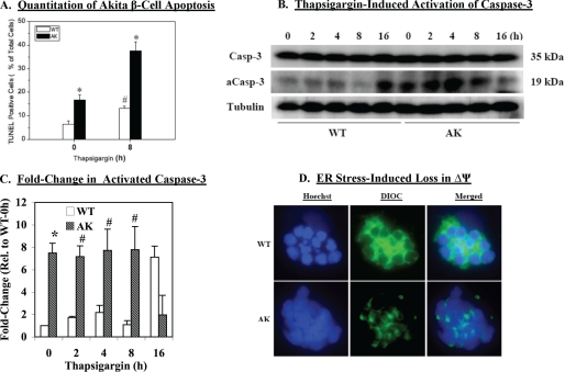
In view of the above findings, we next examined if the Akita cells are more susceptible to apoptosis. TUNEL analyses (Fig. 4A) revealed a higher incidence of basal apoptosis that was coincident with higher levels of activated caspase-3 (Fig. 4, B and C). Exposure to thapsigargin induced activation of caspase-3 in both wild type and Akita with peak activation achieved earlier in the Akita cells than in the wild type cells. This is reflected by a greater -fold increase, relative to basal, in Akita cell apoptosis at 8 h (WT, 1.59 ± 0.09 versus Akita (AK), 2.26 ± 0.16, p = 0.0175).
FIGURE 4.
Assessment of apoptosis and mitochondrial membrane potential (ΔΨ) in Akita β-cells. WT and AK β-cells were exposed to DMSO (control) or thapsigargin (T) (0.50 μm) and harvested at various times for analyses. A, apoptosis quantitation. Total cell number was determined using DAPI nuclear stain, and the mean ± S.E. (n = 3) percentages of TUNEL-positive apoptotic cells are presented. *, AK group significantly different from WT, p < 0.05. #, WT treated group significantly different from WT control group, p < 0.05. B, caspase-3 activation. Cell lysates were prepared and processed for full-length and activated caspase-3 (Casp-3 and aCasp-3, respectively) immunoblotting analyses. Tubulin was used as a loading control. These analyses were done three separate times. C, activated caspase-3 densitometry. The ratio of the activated caspase-3-immunoreactive band to the corresponding tubulin band was determined, and means ± S.E. of -fold change relative to WT at 0 h are presented. * and #, significantly different from corresponding WT; p < 0.01 and p < 0.05, respectively. D, ΔΨ analyses. Following exposure to DMSO or thapsigargin for 8 h, the WT and AK β-cells were loaded with the blue nuclear (Hoechst) (left panels) and green mitochondrial (DiOC6(3)) (DIOC) (middle panels) stains and examined by confocal microscopy. The merged images are shown in the right panels.
ER Stress Amplifies ΔΨ Loss in Akita β-Cells
Because induction of ER stress led to mitochondrial abnormalities in the INS-1 cells, we examined whether a similar affect was evident in the Akita cells. Compared with basal conditions (Fig. 2B), exposure to thapsigargin for 8 h resulted in dispersion of DiOC6(3) staining and loss in nuclear uniformity in the WT cells (Fig. 4D). These outcomes were accelerated in the Akita cells and suggest that activation of the mitochondrial apoptotic pathway contributes to ER stress-induced apoptosis of these cells also.
Expression and ER Stress-induced Activation of iPLA2β and NSMase
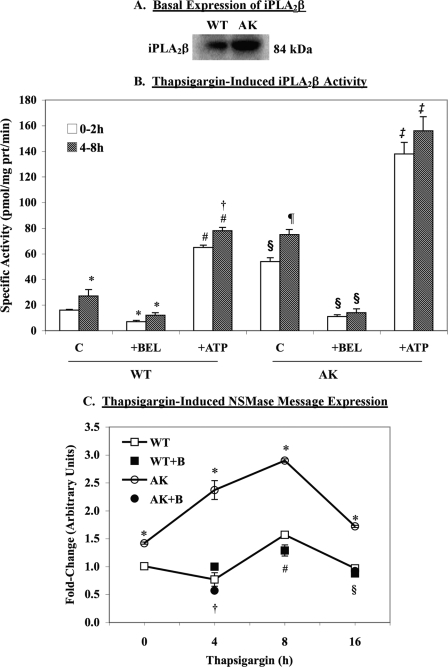
Our collective observations (40–42) indicate that ER stress-induced apoptosis occurs by an iPLA2β-dependent mechanism involving induction of NSMase and activation of the mitochondrial apoptotic pathway. The above findings suggest participation of both NSMase and the mitochondria in Akita β-cell death. We therefore examined the extent of the iPLA2β role in this process. Surprisingly, basal expression of iPLA2β protein was found to be greater in the Akita than in the WT cells (Fig. 5A), and this was reflected by a 3-fold higher activity (16 ± 1 pmol/mg of protein/min for WT versus 54 ± 3 pmol/mg of protein/min for Akita). Also, as shown in Fig. 5B, exposure to thapsigargin promoted iPLA2β activity in both WT and AK β-cells. Consistent with manifestation of an iPLA2β activity, basal and thapsigargin-treated activities were suppressed by BEL and stimulated by ATP (∼3-fold) in WT and AK β-cells.
FIGURE 5.
Basal and ER stress-induced expression of iPLA2β and NSMase. A, iPLA2β protein. Cell lysates were prepared from WT and AK β-cells, and 30-μg protein aliquots were processed for immunoblotting analyses. B, iPLA2β activity. Cytosol was prepared from control or thapsigargin-treated (0.50 μm) AK cells, and iPLA2β catalytic activity was assayed in 25 μg of cytosolic protein in the absence and presence of ATP (10 mm) or BEL (1 μm). Means ± S.E. (n = 6–8) of specific activity (pmol/mg protein/min) are presented. *, WT 4–8 h significantly different from WT 0–2 h and WT with BEL significantly different from corresponding WT without BEL, p < 0.05. #, WT with ATP significantly different from corresponding WT without ATP, p < 0.0001. †, WT with ATP 4–8 h significantly different from WT 0–2 h, p < 0.005. §, AK 0–2 h significantly different from WT 0–2 h and AK with BEL significantly different from corresponding AK without BEL, p < 0.0001. ¶, AK 4–8 h significantly different from AK 0–2 h, p < 0.001. ‡, AK with ATP significantly different from AK without ATP, p < 0.00001. C, NSMase message. Total RNA was isolated from DMSO- or thapsigargin-treated (0–16 h) WT and AK β-cells, and qRT-PCR analyses were performed for NSMase. In some experiments, the cells were treated with BEL (+B) (1 μm) for 30 min, washed, and then exposed to thapsigargin. Means ± S.E. (n = 5–6) of the -fold increase in message are presented. 18 S was used as a housekeeping control. *, AK significantly different from corresponding WT, p < 0.0005. †, AK with BEL significantly different from AK, p < 0.001. #, WT with BEL significantly different from WT, p < 0.05. §, AK with BEL significantly different from AK, p < 0.05.
Because we previously reported iPLA2β-mediated induction of NSMase during ER stress, we compared its message levels in the WT and Akita cells. As shown in Fig. 5C, basal NSMase was higher in the Akita than in the WT cells, and exposure to thapsigargin induced NSMase. The increase in NSMase message occurred earlier and reached a higher -fold increase in the Akita cells, relative to WT cells. Consistent with an iPLA2β-mediated effect, NSMase induction in both groups was inhibited by inactivation of iPLA2β with BEL.
In contrast with the earlier observations in INS-1 cells, in which iPLA2β was overexpressed (41, 42) and the cells were treated with thapsigargin, the responses described here in the Akita β-cells occurred in a spontaneous ER stress model. The present findings therefore strengthen the likelihood that iPLA2β plays a critical role in ER stress-induced β-cell apoptosis.
Evidence for Induction of iPLA2β by SREBP-1 during ER Stress
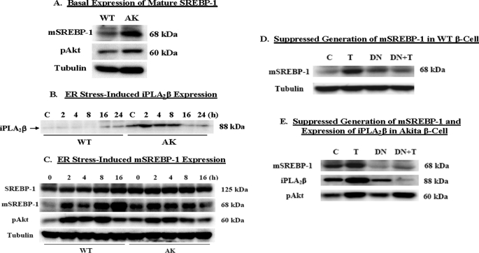
The finding of increased iPLA2β expression in the Akita cells suggests that the β-cell iPLA2β may be subject to regulation, but the mechanism for its induction remains to be elucidated. One potential possibility is suggested by the recent report identifying a sterol-regulatory element (SRE) in the iPLA2β gene (53). That study performed in Chinese hamster ovary cells revealed that binding of SREBPs to SRE leads to iPLA2β transcription. Of relevance to our interests is that SREBPs are known to be induced under stressful conditions and processed to the mature form of SREBPs (mSREBP) in β-cells experiencing ER stress due to glucolipotoxicity and thapsigargin (54). Consistent with these observations, we find that basal expression of mSREBP-1 protein is greater in Akita than in WT cells (Fig. 6A). Further, Akt phosphorylation, which promotes SREBP-1 processing and nuclear accumulation of the mSREBP-1 (55–57), is also increased.
FIGURE 6.
SREBP-1 expression and SREBP-1-induced activation of iPLA2β. WT and AK β-cells were exposed to DMSO (Control) or thapsigargin (T) (0.50 μm) and harvested at various times, and 30-μg protein aliquots were processed for immunoblotting analyses. A, basal mSREBP-1 expression. B, thapsigargin-induced iPLA2β expression. C, thapsigargin-induced mSREBP-1 expression. DN SREBP-1 was expressed in WT and AK β-cells and treated with DMSO or thapsigargin (0.50 μm) for 8 h. Cell lysates were then prepared, and 30-μg protein aliquots were processed for immunoblotting analyses for mSREBP-1 and iPLA2β. D, mSREBP-1 in WT β-cells. E, mSREBP-1 and iPLA2β in Akita β-cells. Tubulin was used as a loading control. These analyses were done 3–5 separate times.
To determine if there is a link between SREBP-1 and iPLA2β expression, cells were treated with thapsigargin and harvested at various times for immunoblotting analyses. Exposure to thapsigargin promoted a temporal increase in iPLA2β expression in both WT and Akita cells (Fig. 6B), with a more pronounced and earlier increase apparent in the Akita cells. Also, as seen in Fig. 6C, corresponding increases in mSREBP-1 and pAkt are also evident. These findings reveal a correlation between iPLA2β and generation of SREBP-1; however, they do not indicate whether the increase in iPLA2β expression is due to SREBP-1 processing.
To address a direct role of SREBP-1 on iPLA2β induction, we expressed a DN form of SREBP-1 in the WT and Akita cells. It has been reported that although the DN form itself is unable to activate transcription, it interferes with the processing of endogenous SREBP-1 and its binding to the SRE element (49). As seen in Fig. 6D, expression of the DN SREBP-1 reduced basal levels of the mSREBP-1 form and its increase following exposure to thapsigargin. In association with the reduction in mSREBP-1, both basal and thapsigargin-induced increases in iPLA2β were suppressed in cells expressing the DN SREBP-1 (Fig. 6E). As expected by the upstream role of pAkt in SREBP-1 activation, phosphorylation of Akt was unaffected by DN SREBP-1 (data not shown). These findings are taken to indicate that SREBP-1 activation can promote iPLA2β expression.
Effects of iPLA2β Knockdown in the Akita Cells
Because basal iPLA2β is increased in the Akita cells that are predisposed to developing ER stress and is induced in both WT and Akita cells following exposure to thapsigargin, we next examined whether suppression of iPLA2β expression prevents the observed outcomes. To address this issue, we used siRNA protocols to knockdown iPLA2β in the WT and Akita cells. The cells were harvested under basal conditions or following an 8-h exposure to thapsigargin. As illustrated in Fig. 7A, both basal and thapsigargin-induced iPLA2β protein expression are reduced in the WT and Akita cells transfected with the iPLA2β siRNA (si), relative to cells transfected with the control siRNA (si*). Comparison of iPLA2β message in the control siRNA-transfected WT and Akita cells revealed nearly 2.5-fold higher basal iPLA2β message in the Akita cells (Fig. 7B). Transfection of the Akita cells with the iPLA2β siRNA reduced basal iPLA2β message levels and prevented iPLA2β induction due to thapsigargin.
Basal NSMase message levels that were higher in non-transfected Akita cells (Fig. 5C) remained greater in the Akita, relative to wild type, cells transfected with the control siRNA (arbitrary units 1.01 ± 0.06 for WT versus 2.09 ± 0.40 for AK, p = 0.0171). Although transfection with the iPLA2β siRNA did not alter basal NSMase message levels, it significantly suppressed thapsigargin-induced increase in NSMase message (Fig. 7C).
To obtain a measure of a role for iPLA2β in promoting loss in mitochondrial ΔΨ, the cells were processed by flow cytometry. This allowed quantitative assessment of the number of cells with compromised ΔΨ. The incidence of loss in ΔΨ under untransfected basal conditions was greater in the Akita, relative to wild type, cells (22 ± 2% for WT versus 33 ± 2% for AK, p = 0.0185). As shown in Fig. 7D, transfection with control or iPLA2β siRNAs did not significantly affect the basal incidence of cells with ΔΨ loss, but iPLA2β siRNA prevented thapsigargin-induced increase in the percentage of Akita cells that lose ΔΨ.
The effect of suppressing iPLA2β expression on the final outcome of apoptosis was tested next using TUNEL analyses. As shown in Fig. 7E, transfection with control siRNA did not suppress the higher incidence of apoptosis in Akita cells, relative to WT cells (see Fig. 4A). However, transfection with iPLA2β siRNA suppressed basal apoptosis and also prevented thapsigargin-induced apoptosis in the Akita cells. These findings are taken as direct evidence of an important role for iPLA2β in the apoptosis process induced by ER stress in β-cells.
iPLA2β, SREBP-1, and NSMase Messages and iPLA2β and SRE-binding Protein in Islets from Akita Mice
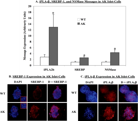
In light of the observations in the Akita β-cells, we considered whether iPLA2β and SREBP-1 expression is altered under in vivo conditions in the Akita mouse model. We therefore isolated islets from Akita and WT littermates and prepared cDNA for qRT-PCR analyses. The mice were sacrificed within a week of weaning (4–5 weeks of age). Even at this early age, the Akita mice exhibited hyperglycemia (∼450 mg/dl), whereas the WT mice were euglycemic. Consistent with the findings in the Akita cells, mRNAs for iPLA2β, SREBP-1, and NSMase were significantly elevated in the Akita islets, relative to WT islets (Fig. 8A).
FIGURE 8.
iPLA2β, SREBP-1, and NSMase message and iPLA2β and SREBP-1 protein expression in Akita mouse islets. Islets were isolated from WT and Akita mice at 4 weeks of age. Total RNA was prepared, and qRT-PCR analyses for iPLA2β, SREBP-1, and NSMase were performed. Means ± S.E. (n = 3–5) of the -fold increase in each message are presented (A). 18 S was used as a control. *, AK group significantly different from corresponding WT group, p < 0.05. B and C, paraffin sections (10 μm) of islets isolated from WT and AK mice at ∼4 weeks of age were prepared and stained for cell nuclei (DAPI) (blue) and SREBP-1 (red in B) or iPLA2β (red in C), and fluorescence was recorded using a Nikon Eclipse TE300 microscope. The insets in B are magnifications of islet cell nuclei.
To substantiate the findings of higher message in the Akita islets, we performed immnocytofluorescence analyses for iPLA2β and SREBP-1. Paraffin sections (10 μm) of islets were prepared and stained for nucleus-associating DAPI (blue) and iPLA2β or SREBP-1 (red). The exposure times for each were the same in WT and AK sections. Whereas diffuse background fluorescence was evident in the WT islets, intense and punctate staining of SREBP-1 (Fig. 8B) and iPLA2β (Fig. 8C) are seen in the Akita islets. These findings are taken as evidence for increased expression of both SREBP-1 and iPLA2β in islets from diabetic Akita mice, relative to islets from age-matched normoglycemic WT mice. Similar to the observations in the Akita β-cells above, the Akita islet cell nuclei were also irregular in shape with some evidence of fragmentation in comparison with the rounded and uniform structure of the nuclei in WT islets (insets in the left panels of Fig. 8B). Our findings in Akita islets thus reveal the potential activation of a pathway linking SREBP-1, iPLA2β, and NSMase in an animal model that develops diabetes as a consequence of β-cell apoptosis resulting from spontaneous development of ER stress in islet β-cells.
DISCUSSION
Both T1DM and T2DM are associated with losses in β-cells due to apoptosis. It is therefore important to determine the underlying mechanisms that contribute to this process. Recent reports in experimental models and in clinical settings suggest that ER stress is a potential cause of β-cell apoptosis during the development of diabetes mellitus (6, 15–22, 24–26). Because β-cells serve a secretory function, they are endowed with a highly developed ER that renders them particularly susceptible to developing ER stress.
We found that ER stress causes INS-1 cell apoptosis, in part, by an iPLA2β-dependent mechanism (40). Subsequent studies revealed a previously unrecognized pathway (41, 42) involving accumulation of ceramides via an iPLA2β-mediated induction of NSMase. The ceramides, generated from NSMase-catalyzed hydrolysis of sphingomyelins, promote mitochondrial abnormalities and amplify the apoptosis outcome. The roles for iPLA2β and NSMase in this process are supported by the findings of suppression of sphingomyelin hydrolysis, ceramide generation, mitochondrial activation, and apoptosis following inactivation of iPLA2β or NSMase (41, 42).
The above proposed schema of iPLA2β involvement in β-cell apoptosis was derived from studies in INS-1 cells in which ER stress was induced with thapsigargin. The plausibility of the pathway is strengthened by the finding that it is amplified by overexpression of iPLA2β and suppressed by inactivation of iPLA2β. However, identification of a similar role for iPLA2β in a spontaneous ER stress model would significantly strengthen the assertion that iPLA2β participates in ER stress-induced β-cell apoptosis. We therefore chose to address this issue in the Akita mouse model. However, because the islet yield from these mice is limiting, we pursued studies in a β-cell line established from the Akita mice (43). The findings in these cells were compared with a β-cell line derived from isogenic WT littermates.
Herein, we find that even under basal conditions, the Akita β-cells are predisposed to developing ER stress, as reflected by the greater induction of pPERK, and to be more prone to undergo apoptosis, as revealed by the greater abundance of TUNEL-positive cells and activated caspase-3, relative to WT β-cells. The greater propensity of the Akita cells to develop ER stress under basal conditions is also revealed by the nearly 2-fold higher basal splicing of XBP1 mRNA in the Akita cells, relative to isogenic WT counterparts (58).
The Akita cells also expressed higher message for NSMase, contained lower abundances of sphingomyelins, and exhibited a higher incidence of loss in mitochondrial membrane potential (ΔΨ). Permeabilization of the outer mitochondrial membrane is considered to be a “point of no return” during apoptosis (59), and PTP opening alters outer mitochondrial membrane permeability. Prolonged activation of PTP leads to loss in ΔΨ and release of cytochrome c, a necessary event in the mitochondrial cell death pathway (60, 61), from the mitochondrial intermembrane space into the cytosol. Following its release into the cytosol, the proapoptotic cytochrome c forms the apoptosome complex with apoptosis protease-activating factor-1 to induce caspases and subsequent apoptosis (62). The present findings therefore are consistent with the triggering of mitochondrial abnormalities as a consequence of increased sphingomyelin hydrolysis in Akita β-cells (41, 42).
Most intriguingly, the Akita cells expressed higher iPLA2β protein than the WT cells. This was reflected by a nearly 3-fold higher basal cytosolic iPLA2β specific activity. These findings strengthen the likelihood that iPLA2β-mediated NSMase activation is a critical factor in ER stress-induced β-cell apoptosis. To further establish this possibility, the effects of accelerating ER stress in the Akita cells were assessed. Exposure to thapsigargin caused elevations in caspase-3 activation and abundance of TUNEL-positive cells that were accompanied by temporal increases in iPLA2β protein and specific activity in both WT and Akita β-cells. However, these outcomes were accelerated in the Akita cells. Thapsigargin also induced NSMase in both WT and Akita cells, and this occurred earlier and the -fold increases in message were greater in the Akita cells. That this was an iPLA2β-dependent effect was evidenced by the suppression in NSMase induction following inactivation of iPLA2β in both WT and Akita cells.
To establish that the outcomes measured in the Akita β-cells require iPLA2β, the Akita cells were transfected with iPLA2β siRNA to knock down iPLA2β message and protein levels. The siRNA protocol resulted in dramatic suppression of not only basal but also thapsigargin-induced increases in iPLA2β message and protein. Further, whereas thapsigargin-induced NSMase induction, mitochondrial membrane potential loss, and apoptosis were unaffected by transfection with control siRNA, they were significantly suppressed in Akita cells transfected with the iPLA2β siRNA. In fact, the higher basal apoptosis was also significantly decreased in these cells. These findings indicate that iPLA2β is a critical factor during the development and progression of ER stress-induced abnormalities in the β-cell.
Our recent finding of higher iPLA2β protein and activity in the ER and mitochondria of INS-1 cells exposed to thapsigargin was suggestive of increased mobilization of iPLA2β by these organelles with the onset and development of ER stress (41, 42). However, the surprising finding of higher basal iPLA2β protein expression in Akita cells in the present study raises the possibility that it may in fact be subject to pre- and post-transcriptional regulation under stressful conditions.
Other than ATP, only two potential candidates have been identified as regulating iPLA2β: CaMKIIβ and SREBP-1. Whereas CaMKIIβ is reported to activate iPLA2β (63), SREBP-1 has been shown to promote transcription of the iPLA2β gene (53). The transcription factor ATF6 plays a key role in the unfolded protein response, and ER stress leads to its cleavage by site 1 and site 2 proteases that are also activated by ER stress (64). These proteases process SREBPs (65) into active mature forms that enter the nucleus and transactivate target genes (66). SREBPs are known to be induced under stressful conditions and are activated in β-cells experiencing ER stress due to glucolipotoxicity or thapsigargin (54). A fascinating extension to these observations is the recent identification of an SRE in the iPLA2β gene (53). That study, performed in Chinese hamster ovary cells that were mutated to constitutively generate mSREBPs, revealed that binding of mSREBPs to SRE leads to iPLA2β transcription. We therefore considered the possibility that SREBP-1 may modulate iPLA2β levels in β-cells that are predisposed to developing ER stress.
We find that processing of SREBP-1 to the mSREBP-1 form under basal conditions is higher in the Akita β-cells than in the WT β-cells. This coincided with higher pAkt levels, which is consistent with previous reports that phosphorylation of Akt stimulates the synthesis and nuclear localization of mSREBP-1 (55–57). Further, exposure to thapsigargin led to temporal increases in mSREBP-1 (and pAkt) in parallel with elevations in iPLA2β protein. These findings provide the first evidence of a correlation between mSREBP-1 and iPLA2β expression with the onset and progression of diminished cellular integrity.
To determine if there is a causal relationship between SREBP-1 activation and iPLA2β expression, a DN SREBP-1 was expressed in WT and Akita β-cells. The DN form itself is unable to activate transcription, but it interferes with the processing of endogenous SREBP-1 and its binding to the SRE element (49). As expected, both basal and thapsigargin-induced increase in mSREBP-1 were suppressed in WT and Akita cells by DN-SREBP-1, whereas phosphorylation of Akt, an event upstream of SREBP-1, remained unaffected. Also, consistent with mSREBP-1-mediated regulation of iPLA2β expression, both basal and induced iPLA2β expression were suppressed by the DN protein.
These observations therefore identify a novel mechanism by which a biological process (stress) can regulate iPLA2β expression in β-cells. Because of accumulating evidence supporting a causative role for ER stress-induced β-cell apoptosis in the development of diabetes, we sought to substantiate our findings in the Akita (Ins2Akita) mouse. These mice develop mild hyperglycemia rapidly after birth, although they continue to live for over 8 months. These mice contain a spontaneous dominant mutation in the insulin 2 gene on a C57BL6 mouse background that results in the replacement of cysteine with tyrosine at position 96, causing disruption of a disulfide bridge required for proper insulin folding. This causes proinsulin to accumulate in the ER and leads to the initiation of the unfolded protein response. The unresolved ER stress subsequently leads to pancreatic islet β-cell apoptosis in the absence of obesity, insulitis, or insulin resistance (45, 67–70). Heterozygous Akita mice become hyperglycemic at 3–4 weeks of age but remain fertile and viable in the absence of exogenous insulin treatment. The diabetic phenotype is more severe and progressive in the Akita male than in the female. We therefore examined islets from male Akita and WT littermates of ∼4 weeks of age.
Consistent with the findings in the Akita β-cells, iPLA2β, SREBP-1, and NSmase messages were significantly increased in the Akita islets, as compared with WT islets. This was supported by the more prominent staining for iPLA2β and SREBP-1 in Akita, relative to the WT, islet sections. It may be argued that the SREBP-1 detected does not appear to be nucleus-associated. However, typically, only a fraction of the total SREBP-1 pool is processed to the mature form that translocates to the nucleus. Although others have demonstrated nuclear translocation by immunocytochemistry (48, 49), these studies were performed with overexpressed truncated SREBP1. As such, it is likely that the immunocytochemistry assay is not sufficiently sensitive to detect translocation of endogenous SREBP1. Nevertheless, collectively, these findings strongly support a link between and a role for SREBP-1, iPLA2β, and NSMase during the development of diabetes mellitus due to ER stress-induced in β-cell apoptosis.
Multiple lines of evidence indicate that the loss of β-cells due to apoptosis contributes to the onset and progression of T1DM and T2DM. It is now recognized that prolonged ER stress can cause β-cell apoptosis and lead to the development of diabetes mellitus, and we recently identified a role for iPLA2β in ER stress-induced β-cell apoptosis. Here, we present evidence for the activation of iPLA2β in a spontaneous ER stress model that leads to the development of diabetes due to β-cell apoptosis. We further identified for the first time that iPLA2β expression is subject to regulation by SREBP-1 during the evolution of a metabolic disorder, the incidence of which continues to rise. As such, our findings contribute to the understanding of iPLA2β biology and may provide insight into developing novel targeted therapeutics to prevent or delay β-cell apoptosis.
Acknowledgments
We acknowledge the advice provided by the Washington University Diabetes Research and Training Center-supported Morphology and β-Cell Morphology cores.
This work was supported, in whole or in part, by National Institutes of Health Grants R01-DK69455, P41-RR00954, P60-DK20579, P30-DK56341, and DK065671. This work was also supported by a grant from the American Diabetes Association (to S. R.) and National Science Foundation Grant NSF-MCB-0544068 (to S. E. B.).
- T1DM
- Type 1 diabetes mellitus
- T2DM
- Type 2 diabetes mellitus
- AK
- Akita
- pAkt
- phosphorylated Akt
- PERK
- pancreatic ER kinase
- pPERK
- phosphorylated PERK
- BEL
- bromoenol lactone suicide inhibitor of iPLA2β
- DN
- dominant negative
- ER
- endoplasmic reticulum
- ESI/MS/MS
- electrospray ionization/tandem mass spectrometry
- GPC
- glycerophosphocholine
- iPLA2β
- β-isoform of group VIA Ca2+-independent phospholipase A2
- NSMase
- neutral sphingomyelinase
- PBS
- phosphate-buffered saline
- PLA2
- phospholipase A2
- mSREBP-1
- mature (or processed) SREBP-1
- TUNEL
- terminal deoxynucleotidyl transferase-mediated (fluorescein) dUTP nick end labeling
- WT
- wild type
- siRNA
- small interfering RNA
- DAPI
- 4′,6-diamidino-2-phenylindole
- qRT-PCR
- quantitative reverse transcription-PCR
- SRE
- sterol-regulatory element
- DiOC6(3)
- 3,3′-dihexyloxacarbocyanine iodide.
REFERENCES
- 1.Klöppel G., Löhr M., Habich K., Oberholzer M., Heitz P. U. (1985) Surv. Synth. Pathol. Res. 4, 110–125 [DOI] [PubMed] [Google Scholar]
- 2.Stefan Y., Orci L., Malaisse-Lagae F., Perrelet A., Patel Y., Unger R. H. (1982) Diabetes 31, 694–700 [DOI] [PubMed] [Google Scholar]
- 3.Butler A. E., Janson J., Bonner-Weir S., Ritzel R., Rizza R. A., Butler P. C. (2003) Diabetes 52, 102–110 [DOI] [PubMed] [Google Scholar]
- 4.Yoon K. H., Ko S. H., Cho J. H., Lee J. M., Ahn Y. B., Song K. H., Yoo S. J., Kang M. I., Cha B. Y., Lee K. W., Son H. Y., Kang S. K., Kim H. S., Lee I. K., Bonner-Weir S. (2003) J. Clin. Endocrinol. Metab. 88, 2300–2308 [DOI] [PubMed] [Google Scholar]
- 5.Araki E., Oyadomari S., Mori M. (2003) Exp. Biol. Med. (Maywood) 228, 1213–1217 [DOI] [PubMed] [Google Scholar]
- 6.Cardozo A. K., Ortis F., Storling J., Feng Y. M., Rasschaert J., Tonnesen M., Van Eylen F., Mandrup-Poulsen T., Herchuelz A., Eizirik D. L. (2005) Diabetes 54, 452–461 [DOI] [PubMed] [Google Scholar]
- 7.Mathis D., Vence L., Benoist C. (2001) Nature 414, 792–798 [DOI] [PubMed] [Google Scholar]
- 8.Tisch R., McDevitt H. (1996) Cell 85, 291–297 [DOI] [PubMed] [Google Scholar]
- 9.Bernard C., Berthault M. F., Saulnier C., Ktorza A. (1999) FASEB J. 13, 1195–1205 [DOI] [PubMed] [Google Scholar]
- 10.Pick A., Clark J., Kubstrup C., Levisetti M., Pugh W., Bonner-Weir S., Polonsky K. S. (1998) Diabetes 47, 358–364 [DOI] [PubMed] [Google Scholar]
- 11.Butler A. E., Janson J., Soeller W. C., Butler P. C. (2003) Diabetes 52, 2304–2314 [DOI] [PubMed] [Google Scholar]
- 12.Diaz-Horta O., Kamagate A., Herchuelz A., Van Eylen F. (2002) Diabetes 51, 1815–1824 [DOI] [PubMed] [Google Scholar]
- 13.Rao R. V., Castro-Obregon S., Frankowski H., Schuler M., Stoka V., del Rio G., Bredesen D. E., Ellerby H. M. (2002) J. Biol. Chem. 277, 21836–21842 [DOI] [PubMed] [Google Scholar]
- 14.Kaufman R. J. (1999) Genes Dev. 13, 1211–1233 [DOI] [PubMed] [Google Scholar]
- 15.Oyadomari S., Araki E., Mori M. (2002) Apoptosis 7, 335–345 [DOI] [PubMed] [Google Scholar]
- 16.Oyadomari S., Koizumi A., Takeda K., Gotoh T., Akira S., Araki E., Mori M. (2002) J. Clin. Invest. 109, 525–532 [DOI] [PMC free article] [PubMed] [Google Scholar]
- 17.Socha L., Silva D., Lesage S., Goodnow C., Petrovsky N. (2003) Ann. N.Y. Acad. Sci. 1005, 178–183 [DOI] [PubMed] [Google Scholar]
- 18.Harding H. P., Zeng H., Zhang Y., Jungries R., Chung P., Plesken H., Sabatini D. D., Ron D. (2001) Mol. Cell 7, 1153–1163 [DOI] [PubMed] [Google Scholar]
- 19.Delépine M., Nicolino M., Barrett T., Golamaully M., Lathrop G. M., Julier C. (2000) Nat. Genet. 25, 406–409 [DOI] [PubMed] [Google Scholar]
- 20.Takeda K., Inoue H., Tanizawa Y., Matsuzaki Y., Oba J., Watanabe Y., Shinoda K., Oka Y. (2001) Hum. Mol. Genet. 10, 477–484 [DOI] [PubMed] [Google Scholar]
- 21.Fonseca S. G., Fukuma M., Lipson K. L., Nguyen L. X., Allen J. R., Oka Y., Urano F. (2005) J. Biol. Chem. 280, 39609–39615 [DOI] [PubMed] [Google Scholar]
- 22.Oyadomari S., Takeda K., Takiguchi M., Gotoh T., Matsumoto M., Wada I., Akira S., Araki E., Mori M. (2001) Proc. Natl. Acad. Sci. U.S.A. 98, 10845–10850 [DOI] [PMC free article] [PubMed] [Google Scholar]
- 23.Kröncke K. D., Brenner H. H., Rodriguez M. L., Etzkorn K., Noack E. A., Kolb H., Kolb-Bachofen V. (1993) Biochim. Biophys. Acta 1182, 221–229 [DOI] [PubMed] [Google Scholar]
- 24.Corbett J. A., McDaniel M. L. (1992) Diabetes 41, 897–903 [DOI] [PubMed] [Google Scholar]
- 25.Mandrup-Poulsen T. (1996) Diabetologia 39, 1005–1029 [DOI] [PubMed] [Google Scholar]
- 26.Yamada T., Ishihara H., Tamura A., Takahashi R., Yamaguchi S., Takei D., Tokita A., Satake C., Tashiro F., Katagiri H., Aburatani H., Miyazaki J., Oka Y. (2006) Hum. Mol. Genet. 15, 1600–1609 [DOI] [PubMed] [Google Scholar]
- 27.Schaloske R. H., Dennis E. A. (2006) Biochim. Biophys. Acta 1761, 1246–1259 [DOI] [PubMed] [Google Scholar]
- 28.Ma Z., Ramanadham S., Wohltmann M., Bohrer A., Hsu F. F., Turk J. (2001) J. Biol. Chem. 276, 13198–13208 [DOI] [PubMed] [Google Scholar]
- 29.Mancuso D. J., Jenkins C. M., Gross R. W. (2000) J. Biol. Chem. 275, 9937–9945 [DOI] [PubMed] [Google Scholar]
- 30.Tanaka H., Takeya R., Sumimoto H. (2000) Biochem. Biophys. Res. Commun. 272, 320–326 [DOI] [PubMed] [Google Scholar]
- 31.Ma Z., Ramanadham S., Kempe K., Chi X. S., Ladenson J., Turk J. (1997) J. Biol. Chem. 272, 11118–11127 [PubMed] [Google Scholar]
- 32.Boilard E., Surette M. E. (2001) J. Biol. Chem. 276, 17568–17575 [DOI] [PubMed] [Google Scholar]
- 33.Isenović E., LaPointe M. C. (2000) Hypertension 35, 249–254 [DOI] [PubMed] [Google Scholar]
- 34.Maggi L. B., Jr., Moran J. M., Scarim A. L., Ford D. A., Yoon J. W., McHowat J., Buller R. M., Corbett J. A. (2002) J. Biol. Chem. 277, 38449–38455 [DOI] [PubMed] [Google Scholar]
- 35.Tithof P. K., Olivero J., Ruehle K., Ganey P. E. (2000) Toxicol. Sci. 53, 40–47 [DOI] [PubMed] [Google Scholar]
- 36.Williams S. D., Ford D. A. (2001) Am. J. Physiol. Heart Circ. Physiol. 281, H168–H176 [DOI] [PubMed] [Google Scholar]
- 37.Moran J. M., Buller R. M., McHowat J., Turk J., Wohltmann M., Gross R. W., Corbett J. A. (2005) J. Biol. Chem. 280, 28162–28168 [DOI] [PubMed] [Google Scholar]
- 38.Ramanadham S., Hsu F. F., Bohrer A., Ma Z., Turk J. (1999) J. Biol. Chem. 274, 13915–13927 [DOI] [PubMed] [Google Scholar]
- 39.Ramanadham S., Song H., Hsu F. F., Zhang S., Crankshaw M., Grant G. A., Newgard C. B., Bao S., Ma Z., Turk J. (2003) Biochemistry 42, 13929–13940 [DOI] [PMC free article] [PubMed] [Google Scholar]
- 40.Ramanadham S., Hsu F. F., Zhang S., Jin C., Bohrer A., Song H., Bao S., Ma Z., Turk J. (2004) Biochemistry 43, 918–930 [DOI] [PMC free article] [PubMed] [Google Scholar]
- 41.Lei X., Zhang S., Bohrer A., Bao S., Song H., Ramanadham S. (2007) Biochemistry 46, 10170–10185 [DOI] [PMC free article] [PubMed] [Google Scholar]
- 42.Lei X., Zhang S., Bohrer A., Ramanadham S. (2008) J. Biol. Chem. 283, 34819–34832 [DOI] [PMC free article] [PubMed] [Google Scholar]
- 43.Nozaki J., Kubota H., Yoshida H., Naitoh M., Goji J., Yoshinaga T., Mori K., Koizumi A., Nagata K. (2004) Genes Cells 9, 261–270 [DOI] [PubMed] [Google Scholar]
- 44.Kayo T., Koizumi A. (1998) J. Clin. Invest. 101, 2112–2118 [DOI] [PMC free article] [PubMed] [Google Scholar]
- 45.Yoshioka M., Kayo T., Ikeda T., Koizumi A. (1997) Diabetes 46, 887–894 [DOI] [PubMed] [Google Scholar]
- 46.Miyazaki J., Araki K., Yamato E., Ikegami H., Asano T., Shibasaki Y., Oka Y., Yamamura K. (1990) Endocrinology 127, 126–132 [DOI] [PubMed] [Google Scholar]
- 47.Desagher S., Osen-Sand A., Nichols A., Eskes R., Montessuit S., Lauper S., Maundrell K., Antonsson B., Martinou J. C. (1999) J. Cell Biol. 144, 891–901 [DOI] [PMC free article] [PubMed] [Google Scholar]
- 48.Sato R., Yang J., Wang X., Evans M. J., Ho Y. K., Goldstein J. L., Brown M. S. (1994) J. Biol. Chem. 269, 17267–17273 [PubMed] [Google Scholar]
- 49.Kim J. B., Spiegelman B. M. (1996) Genes Dev. 10, 1096–1107 [DOI] [PubMed] [Google Scholar]
- 50.Salvalaggio P. R., Deng S., Ariyan C. E., Millet I., Zawalich W. S., Basadonna G. P., Rothstein D. M. (2002) Transplantation 74, 877–879 [DOI] [PubMed] [Google Scholar]
- 51.Yoshida H. (2007) FEBS J. 274, 630–658 [DOI] [PubMed] [Google Scholar]
- 52.Hsu F. F., Turk J. (2000) J. Am. Soc. Mass Spectrom. 11, 437–449 [DOI] [PubMed] [Google Scholar]
- 53.Seashols S. J., del Castillo Olivares A., Gil G., Barbour S. E. (2004) Biochim. Biophys. Acta 1684, 29–37 [DOI] [PubMed] [Google Scholar]
- 54.Wang H., Kouri G., Wollheim C. B. (2005) J. Cell Sci. 118, 3905–3915 [DOI] [PubMed] [Google Scholar]
- 55.Furuta E., Pai S. K., Zhan R., Bandyopadhyay S., Watabe M., Mo Y. Y., Hirota S., Hosobe S., Tsukada T., Miura K., Kamada S., Saito K., Iiizumi M., Liu W., Ericsson J., Watabe K. (2008) Cancer Res. 68, 1003–1011 [DOI] [PubMed] [Google Scholar]
- 56.Menendez J. A., Lupu R. (2007) Nat. Rev. Cancer 7, 763–777 [DOI] [PubMed] [Google Scholar]
- 57.Porstmann T., Griffiths B., Chung Y. L., Delpuech O., Griffiths J. R., Downward J., Schulze A. (2005) Oncogene 24, 6465–6481 [DOI] [PubMed] [Google Scholar]
- 58.Han D., Lerner A. G., Vande Walle L., Upton J. P., Xu W., Hagen A., Backes B. J., Oakes S. A., Papa F. R. (2009) Cell 138, 562–575 [DOI] [PMC free article] [PubMed] [Google Scholar]
- 59.Chipuk J. E., Bouchier-Hayes L., Green D. R. (2006) Cell Death Differ. 13, 1396–1402 [DOI] [PubMed] [Google Scholar]
- 60.Garrido C., Galluzzi L., Brunet M., Puig P. E., Didelot C., Kroemer G. (2006) Cell Death Differ. 13, 1423–1433 [DOI] [PubMed] [Google Scholar]
- 61.Korsmeyer S. J., Wei M. C., Saito M., Weiler S., Oh K. J., Schlesinger P. H. (2000) Cell Death Differ. 7, 1166–1173 [DOI] [PubMed] [Google Scholar]
- 62.Schafer Z. T., Kornbluth S. (2006) Dev. Cell 10, 549–561 [DOI] [PubMed] [Google Scholar]
- 63.Wang Z., Ramanadham S., Ma Z. A., Bao S., Mancuso D. J., Gross R. W., Turk J. (2005) J. Biol. Chem. 280, 6840–6849 [DOI] [PMC free article] [PubMed] [Google Scholar]
- 64.Werstuck G. H., Lentz S. R., Dayal S., Hossain G. S., Sood S. K., Shi Y. Y., Zhou J., Maeda N., Krisans S. K., Malinow M. R., Austin R. C. (2001) J. Clin. Invest. 107, 1263–1273 [DOI] [PMC free article] [PubMed] [Google Scholar]
- 65.Ye J., Rawson R. B., Komuro R., Chen X., Davé U. P., Prywes R., Brown M. S., Goldstein J. L. (2000) Mol. Cell 6, 1355–1364 [DOI] [PubMed] [Google Scholar]
- 66.Brown M. S., Goldstein J. L. (1997) Cell 89, 331–340 [DOI] [PubMed] [Google Scholar]
- 67.Izumi T., Yokota-Hashimoto H., Zhao S., Wang J., Halban P. A., Takeuchi T. (2003) Diabetes 52, 409–416 [DOI] [PubMed] [Google Scholar]
- 68.Mathews C. E., Langley S. H., Leiter E. H. (2002) Transplantation 73, 1333–1336 [DOI] [PubMed] [Google Scholar]
- 69.Ron D. (2002) J. Clin. Invest. 109, 443–445 [DOI] [PMC free article] [PubMed] [Google Scholar]
- 70.Schmidt R. E., Green K. G., Snipes L. L., Feng D. (2009) Exp. Neurol. 216, 207–218 [DOI] [PMC free article] [PubMed] [Google Scholar]


