Abstract
Analysis of parasite-host interactions can reveal the intricacies of immunity and identify ways to modulate immunopathological reactions. We assessed the ability of a phosphate-buffered saline-soluble extract of adult Hymenolepis diminuta to suppress macrophage (human THP-1 cell line, murine peritoneal macrophages) activity in vitro and the impact of treating mice with this extract on colitis induced by dinitrobenzene sulfonic acid (DNBS). A high-molecular-mass fraction of adult H. diminuta (HdHMW) or excretory/secretory products reduced macrophage activation: lipopolysaccharide (LPS)-induced interleukin-1β (IL-1β), IL-6, and tumor necrosis factor alpha (TNF-α) and poly(I:C)-induced TNF-α and IL-6 were suppressed by HdHMW. The active component in the HdHMW extract was minimally sensitive to boiling and trypsin digestion, whereas the use of sodium metaperiodate, as a general deglycosylation strategy, indicated that the immunosuppressive effect of HdHMW was at least partially dependent on a glycan: treating the HdHMW with neuraminidase and α-mannosidase failed to inhibit its blockade of LPS-induced TNF-α production by THP-1 macrophages. Mice treated with DNBS developed colitis, as typified by wasting, shortening of the colon, macroscopic and microscopic tissue damage, and an inflammatory infiltrate. Mice cotreated with HdHMW (three intraperitoneal injections) displayed significantly less inflammatory disease, and this was accompanied by reduced TNF-α production and increased IL-10 and IL-4 production by mitogen-stimulated spleen cells. However, cotreatment of mice with neutralizing anti-IL-10 antibodies had only a minor impact on the anticolitic effect of the HdHMW. We speculate that purification of the immunosuppressive factor(s) from H. diminuta has the potential to lead to the development of novel immunomodulatory drugs to treat inflammatory disease.
Epidemiological studies suggest that ∼30% of humans are infected with one or more species of parasitic helminth. These parasites, ranging from flukes to tapeworms to abrasive and tissue-damaging nematodes, evoke stereotypic immune responses in their human hosts: responses dominated by T helper type 2 (TH2) events. Although parasitism by definition is a malevolent condition, it is also clear that parasitic helminths are masters of immune regulation (25) since they evade or dampen the immune response of their hosts, affording them the opportunity to reach maturity, reproduce, and complete their life cycle. Consequently, much can be learned from host-parasite relationships, particularly when the parasite is used as a tool to assess host immunity (46).
In this regard, analyses of permissive and nonpermissive systems in which the host expels the parasite, such as the case of mice infected with the rat tapeworm, Hymenolepis diminuta (28), have contributed directly to our knowledge of how the mammalian immune system is mobilized to combat metazoan parasites. Alternatively, defining how the parasite overcomes its host's immune response has shed light on anti-inflammatory and immunosuppressive mechanisms, and recently this has evolved to a point where data from animal models, and a lesser number of clinical observations, suggest that infection with helminth parasites may reduce the severity of multiple sclerosis (23), type I diabetes mellitus (49), asthma (47), inflammatory bowel disease (8, 9, 30, 34), and perhaps cardiovascular disorders (24). However, although medical therapy based on infective organisms can, in some circumstances, be effective (42, 43), this comes with the caveat that unpredicted side effects could offset any therapeutic benefits. This concern is negated if immunoregulatory molecules are isolated from the parasite and are used directly or serve as blueprints for drug development (14). In this context, a number of immunoregulatory molecules have been partially or wholly identified from helminth parasites and exhibit a range of bioactivities that include the induction of eosinophil apoptosis (3, 40), the induction of TH2 events (16), and the induction of alternatively activated macrophages (7).
We previously demonstrated that a crude extract of H. diminuta significantly reduced proliferation and cytokine production by human and murine T cells (45). Reasoning that the immunosuppressive effect need not be restricted to T cells, the present study assessed the ability of a high-molecular-mass fraction from adult H. diminuta (HdHMW) and excreted and/or secreted products (HdES) to block macrophage activation, as assessed mainly by tumor necrosis factor alpha (TNF-α) production. The data show that a phosphate-buffered saline (PBS)-soluble, high-molecular-mass (>50-kDa) fraction of adult H. diminuta blocks TNF-α production evoked by exposure to lipopolysaccharide (LPS) or poly(I:C). Furthermore, mice treated with this H. diminuta extract were protected against colitis induced by intrarectal instillation of dinitrobenzene sulfonic acid (DNBS). Complete identification of the H. diminuta product(s) that exert these immunosuppressive and anti-inflammatory effects has the potential to yield a number of novel drug candidates that can be used to ameliorate immunopathology.
MATERIALS AND METHODS
Maintenance of H. diminuta.
The life cycle of H. diminuta was maintained in the laboratory by cyclical passage through the intermediate invertebrate host, the flour beetle, Tribolium confusum, and use of Sprague-Dawley rats (Charles River Laboratories, Manitoba, Canada) as the definitive host (28). Briefly, beetles were starved for 48 h and then exposed to gravid proglottids from mature H. diminuta for 48 h. Beetles were returned to flour cultures, and 2 weeks later they were mechanically separated and infective cysticercoids collected. Rats were infected with 10 cysticercoids in 500 μl of sterile 0.9% NaCl by oral gavage.
Helminth extracts and fractionations.
Adult H. diminuta worms were flushed from the small intestine of rats and rinsed (four times) in normal saline at room temperature (45). Then, 20-g (wet weight) portions of H. diminuta were transferred into 20-ml portions of sterile PBS and homogenized on ice at maximum speed for 5 min by using a Polytron PT1200 homogenizer (Kinematica, Inc., New York, NY). The homogenate was centrifuged at 4,000 rpm for 30 min at 4°C, the pelleted material was discarded, and the PBS-soluble supernatant was collected and subjected to two additional rounds of centrifugation. The supernatant was collected, the protein concentration was determined by the Bradford assay, and aliquots of this “crude extract” were stored at −80°C. Samples of the PBS-soluble crude extract were transferred to Vivaspin 20 spin concentrator columns with a molecular mass cutoff (MWCO) of 50 kDa (Sartorius Mechantronics, Ontario, Canada). The column eluent was collected and labeled as an H. diminuta low-molecular-mass fraction (HdLMW; <50 kDa), and the >50-kDa retained fraction is referred to as the H. diminuta high-molecular-mass fraction (HdHMW). Previous analyses showed that preparation of the H. diminuta extract in this manner did result is some LPS contamination, at three to five endotoxin units/ml (45). The data presented here are the aggregate of experiments using five different preparations of H. diminuta to generate five different HdHMW preparations. Based on the antigenic complexity of H. diminuta (38), crude homogenates from entire adult worms were compared to that from worms lacking the anterior 2 cm, which comprises the scolex and neck region.
HdES products.
Ten live adult H. diminuta worms were transferred to a petri dish and washed thoroughly with PBS to remove intestinal debris. Worms were then incubated at 37°C in 20 ml of serum-free RPMI 1640 medium containing penicillin and streptomycin (Gibco, New York, NY) for 4 h, and the conditioned medium containing the excreted/secreted (HdES) products was collected, centrifuged at 4,000 rpm for 30 min to pellet debris, and then fractionated into low- and high-molecular-weight fractions by using 50-kDa MWCO Vivaspin 20 spin columns.
Macrophage activation.
The human monocyte cell line THP-1 (a gift from K. Chadee, University of Calgary) was maintained in RPMI 1640 medium containing penicillin-streptomycin and 10% fetal bovine serum (PAA Laboratories, Inc., Ontario, Canada) at 37°C. Cells (2.5 × 10⁵ cells/well) were seeded into sterile 12-well culture plates, differentiated into macrophages by the addition of phorbol-12-myristate-13-acetate (20 nM; Sigma Chemical Co.), and cultured for 48 or 72 h at 37°C. Phase-contrast microscopy at this point revealed adherent cells with pseudopodial extensions and morphology typical of macrophages. After a 24- to 48-h rest period, the cells were activated with (i) 1 μg of E. coli LPS (or fluorescein isothiocyanate [FITC]-LPS)/ml, (ii) 1 μg of poly(I:C)/ml (all from Sigma), (iii) 1 μg of E. coli flagellin (kindly provided by P. M. Sherman, Hospital for Sick Children, University of Toronto)/ml, or (iv) 1 μg of protein/ml of E. coli sonicate (109 nonpathogenic E. coli strain HB101) sonicated for 1 min at a maximum setting by using a Misonix Microson Ultrasonic cell disruptor (Cole-Parmer Canada, Inc., Montreal, Quebec, Canada). Macrophages were treated with one of these bacterial products with or without concomitant exposure to H. diminuta crude extract, HdHMW, HdLMW, or the high-molecular-weight fraction of the HdES products, and the conditioned medium was collected 48 h later. Similar experiments were conducted with the murine J774 macrophage cell line (provided by B. Winston, University of Calgary).
When we examined the putative effect of the worm antigen on LPS-induced TNF-α production by macrophages, the time and dose responses were also examined, along with a consideration of released versus intracellular (i.e., liberated after the cells were lysed with 0.1% Triton X-100) TNF-α levels and the impact on cells in which protein synthesis had been inhibited by cycloheximide treatment (30 min pretreatment, 1 μg/ml; Sigma).
Murine peritoneal macrophage isolation.
Portions (10 ml) of ice-cold, sterile PBS were injected into the peritoneal cavities of anesthetized 7- to 8-week-old male BALB/c mice (Charles River Laboratories, Manitoba, Canada), and the abdomens were gently massaged. An 18-gauge needle was then inserted along the midline, and lavage fluid was withdrawn. Depending on the yield, either 1 × 10⁵ or 2.5 × 10⁵ cells were seeded into 12-well plates and allowed to adhere for 4 h or overnight at 37°C. Nonadherent cells were removed, and adhered cells were treated with 1 μg of LPS/ml with or without 100 μg of HdHMW or 20 μg of HdES/ml for 48 h, and the supernatants were retained for cytokine analysis. In some studies, macrophages from IL-10-deficient mice were used (provided by C. Waterhouse, University of Calgary).
Cytokine ELISA.
The levels of human IL-1β, IL-6, IL-10, and tumor TNF-α and of murine IL-10, IL-4, IFN-γ, and TNF-α were determined by enzyme-linked immunosorbent assay (ELISA) using paired antibodies and according to the manufacturer's instructions (R&D, Minneapolis, MN). The ELISAs had detection limits of 2 to 9 pg/ml. Cytokine concentrations were determined in duplicate and at a dilution that fell in the middle of the standard curve as defined in preliminary dilution analysis studies. In pilot studies, standard curves for TNF-α were conducted in the presence of 100 μg of the crude extract of H. diminuta/ml to determine whether the extract interfered with TNF-α binding.
Immunolocalization of LPS binding and TLR3 and TLR4 expression.
Macrophages cultured on microscope slides were treated with FITC-LPS (1 μg/ml) with or without (±) HdHMW (10 or 100 μg/ml) for 15 to 30 min. The cells were fixed in 10% formalin (10 min at room temperature), rinsed with PBS, and then assessed in a blinded-fashion by confocal scanning laser microscopy. Alternatively, standard flow cytometry protocols were applied to nonpermeabilized or permeabilized THP-1 cells 30 min after treatment with LPS (1 μg/ml) ± HdHMW (100 μg/ml) for the detection of TLR4 using FITC-conjugated antibodies or TLR3-PE conjugated antibodies (Imgenex).
Bacterial growth and phagocytosis.
E. coli (106; strain HB101) was cultured with or without HdHMW (100 μg/ml) in 10 ml of Luria-Bertani (LB) broth, and bacterial growth was measured by determining the optical density over a 24-h period. In additional studies, THP-1 macrophages were treated with E. coli ± HdHMW for 6 h at 37°C in a six-well plate. Supernatant was collected and inoculated onto LB agar plates in a dilution series, and colonies were counted after overnight culture at 37°C. Macrophages were washed with PBS and treated with 200 μg of gentamicin (Invitrogen)/ml for 2 h. Macrophages were gently washed twice with PBS and lysed with 500 μl of 0.75% Triton X-100 (20 min at 4°C), and the number of internalized bacteria was counted after culture (24 h, 37°C) on LB agar plates. The data were calculated as the percentage of internalized or phagocytosed bacteria compared to extracellular bacteria (32).
RT-PCR.
Treated macrophages (human THP-1 cells or murine macrophages) were lysed with 0.5 ml of TRIzol reagent or lysis buffer from Pure Link total RNA purification system (both from Invitrogen), and the RNA was extracted and purified according to the manufacturer's instructions. A 1-μg portion of RNA was used for reverse transcription (RT) using an iSCRIPT cDNA synthesis kit (Bio-Rad, Ontario, Canada). For each 30-μl PCR, 1 μg of cDNA was used in combination with gene-specific primers (Table 1) and Platinum Taq (Invitrogen). PCR parameters were as follows: 1 cycle of denaturation at 95°C for 3 min; 40 cycles of denaturation, annealing, and extension consisting of 95°C for 30 s, 55°C for 20 s, and 72°C for 12 s; with a final cycle of extension at 72°C for 3 min. Primers were used at a final concentration of 0.2 μM. Amplicons were visualized on a 2% agarose gel containing 0.5 mg of ethidium bromide (Sigma)/ml, and densitometry was calculated by using ImageJ software (National Institutes of Health, Bethesda, MD).
TABLE 1.
Gene-specific primers used for RT-PCR in this study
| Genea | Primer |
|
|---|---|---|
| Orientation | Sequence (5′-3′) | |
| Human | ||
| MMR | Forward | GGC GGT GAC CTC ACA AGT AT |
| Reverse | ACG AAG CCA TTT GGT AAA CG | |
| ARG1 | Forward | AAA ACC AAG TGG GAG CAT TG |
| Reverse | CCA CTT GTG GTT GTC AGT GG | |
| TLR4 | Forward | CAA CAA AGG TGG GAA TGC TT |
| Reverse | TGC CAT TGA AAG CAA CTC TG | |
| TLR5 | Forward | CGC TTC TCC TCC TGT AGT GG |
| Reverse | AAG AGG GAA ACC CCA GAG AA | |
| β-Actin | Forward | GGA CTT CGA GCA AGA GAT GG |
| Reverse | AGC ACT GTG TTG GCG TAC AG | |
| Mouse | ||
| iNOS | Forward | AGA CCT CAA CAG CCC TCA |
| Reverse | GCA GCC TCT TGT CTT TGA CC | |
| ARG1 | Forward | AAC ACT CCC CTG ACA ACC AG |
| Reverse | CCA GCA GGT AGC TGA AGG TC | |
| YM1 | Forward | TGG AGG ATG GAA GTT GGA C |
| Reverse | AAT GAT TCC TGC TCC TGT GG | |
MMR, human macrophage mannose receptor; ARG1, arginase 1; TLR, Toll-like receptor; iNOS, inducible nitric oxide synthase.
Inhibition of prostaglandin synthesis.
THP-1 cells were activated by LPS in the presence of HdHMW ± indomethacin (10−6 M; Sigma) to block cyclooxygenase activity and hence prostaglandin (PG) synthesis. Culture medium supernatants were collected 48 h later for TNF-α determinations.
Characterization of the active component of H. diminuta.
Three types of experiments were conducted. To investigate whether the immunosuppressive component(s) was a classical protein, worm extracts (100 μg/ml) were treated with 1 mg of either proteinase K or trypsin (Invitrogen)/ml for 3 h at 37°C and then boiled for 20 min and assessed for their ability to reduce LPS-induced TNF-α production by THP-1 cells. To determine whether the extracts possessed glycan motifs, which may be involved in immunosuppression, lectin affinity chromatography was performed by using a DIG glycan differentiation kit (Roche-Applied Sciences, Quebec, Canada) according to the manufacturer's instructions. The lectins used bind to specific carbohydrate moieties (Table 2). After treatment, extracts were run on sodium dodecyl sulfate (SDS)-polyacrylamide gels. To determine whether the bioactivity of the component(s) of H. diminuta were carbohydrate dependent, extracts were treated with sodium metaperiodate (10 mM, followed by desalting through a G-25 Sephadex spin column). In additional studies, the HdHMW extract was treated with neuraminidase (0.2 to 5.0 U/100 μg of HdHMW or 20 μg of HdES; this treatment removes sialic acid residues) or α-mannosidase (1 U/100 μg of HdHMW or 20 μg of HdES; this treatment removes mannose residues) overnight at 37°C (all from Sigma). After 20 min of boiling, the effect of the treated H. diminuta products on LPS-induced TNF-α production by THP-1 cells was assessed.
TABLE 2.
Lectins and their carbohydrate-binding specificity used in this study
| Lectin | Abbreviation | Specificitya |
|---|---|---|
| Galanthus nivalis agglutinin | GNA | Terminal N-α mannose (1,3), (1,6), or (1,2) linked to mannose |
| Sambucus nigra agglutinin | SNA | Sialic acid linked (2,6) to galactose |
| Maackia amurensis agglutinin | MAA | Sialic acid linked (2,3) to galactose |
| Peanut agglutinin | PNA | Core disaccharide galactose (1,3) N-acetylgalactosamine |
| Datura stramonium agglutinin | DSA | Galactose-linked (1,4) to N-acetylglucosamine |
Specificity source (Roche Applied Sciences, Mississauga, Ontario, Canada).
Assessment of Erk MAPK.
THP-1 or J774 cells were treated with LPS ± U0126 (10 μM; Calbiochem), an inhibitor of mitogen-activated protein kinase kinase (MAPKK), the enzyme immediately upstream of Erk MAP, and TNF-α production was measured. Subsequently, whole-cell protein extracts of THP-1 cells treated for 30 min with LPS (1 μg/ml) ± HdHMW (100 μg/ml) were examined by immunoblotting, and gels were probed for phosphorylated Erk using a mouse anti-human antibody (1:1,000; catalog no. sc-7383; Santa Cruz Biotechnology, Inc.). Blots were stripped and reprobed with an antibody directed against total Erk (1:1,000; catalog no. sc-94; Santa Cruz Biotech) to assess equal loading in each of the samples. After a washing step, immunoreactive proteins were visualized by enhanced chemiluminescence (Amersham Pharmacia, Piscataway, NJ) and exposed to Kodak XB film (Eastman Kodak Co., Rochester, NY). The blots were scanned, and the density of the bands was calculated by using ImageJ software.
Induction and assessment of murine colitis.
Male, 7- to 9-week-old BALB/c mice were divided into four groups: (i) 50% ethanol (vehicle control), (ii) ethanol plus HdHMW, (iii) DNBS, and (iv) DNBS plus HdHMW. Colitis was induced in anesthetized mice by intrarectal administration (3-cm depth) of 3 mg of DNBS in 100 μl of 50% ethanol via a lubricated polyethylene catheter (17). HdHMW was administered by intraperitoneal (i.p.) injection in three equal doses each of 1 mg/200 μl of sterile PBS (a dose used in our earlier in vivo analysis [45]), 24 h before the administration of DNBS, 30 min before DNBS treatment, and 24 h after DNBS (a pilot study used one dose of HdHMW given 30 min before DNBS). Mice were sacrificed at 72 h post-DNBS, and disease severity and colitis were assessed by calculation of a disease activity score (maximum score = 5 [17]). The entire colon was removed, measured, photographed, and divided based on the percentage length (colitis causes shortening of the colon [17]), such that the terminal 20% was snap-frozen for the determination on myeloperoxidase (MPO) levels, and the 10% located proximal to this was used for histological analysis (maximum score = 12 [17]). Splenocytes were isolated, 5 × 10⁶ cells were activated with the T-cell mitogen concanavalin A (ConA; 2 μg/ml) for 48 h, and the supernatant collected for cytokine determined by ELISA (44).
In additional experiments, some mice received a total of 200 μg of anti-IL-10 antibodies (BD Biosciences) given as 50-, 100-, and 50-μg doses i.p. immediately after the first, second, and third HdHMW treatments, respectively (17).
MPO assay.
Colon tissues were transferred to 5-ml glass tubes and homogenized on ice for 1 min in 1 ml of 0.5% HTAB buffer (hexadecyltrimethylammonium bromide; Sigma) by using a Polytron PT1200 homogenizer. MPO activity was determined by a kinetic assay in which H2O2 catabolism is measured (5), where 1 U of MPO activity in the tissue sample is the amount of enzyme required to degrade 1 μM H2O2 per min.
Colonic histology.
Colon tissue was fixed for 72 h in 10% formalin, dehydrated through a graded series of alcohols, and embedded in paraffin wax. Sections (5 μm) were collected on coded microscope slides, stained with hematoxylin and eosin, cover slipped, and assessed by an individual who was blinded to the coding system. A histological damage score was calculated based on published criteria (17).
Data presentation and analysis.
Data are presented as mean ± the standard error of the mean (SEM), and n values are defined as the number of macrophage preparations from a specified number of experiments or as the number of mice. Two group comparisons were performed with Student t test, and multiple group comparisons were performed by using one-way analysis of variance, followed by post hoc pairwise statistical analyses (e.g., Tukey's t test); P < 0.05 was accepted as a statistically significant difference.
RESULTS
Coomassie blue staining after SDS-PAGE revealed very similar protein expression profiles in three individual preparations of HdHMW. Standard curves (n = 3) for TNF-α conducted in the presence of 100 μg of H. diminuta crude extract only/ml were virtually identical to those performed in the absence of worm antigen, allowing the conclusion that the worm extracts do not interfere with the TNF-α ELISA. It was feasible that the HdHMW might bind to the LPS or block its access to TLR-4. However, immunostaining with FITC-LPS revealed no obvious differences between control THP-1 cells and those treated with HdHMW (data not shown), and this was corroborated by flow cytometry: 78 and 77% of viable THP-1 cells and THP-1 cells exposed to HdHMW (100 μg/ml) were positive for FITC-LPS, respectively (one representative of two experiments).
H. diminuta extracts significantly inhibit the macrophage response to bacterial products.
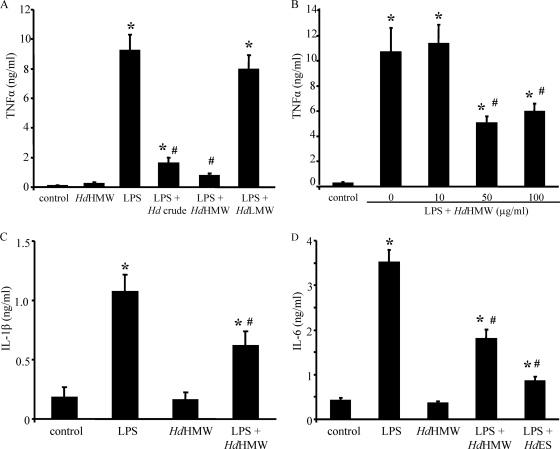
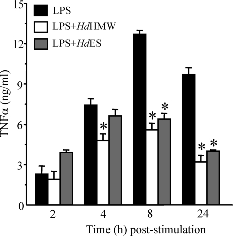
Treatment of THP-1 cells with the crude, PBS-soluble extract of adult H. diminuta significantly reduced LPS-induced TNF-α production (Fig. 1A). Fractionation of whole H. diminuta using the <50-kDa MWCO columns revealed that fractions of <50 kDa did not affect the THP-1 cell response to LPS, whereas in more than 20 experiments the >50-kDa fraction (i.e., HdHMW) reduced (range, 18 to 80% inhibition) LPS-induced TNF-α production (Fig. 1A). In addition, extracts made from H. diminuta that lacked the scolex and neck regions were as effective at blocking TNF-α production as HdHMW from intact worms (data not shown), indicating that the active component was not restricted to the scolex, which contains a number of proteins and/or glycoproteins that are not found in the remainder of the tape (39). Figure 1B shows the dose dependency of the HdHMW effect. In addition, the HdHMW significantly reduced THP-1 cell production of IL-1β and IL-6 in response to LPS challenge (Fig. 1C and D). Analysis of the time dependence of HdHMW revealed that supernatant levels of TNF-α at 4 to 24 h post-LPS, but not at 2 h post-LPS, were statistically significantly reduced (Fig. 2). The reduced amounts of TNF-α in culture medium after HdHMW treatment are unlikely to be due to an inability to release the cytokine since the intracellular levels from THP-1cell lysates 48 h after LPS ± HdHMW were not significantly different (all < 0.2 ng/ml). Similarly, the TNF-α released was synthesized because treatment with cycloheximide resulted in no detectable TNF-α in culture medium obtained 2 to 6 h post-LPS ± HdHMW (n = 2).
FIG. 1.
(A) The crude and high-molecular-mass (HdHMW, >50 kDa) extracts of H. diminuta significantly reduce LPS (1 μg/ml, 48 h)-induced TNF-α production by THP-1 macrophages. The low-molecular-mass (HdLMW, i.e., <50 kDa) preparation was ineffective. (B) Dose dependency of HdHMW in reducing LPS-induced TNF-α (n = 9 THP-1 preparations from three experiments). (C and D) LPS-induced IL-1β and IL-6 production is reduced by concomitant treatment with HdHMW (100 μg/ml) or excretory/secretory products from H. diminuta (HdES, 20 μg/ml) (n = 9 to 18 THP-1 cell preparations/group from three to six experiments; mean ± the SEM; * and #, P < 0.05 compared to the control and LPS, respectively).
FIG. 2.
Bar graph showing that THP-1 cells exposed to LPS (1 μg/ml) and cotreated with either HdHMW (100 μg/ml) or HdES products (20 μg/ml) produced significantly less TNF-α by 4 to 24 h poststimulation (n = 6 to 9 THP-1 cell preparations/group from two to three experiments; mean ± the SEM [*, P < 0.05 compared to the time-matched LPS group]).
Like THP-1 cells, LPS-induced TNF-α and IL-6 production by the murine J774 macrophage cell line and peritoneal macrophages was significantly reduced by cotreatment with HdHMW (Table 3).
TABLE 3.
Murine macrophage cytokine responses to LPS
| Macrophage typea | Mean level (ng/ml) ± SEMb |
|||
|---|---|---|---|---|
| TNF-α |
IL-6 |
|||
| LPS | LPS + HdHMW | LPS | LPS + HdHMW | |
| Murine peritoneal macrophages (n = 4 to 5) | 0.26 ± 0.03 | 0.15 ± 0.02* | 0.42 ± 0.09 | 0.22 ± 0.03* |
| J774 (murine) (n = 12) | 31.8 ± 3.6 | 17.2 ± 2.8* | 15.6 ± 0.7 | 8.9 ± 0.4* |
n, Number of mice or macrophage preparations from four J774 experiments.
*, P < 0.05 compared to the corresponding LPS-only group. LPS, 1 μg/ml; HdHMW, 100 μg/ml. Cytokines were measured by ELISA at 24 h after the addition of LPS.
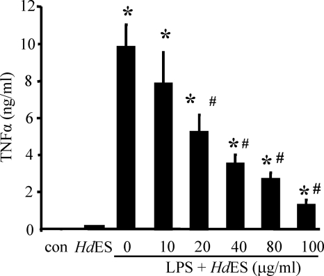
Analysis of HdES products (>50 kDa) showed that this material significantly reduced LPS-induced TNF-α production in a dose-dependent manner (Fig. 3). TNF-α production by LPS-treated macrophages was inhibited by ∼55% by HdES (20 μg/ml; n = 9 to 21 THP-1 cell preparations from three to seven experiments). IL-6 production by LPS-stimulated THP-1 cells was inhibited by HdES (Fig. 1D). Importantly, H. diminuta extracts or the HdES products alone did not evoke TNF-α production from THP-1 cells (Fig. 1 and 3). Suppression of TNF-α release from THP-1 cells by HdES was statistically significant at 8 and 24 h post-LPS challenge (Fig. 2).
FIG. 3.
Bar graph showing the dose dependency of HdES inhibition of LPS-induced TNF-α production by THP-1 cells (n = 3 macrophage preparations; mean ± the SEM [* and #, P < 0.05 compared to the control and LPS, respectively]).
TNF-α and IL-6 production by THP-1 macrophages evoked by poly(I:C) or a sonicate of whole E. coli were reduced in cells cotreated with HdHMW; however, E. coli flagellin-induced TNF-α and IL-6 were not statistically significantly reduced (Table 4).
TABLE 4.
Effect of H. diminuta extracts and excreted/secreted products on cytokine production by stimulated THP-1 macrophages
| Treatment | Mean level (ng/ml) ± SEMa |
|
|---|---|---|
| TNF-α | IL-6 | |
| Poly(I:C) (1 μg/ml) | 5.7 ± 0.6 | 3.6 ± 0.2 |
| Poly(I:C) + HdHMW | 2.6 ± 0.3* | 1.9 ± 0.1* |
| Flagellin (1 μg/ml) | 7.9 ± 1.0 | 3.5 ± 0.6 |
| Flagellin + HdHMW | 7.2 ± 1.2 | 2.6 ± 0.5 |
| Flagellin + HdES | 7.6 ± 1.2 | 2.2 ± 0.2 |
| E. coli sonicate (1 μg of protein/ml) | 9.7 ± 1.2 | NT |
| E. coli + HdHMW | 5.9 ± 1.1* | NT |
| E. coli + HdES | 1.4 ± 0.1* | NT |
n = 3 to 15 macrophage preparations from one to five experiments. Cytokine levels were determined by ELISA at 48 h post treatment. HdHMW, 100 μg/ml; HdES, 20 μg/ml. *, P < 0.05 compared to appropriate positive control stimulus. NT, not tested.
Extracts of H. diminuta are not toxic to macrophages and do not alter TLR4 expression.
Reduced cytokine production could reflect a toxic effect of the HdHMW on the macrophages. This is unlikely since THP-1 viability (based on trypan blue staining) was 96 and 97% 48 h after exposure to LPS or LPS + HdHMW, respectively (n = 6 experiments).
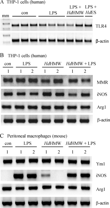
Reduced expression of TLR4 would explain a reduced responsiveness to LPS; however, RT-PCR of TLR4 mRNA does not support this possibility (Fig. 4 [confirmed by densitometry]). In addition, flow cytometry using TLR4 specific antibodies revealed no differences in receptor expression on THP-1 cells treated with LPS (1 μg/ml) ± HdHMW (100 μg/ml). Normalizing the TLR4 expression of LPS-treated cells to 100% revealed a slight increase in the expression of 42% (gated cells) in the LPS + HdHMW group, and the mean fluorescence intensity was not significantly different (one representative experiment of two). Similar observations were made when TLR3 expression was examined by flow cytometry (data not shown).
FIG. 4.
(A) A 48-h exposure to HdHMW (>50 kDa; 100 μg/ml) or excretory/secretory products from H. diminuta (HdES, 20 μg/ml) does not significantly alter expression of the Toll-like receptor 4 (TLR4) mRNA. β-Actin was used as a loading control (n = 3; six to nine preparations per group; mm, molecular marker). (B) Representative gels of semiquantitative RT-PCR products demonstrating that 3 h of treatment with LPS (1 μg/ml) or HdHMW (100 μg/ml) does not appreciably alter the expression of markers indicative of an alternatively activated macrophage (i.e., the mannose receptor (MMR or CD206) in humans, YM1 in mice, or Arg-1. Indeed, untreated THP-1 macrophages showed constitutive expression of mRNA for these molecules. (C) iNOS mRNA was induced in peritoneal macrophages by a 48-h treatment with LPS, but not HdHMW, which also does not affect Ym1 or Arg-1 levels (n = 3 macrophage preparations).
Alternatively activated macrophages (AAM) are hyporesponsive to LPS (13). Using the expression of the mannose receptor (CD206) and arginase-1 (Arg-1) as markers of the AAM phenotype, we found that THP-1 macrophages exposed to LPS, HdHMW, or LPS + HdHMW for 3 h all had similar levels of molecules indicative of AAM, as revealed by RT-PCR, and indeed this was not appreciably different from the nontreated, time-matched macrophages (Fig. 4B). Densitometry of the mRNA product bands revealed no statistical differences. Similarly, a 48-h exposure of murine peritoneal macrophages to HdHMW ± LPS did not upregulate Ym1 or Arg-1 mRNA expression (Fig. 4C), although the presence of LPS did enhance iNOS expression. Thus, the induction of an AAM phenotype is unlikely to be responsible for the reduced responses to LPS observed in HdHMW-treated macrophages.
Extracts of H. diminuta do not reduce macrophage phagocytosis of E. coli.
Assessing phagocytosis, we observed no significant difference in the ability of THP-1 cells ± HdHMW to phagocytose E. coli (control [1.8% ± 0.43%] and HdHMW [1.6% ± 0.21%] of extracellular bacterial counts [n = 4 experiments with three macrophage preparations/condition/experiment]). Also, HdHMW was neither bacteriostatic nor bactericidal based on E. coli 6-h growth curves (n = 3). These data provide additional support in favor of HdHMW not reducing macrophage viability.
Immunosuppressive effect of HdHMW is not via PG or IL-10 production.
Prostaglandins (PGs) can suppress immune activity (48). We postulated that HdHMW induced PGs could contribute to the reduced TNF-α production. However, concomitant inhibition of PG synthesis with indomethacin did not ameliorate the ability of HdHMW to reduce LPS-induced TNF-α production by THP-1 cells (LPS = 14.8 ± 2.9; LPS + HdHMW [100 μg/ml] = 6.4 ± 1.3*; LPS + HdHMW + indomethacin [10−6 M] = 8.5 ± 1.9* ng of TNF-α/ml; n = 9 THP1 preparations from three experiments [*, P < 0.05 compared to LPS-only-treated THP-1 cells]). Another possibility was that treatment with HdHMW extracts induced IL-10 that fed back onto the THP-1 cell to reduce the effects of LPS. However, measurement of IL-10 revealed no significant differences between THP-1 cells treated with LPS, HdHMW or both agents (data not shown). Also, HdHMW reduced TNF-α production from LPS-stimulated peritoneal macrophages from IL-10 knockout mice by 45% (LPS = 165 ± 57 versus LPS + HdHMW = 92 ± 32 pg of TNF-α/ml [n = 4]).
Immunosuppressive components are not heat-sensitive proteins.
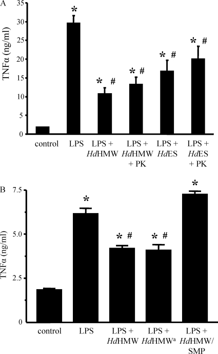
The HdHMW and HdES preparations were treated with proteinase K or trypsin for 3 h at 37°C and then boiled for 20 min to destroy enzymatic activities and disrupt proteins in the extracts. These treatments had, at best, a small effect on the immunosuppressive of the HdHMW (Fig. 5A). In eight experiments, LPS + HdHMW versus LPS + HdHMW-boiled reduced TNF-α production by THP-1 cells by 49 and 32%, respectively, compared to LPS-only-treated cells. Using a paired t test to negate interexperiment variation, there was a statistically significant effect of boiling (P = 0.03), but the overall immunological significance of this is likely minimal.
FIG. 5.
(A) Treating the high-molecular-mass (>50 kDa; 100 μg/ml) extract of H. diminuta (HdHMW) or excretory/secretory products from H. diminuta (HdES, 20 μg/ml) with proteinase K (PK, 1 mg/ml; 3 h), followed by boiling (20 min), results in a small reduction in the ability of either preparation to inhibit LPS (1 μg/ml, 48 h)-induced TNF-α production by THP-1 macrophages (n = 15 to 27 THP-1 cell preparations/group from five to eight experiments). (B) Bar graph showing that deglycosylation of HdHMW with sodium metaperiodate (SMP) significantly reduces its ability to inhibit TNF-α production by THP-1 cells stimulated with LPS (mean ± the SEM; n = 3 to 4 THP-1 cell preparations [* and #, P < 0.05 compared to the control and LPS, respectively]; the superscript “a” indicates that the active component of HdHMW was recovered from G-25 Sephadex desalting columns that were used to remove the SMP from the HdHMW + SMP preparation).
H. diminuta possesses carbohydrates which react with specific lectins.
We investigated the carbohydrate content of HdHMW under reducing conditions by using lectins with affinities for specific oligosaccharide modifications (Table 2). HdHMW displayed reactivity with the lectin SNA (indicating the presence of sialic acid linked α-2,6 to galactose) to produce a prominent band at ∼38 kDa and also reacted with MAA (specific for sialic acid linked α-2,3 to galactose) to generate bands between 20 and 50 kDa. HdHMW reactivity to DSA (indicates the presence of galactose linked 1,4 to N-acetylglucosamine) revealed a high-molecular-mass band at ∼170 kDa and an ∼37-kDa product. In contrast to previous findings (39), HdHMW reacted with PNA (a lectin specific for core galactose linked 1,3 to N-acetylgalactosamine) to reveal a cluster of poorly resolved bands with molecular masses of >170 kDa and a prominent product at 85 to 90 kDa. Of the five lectins used, HdHMW displayed the broadest reactivity to GNA (indicating terminal mannose), generating bands ranging from 35 to >170 kDa.
Enzymatic degradation of glycoproteins reduces the immunosuppressive effect of HdHMW.
Treatment with sodium meta-periodate, to accomplish broad deglycosylation, significantly reduced the ability of HdHMW to inhibit LPS stimulation of THP-1 cells (Fig. 5B). Although HdHMW displayed extensive reactivity for mannose, α-mannosidase treatment did not affect the reduction in TNF-α (e.g., LPS = 7.0 ± 1.0, LPS + HdHMW = 3.2 ± 0.7*, and LPS + HdHMW + mannosidase = 1.7 ± 0.4* ng of TNF-α/ml; n = 5 experiments [*, P < 0.05 compared to LPS-only-treated THP-1 cells]). SNA lectin binding detected a prominent ∼38-kDa band in the HdHMW extract that was ablated by neuraminidase treatment. Investigating the possibility that a neuraminidase-sensitive glycoprotein could be the active ingredient in HdHMW immunosuppression, we noted higher TNF-α levels in LPS + HdHMW + neuraminidase compared to LPS + HdHMW only. However, neuraminidase and boiled neuraminidase evoked the release of ∼1.0 to 1.5 ng of TNF-α/ml from THP-1 cells (n = 4), and this is likely due to a contaminant in the enzyme preparation that is produced in Clostridium perfringens. Thus, the immunosuppressive glycan in the HdHMW is likely insensitive to neuraminidase treatment.
HdHMW does not function via inhibition of Erk MAPK.
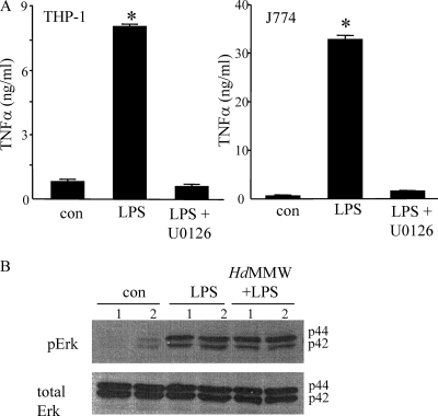
Pharmacological inhibition of Erk MAPK activity, as expected, almost completely abolished LPS-induced TNF-α production by THP-1 and J774 macrophages (Fig. 6A). However, Erk activation, as gauged by phosphorylation on immunoblots, was not affected by HdHMW (Fig. 6B), indicating that the site of HdHMW activity is either downstream of Erk MAPK or is via a different signaling pathway.
FIG. 6.
(A) LPS (1 μg/ml)-induced TNF-α production by human (THP-1) and murine (J774) macrophage cell lines is inhibited by treatment with the MAPKK inhibitor, U0126 (10 μM) (mean ± the SEM; n = 20 THP-1 preparations from six experiments and n = 12 J774 preparations from four experiments [*, P < 0.05 compared to the control “con”]). (B) Representative immunoblot (n = 3) showing that LPS (30 min) evokes Erk activation (i.e., increased levels of phosphorylated Erk [pErk]) in THP-1 cells and that this is not affected by HdHMW (100 μg/ml) cotreatment (total Erk expression is shown as a loading control; two separate THP-1 preparations are shown per condition).
HdHMW reduces murine DNBS colitis.
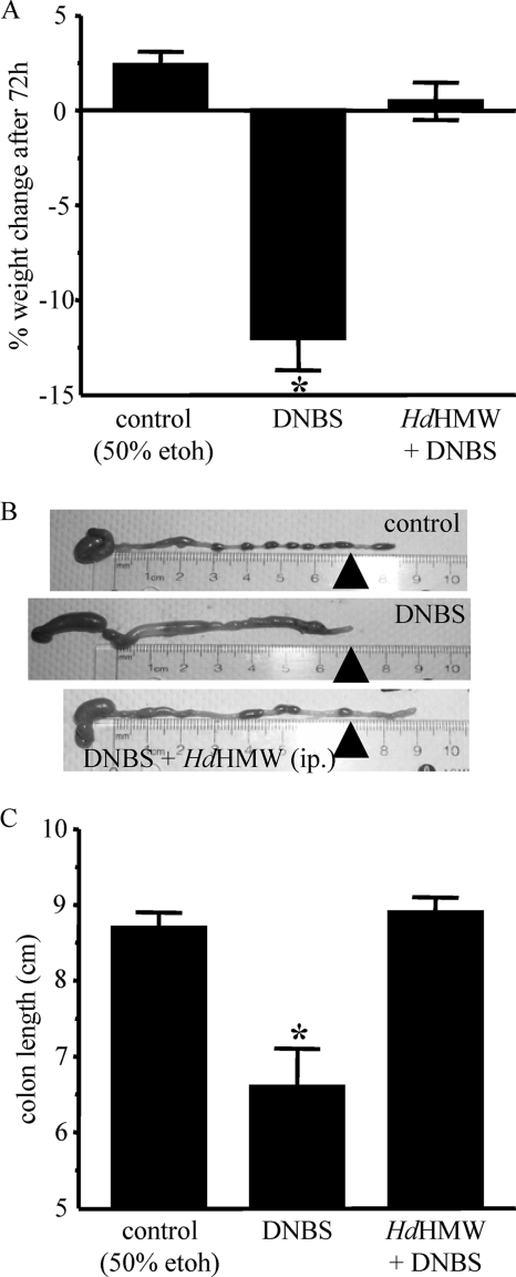
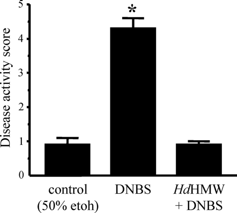
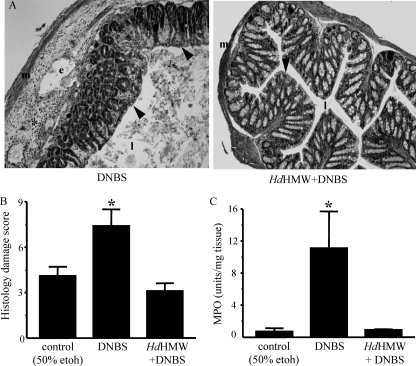
Consistent with previous reports (10, 17), direct instillation of 3 mg of DNBS in a 50% ethanol solution into the colon of BALB/c mice resulted in an acute inflammatory response characterized by weight loss, shortening of the colon, and evidence of ulceration (Fig. 7). Three of twelve mice in the DNBS group reached a predetermined disease endpoint and were humanely sacrificed prior to reaching the 72-h post-DNBS time point. A single i.p. injection of 1 mg of HdHMW, given 30 min prior to DNBS, did not affect the severity of the colitis. However, when HdHMW was administered over a 3-day period (1 mg/i.p. injection/day), this resulted in a significant diminution of DNBS-colitis as assessed by weight loss, colon length (Fig. 7), disease activity score (Fig. 8), histological assessment, and colonic MPO activity (Fig. 9) (0/11 mice reached a disease severity that required euthanasia). In addition, the spleens of DNBS + HdHMW-treated mice (160 ± 20 mg) were statistically significantly larger than those of control (100 ± 10 mg) and DNBS-only (89 ± 10 mg) mice (n = 4). Analysis of spleen cell cytokine production in response to ConA stimulation showed that cells from DNBS + HdHMW-treated mice produced less TNF-α and substantially more IL-10 and IL-4 than DNBS or control mice (Table 5). Mice cotreated with HdHMW + anti-IL-10 neutralizing antibodies had a degree of protection from DNBS-induced colitis similar to that of HdHMW-treated mice, as gauged by disease activity scores and MPO, whereas histological assessment revealed slightly worse tissue damage and inflammatory cell infiltrate (Table 6).
FIG. 7.
Bar graphs illustrating reduced severity of colitis in male BALB/c mice induced by intrarectal instillation of DNBS (3 mg in 50% ethanol) with three i.p. injections of the high-molecular-mass (>50 kDa; 1 mg of HdHMW in 200 μl of PBS) extract of H. diminuta (HdHMW) as gauged by changes in body weight (A), representative images of colon length (arrowheads indicate a 7-cm length) (B), and colon length measurements (mean ± the SEM; n = 5 to 11 mice from three experiments; *, P < 0.05 compared to the control) (C).
FIG. 8.
Bar graph illustrating reduced severity of colitis in male BALB/c mice induced by intrarectal instillation of DNBS (3 mg in 50% ethanol) with three i.p. injections of the high-molecular-mass (>50 kDa; 1 mg of HdHMW in 200 μl of PBS) extract of H. diminuta, as gauged by disease activity score (mean ± the SEM; n = 5 to 11 mice from three experiments [*, P < 0.05 compared to the control]).
FIG. 9.
Treatment with the high-molecular-mass (>50 kDa; 1 mg of HdHMW in 200 μl of PBS) extract of H. diminuta reduced DNBS-induced colonic histopathology, as shown by representative hematoxylin-and-eosin-stained images (m, muscle; e, edema; l, lumen of the colon; *, inflammatory infiltrate; arrowhead, epithelium [original magnification, ×400]) (A) and the calculation of histological damage scores (B) and is corroborated by reductions in MPO activity (C) (mean ± the SEM; n = 5 to 11 mice from three experiments [*, P < 0.05 compared to the control]).
TABLE 5.
Spleen cell cytokine production in response to ConA treatment
| Treatment | Mean level (pg/ml) ± SEM |
||
|---|---|---|---|
| IL-4 | IL-10 | TNF-α | |
| Control (i.e., 50% ethanol) | 175 ± 36 (4) | 32 ± 17 (7) | 783 ± 430 (3) |
| DNBSa | 113 ± 24 (4) | 21 ± 14 (7) | 1,535 ± 259 (3) |
| DNBS + HdHMW (three 1-mg i.p. doses) | 771 ± 268* (4) | 1,818 ± 423* (8) | 50 ± 25* (3) |
DNBS, 3 mg in 100 μl of 50% ethanol. Animals were assessed 72 h later. Spleen cells (5 × 10⁶) were stimulated with 2 μg of ConA/ml, and supernatants were collected 24 h later. n values are given in parentheses. *, P < 0.05 compared to control and DNBS.
TABLE 6.
In vivo use of IL-10 neutralizing antibodies does not affect the anticolitic effect of treatment with HdHMW
| Treatment | Mean ± SEMa |
||||
|---|---|---|---|---|---|
| Disease activity score | MPO activity (U/mg of tissue) | Histology damage score | Spleen wt (mg) | Splenic IL-10 (pg/ml) | |
| Control (50% ethanol) | 0.4 ± 0.1 | 4.8 ± 0.6 | 1.4 ± 0.2 | 73 ± 3 | 81 ± 18 |
| DNBS (3 mg, 72 h) | 3.8 ± 0.3* | 34.3 ± 5.0* | 10.4 ± 1.0* | 77 ± 3* | 72 ± 27 |
| DNBS + HdHMW | 1.8 ± 0.2*# | 13 ± 3.0*# | 4.1 ± 1.0*# | 153 ± 10*# | 1207 ± 340*# |
| DNBS + HdHMW + anti-IL-10 | 2.7 ± 0.4*#§ | 8.9 ± 1.2*# | 7.2 ± 0.8*#† | 137 ± 7*# | 968 ± 138*# |
Data are means from eight mice in two experiments. HdHMW, three 1-mg i.p. doses. Anti-IL-10 antibodies were given as a total of 200 μg/mouse in three i.p. injections. *, #, and †, P < 0.05 compared to the control, DNBS, and DNBS + HdHMW groups, respectively. §, P = 0.062 compared to DNBS + HdHMW. Spleen cells (5 × 10⁶) were stimulated with ConA (2 μg/ml) for 48 h. The HdHMW preparations in these experiments had substantial anticolitic effects, although some inflammatory disease was still apparent.
DISCUSSION
Assessing parasite-host interactions has yielded key insights into immunity (2, 15, 35). All classes of parasitic helminth influence immune reactions in their host via the release of immunomodulatory molecules (14, 21, 33, 44). Here, we show that (i) a PBS-soluble, heat-stable fraction of adult H. diminuta and ES products significantly reduces LPS-stimulated TNF-α production by macrophages and (ii) systemic delivery of a high-molecular-weight extract of H. diminuta reduces the severity of colitis induced in mice by intrarectal delivery of DNBS.
As a source of TNF-α, IL-1β, and IL-6, macrophages are often considered proinflammatory (37). Conversely, macrophage subtypes have been identified, some producing IL-10, that serve anti-inflammatory or tissue reparative roles (22). For example, macrophages were implicated in the resolution of dextran-sodium sulfate-induced colitis in mice infected with Schistosoma mansoni (41). Given the possibility that helminth-derived molecules might induce anti-inflammatory type macrophages (7, 11), we tested the effect of an extract of H. diminuta and ES products on macrophage function. Analysis of three macrophage types revealed that HdHMW and HdES significantly inhibited macrophage activation by LPS and poly(I:C), as measured by cytokine production. These novel findings in relation to H. diminuta add to a substantial literature showing that helminth-derived products can reduce the LPS activation of macrophages and dendritic cells as assessed by cytokine, and/or nitric oxide production and the induction of major histocompatibility complex class II and B7 expression (6, 12, 20).
The data presented here raise a number of questions. What is the mechanism underlying the immunosuppressive effect of the H. diminuta extract? Can the immunosuppressive molecule(s) from H. diminuta be identified? Will the H. diminuta extract suppress inflammatory disease?
We found that HdHMW neither reduced the binding of FITC-LPS to THP-1 cells, nor did it reduce THP-1 cell viability, a finding corroborated by normal levels of phagocytosis of E. coli in the HdHMW-treated cells. The latter indicates that whereas HdHMW-treated macrophages have a reduced proinflammatory potential (i.e., they generate less TNF-α, IL-1β, and IL-6 in response to LPS), their ability to phagocytose bacteria remains intact. It was feasible that the reduced responsiveness to LPS was due to loss of TLR-4 expression or the induction of an AAM-like phenotype characterized upregulation of CD206, Ym1, and/or Arg-1 by HdHMW (1, 7); neither possibility is supported by flow cytometric studies and/or RT-PCR assessment of mRNA for these molecules. Subsequent experiments also argued against the induction of PGs or IL-10 by HdHMW that would feed back onto the macrophage and inhibit its responsiveness to LPS. Similarly, inhibition of dendritic cell cytokine production by an extract of Nippostrongylus brasiliensis was not dependent on IL-10 (26), whereas inhibition of LPS-induced IL-12 production by dendritic cells by S. mansoni egg antigen was shown to occur via IL-10 (20). The possibility that HdHMW did not in fact block TNF-α production, but rather prevented its release from the cells, was considered: however, the intracellular levels of TNF-α were not significantly different 48 h after treatment with LPS ± HdHMW.
The HdHMW could affect a number of cellular processes to block TNF-α production including, but not restricted to, inhibition of activation or degradation of adaptor (e.g., TRIF and MyD88) or signaling molecules (e.g., IRF3, NFκB, and MAPK) and inhibition of transcription factor nuclear localization (4, 6, 12, 20). Inhibition of MAPKs in macrophages by helminth-derived products has been reported (6), and pharmacological blockade of Erk MAPK reduced LPS-induced TNF-α from THP-1 and J774 cells (Fig. 6). However, LPS-induced Erk activation in THP-1 cells was at best subtly reduced (i.e., the p42 form) by HdHMW cotreatment, indicating that the immunosuppressive effect of HdHMW is unlikely to be Erk-MAPK dependent. Activation NFκB is important in TLR4-LPS driven TNF-α production, and helminth products have been found to reduce NFκB activation (12). To date, our preliminary studies have not shown any consistent inhibition of IκB phosphorylation, which would reduce NFκB activation, by HdHMW. Finally, it is noteworthy that the release of TNF-α 2 h after LPS exposure was unaffected by HdMHW, implying that the inhibitory effect of HdHMW may be downstream of very rapidly mobilized signals (e.g., MAPK and NF-κB).
Boiling and protease digestion had minimal effects on the ability of either HdHMW or HdES to block LPS activation of THP-1 cells. These data suggest that the active component(s) of the H. diminuta extract is not a heat-sensitive protein (i.e., with an intact, folded conformation). A plethora of studies have shown that many immunosuppressive molecules from helminths are glycoproteins (19, 29, 33, 44) and, to a lesser extent, lipid-based molecules (19). We confirmed the presence of multiple glycoproteins in H. diminuta and, in addition, (i) we found GalNAc-containing glycoproteins in homogenized adults, but not in the ES products, suggesting that these glycoproteins are not secreted, and, in contrast to Schmidt (39), (ii) we detected GlcNAc glycoproteins in somatic extracts from adult worms in which the scolex had been removed. Using a broad deglycosylation strategy, sodium metaperiodate treatment of HdHMW significantly reduced its ability to inhibit LPS-induced TNF-α production by THP-1 cells. Abundant mannose modifications of glycoproteins occur in HdHMW but treatment of the extract with α-mannosidase affected neither the inhibition of TNF-α nor IL-6 production in LPS + HdHMW-treated THP-1 cells. A number of glycoproteins with α-2,6 sialic acid modifications were ablated by neuraminidase treatment. However, whereas treatment of HdHMW with neuraminidase resulted in increased TNF-α from LPS-treated THP-1 cells, we speculate that this was likely the result of a C. perfringens-derived contaminant in the enzyme preparation. Thus, considerable research will be required to identify the glycoproteins in HdHMW (and likely HdES also) that inhibit macrophage activation by LPS. Finally, the HdHMW will have some degree of contamination with bacterial components (we previously identified trace amounts of LPS [45]); however, these would be expected to be additive and/or synergistic with LPS activation of the macrophage. Although we feel it is unlikely, we cannot, at this stage, unequivocally rule out the possibility that an immunosuppressive product from the enteric flora in the HdHMW contributes to the inhibition of macrophage activity.
The ability of HdHMW to inhibit the activation of murine macrophages raises the question of whether in vivo administration would affect the outcome of disease in murine model systems. Since proinflammatory macrophages have been implicated in the pathogenesis or pathophysiology of IBD, we hypothesized that HdHMW would reduce disease in murine models of colitis. Although a single dose of HdHMW failed to alleviate DNBS-induced colitis, a three-dose regimen resulted in significant amelioration of colitis, as gauged by body weight, colon length, macroscopic appearance, tissue MPO levels, and colonic histopathology. This in vivo effect of HdHMW was accompanied by splenomegaly and skewing of mitogen-evoked cytokine production by spleen cells from HdHMW + DNBS-treated mice toward production of more IL-10 and IL-4 and less TNF-α than those from DNBS-only-treated mice. We have shown that H. diminuta-evoked IL-10 results in less severe colitis, and the HdHMW-induced IL-10 is compatible with this (17); however, in the context of systemic delivery of HdHMW, concomitant administration of anti-IL-10 antibodies, with the exception of an improvement in histopathology, did not ablate the anticolitic effect, indicating that (i) in this instance IL-10 may be only one component of the anti-inflammatory effect and that (ii) the anti-inflammatory effects of IL-10 are contextual and may be most effective as a prophylactic or adjuvant therapy. In accordance with the in vitro data, HdHMW suppression of macrophage reactivity could account, at least in part, for the reduced colitis (noting that splenocyte TNF-α production was reduced), although T cells (and other immune cells) are also likely targets for the in vivo effects of the helminth extract (45). This possibility needs to be tested. So while the precise mechanism of action of HdHMW is unclear, this is, to our knowledge, the first demonstration of the ability of a cestode-derived extract to inhibit murine colitis. As such it complements recent studies showing that undefined extracts of Trichinella spiralis and S. mansoni reduce the severity of trinitrobenzene sulfonic acid (TNBS)-induced colitis in mice (30, 31) and those showing that that S. mansoni soluble egg antigen, Acanthocheilonema viteae ES-62, and a PBS-soluble extract of Ascaris suum can inhibit colitis, arthritis, and asthma in murine model systems (8, 18, 27, 36).
In summary, we have shown that a PBS-soluble extract and ES products from H. diminuta dose and time dependently inhibit LPS-induced TNF-α production from macrophages in vitro without affecting cell viability or the phagocytosis of E. coli. The active immunosuppressive activity of the HdHMW is due, at least partially, to a mannosidase- and neuraminidase-insensitive glycoprotein. We conclude that H. diminuta, and parasitic helminths in general, are an untapped source of immunomodulatory drugs (or molecules that can serve as blueprints for novel drugs [14, 19]) and support this postulate with data showing that systemic delivery of HdHMW to mice protects them against DNBS-induced colitis.
Acknowledgments
Funding for this research was provided by a Natural Science and Engineers Research Council of Canada (NSERC) to D.M.M. Original observations were based on work funded by the Crohn's and Colitis Foundation of Canada (CCFC). M.J.G.J. was supported by CCFC/Canadian Institutes of Health Research (CIHR)/Canadian Association of Gastroenterology (CAG) postdoctoral fellowship, and M.E.D.C. was supported by a CAG/CCFC/CIHR summer studentship. D.M.M. holds a Canada Research Chair (Tier 1) and is an Alberta Heritage Foundation for Medical Research (AHFMR) Scientist. J.A.M. holds a Canada Research Chair (Tier 2) and is an AHFMR Senior Scholar.
We thank S. Cho for help with the ELISA and J. Coorssen for helpful discussions (both from the University of Calgary).
Editor: J. F. Urban, Jr.
Footnotes
Published ahead of print on 22 December 2009.
REFERENCES
- 1.Atochina, O., A. A. Da'dara, M. Walker, and D. A. Harn. 2008. The immunomodulatory glycan LNFPIII initiates alternative activation of murine macrophages in vivo. Immunology 125:111-121. [DOI] [PMC free article] [PubMed] [Google Scholar]
- 2.Cliffe, L. J., N. E. Humphreys, T. E. Lane, C. S. Potten, C. Booth, and R. K. Grencis. 2005. Accelerated intestinal epithelial cell turnover: a new mechanism of parasite expulsion. Science 308:1463-1465. [DOI] [PubMed] [Google Scholar]
- 3.Culley, F. J., A. Brown, D. M. Conroy, I. Sabroe, D. I. Pritchard, and T. J. Williams. 2000. Eotaxin is specifically cleaved by hookworm metalloproteases preventing its action in vitro and in vivo. J. Immunol. 165:6447-6453. [DOI] [PubMed] [Google Scholar]
- 4.Deehan, M. R., M. M. Harnett, and W. Harnett. 1997. A filarial nematode secreted product differentially modulates expression and activation of protein kinase C isoforms in B lymphocytes. J. Immunol. 159:6105-6111. [PubMed] [Google Scholar]
- 5.Diaz-Granados, N., K. Howe, J. Lu, and D. M. McKay. 2000. Dextran sulfate sodium-induced colonic histopathology, but not altered epithelial ion transport, is reduced by inhibition of phosphodiesterase activity. Am. J. Pathol. 156:2169-2177. [DOI] [PMC free article] [PubMed] [Google Scholar]
- 6.Dirgahayu, P., S. Fukumoto, K. Miura, and K. Hirai. 2002. Excretory/secretory products from plerocercoids of Spirometra erinaceieuropaei suppress the TNF-α gene expression by reducing phosphorylation of ERK1/2 and p38 MAPK in macrophages. Int. J. Parasitol. 32:1155-1162. [DOI] [PubMed] [Google Scholar]
- 7.Donnelly, S., S. M. O'Neill, M. Sekiya, G. Mulcahy, and J. P. Dalton. 2005. Thioredoxin peroxidase secreted by Fasciola hepatica induces the alternative activation of macrophages. Infect. Immun. 73:166-173. [DOI] [PMC free article] [PubMed] [Google Scholar]
- 8.Elliott, D. E., J. Li, A. Blum, A. Metwali, K. Qadir, J. F. Urban, Jr., and J. V. Weinstock. 2003. Exposure to schistosome eggs protects mice from TNBS-induced colitis. Am. J. Physiol. Gastrointest. Liver Physiol. 284:G385-G391. [DOI] [PubMed] [Google Scholar]
- 9.Elliott, D. E., T. Setiawan, A. Metwali, A. Blum, J. F. Urban, Jr., and J. V. Weinstock. 2004. Heligmosomoides polygyrus inhibits established colitis in IL-10-deficient mice. Eur. J. Immunol. 34:2690-2698. [DOI] [PubMed] [Google Scholar]
- 10.Ghia, J. E., F. Galeazzi, D. C. Ford, C. M. Hogaboam, B. A. Vallance, and S. M. Collins. 2008. Role of M-CSF-dependent macrophages in colitis is driven by the nature of the inflammatory stimulus. Am. J. Physiol. Gastrointest. Liver Physiol. 294:G770-G777. [DOI] [PubMed] [Google Scholar]
- 11.Gomez-Garcia, L., I. Rivera-Montoya, M. Rodriguez-Sosa, and L. I. Terrazas. 2006. Carbohydrate components of Taenia crassiceps metacestodes display Th2-adjuvant and anti-inflammatory properties when co-injected with bystander antigen. Parasitol. Res. 99:440-448. [DOI] [PubMed] [Google Scholar]
- 12.Goodridge, H. S., E. H. Wilson, W. Harnett, C. C. Campbell, M. M. Harnett, and F. Y. Liew. 2001. Modulation of macrophage cytokine production by ES-62, a secreted product of the filarial nematode Acanthocheilonema viteae. J. Immunol. 167:940-945. [DOI] [PubMed] [Google Scholar]
- 13.Gordon, S. 2003. Alternative activation of macrophages. Nat. Rev. Immunol. 3:23-35. [DOI] [PubMed] [Google Scholar]
- 14.Harnett, W., and M. M. Harnett. 2006. Molecular basis of worm-induced immunomodulation. Parasite Immunol. 28:535-543. [DOI] [PubMed] [Google Scholar]
- 15.Hayes, K. S., A. J. Bancroft, and R. K. Grencis. 2004. Immune-mediated regulation of chronic intestinal nematode infection. Immunol. Rev. 201:75-88. [DOI] [PubMed] [Google Scholar]
- 16.Holland, M. J., Y. M. Harcus, A. Balic, and R. M. Maizels. 2005. Th2 induction by Nippostrongylus secreted antigens in mice deficient in B cells, eosinophils or MHC class I-related receptors. Immunol. Lett. 96:93-101. [DOI] [PubMed] [Google Scholar]
- 17.Hunter, M. M., A. Wang, C. L. Hirota, and D. M. McKay. 2005. Neutralizing anti-IL-10 antibody blocks the protective effect of tapeworm infection in a murine model of chemically induced colitis. J. Immunol. 174:7368-7375. [DOI] [PubMed] [Google Scholar]
- 18.Itami, D. M., T. M. Oshiro, C. A. Araujo, A. Perini, M. A. Martins, M. S. Macedo, and M. F. Edo-Soares. 2005. Modulation of murine experimental asthma by Ascaris suum components. Clin. Exp. Allergy 35:873-879. [DOI] [PubMed] [Google Scholar]
- 19.Johnston, M. J. G., J. A. MacDonald, and D. M. McKay. 2009. Parasitic helminths: a pharmacopeia of anti-inflammatory molecules. Parasitology 136:125-147. [DOI] [PubMed] [Google Scholar]
- 20.Kane, C. M., L. Cervi, J. Sun, A. S. McKee, K. S. Masek, S. Shapira, C. A. Hunter, and E. J. Pearce. 2004. Helminth antigens modulate TLR-initiated dendritic cell activation. J. Immunol. 173:7454-7461. [DOI] [PubMed] [Google Scholar]
- 21.Ko, A. I., U. C. Drager, and D. A. Harn. 1990. A Schistosoma mansoni epitope recognized by a protective monoclonal antibody is identical to the stage-specific embryonic antigen 1. Proc. Natl. Acad. Sci. U. S. A. 87:4159-4163. [DOI] [PMC free article] [PubMed] [Google Scholar]
- 22.Kreider, T., R. M. Anthony, J. F. Urban, Jr., and W. C. Gause. 2007. Alternatively activated macrophages in helminth infections. Curr. Opin. Immunol. 19:448-453. [DOI] [PMC free article] [PubMed] [Google Scholar]
- 23.La Flamme, A. C., K. Ruddenklau, and B. T. Backstrom. 2003. Schistosomiasis decreases central nervous system inflammation and alters the progression of experimental autoimmune encephalomyelitis. Infect. Immun. 71:4996-5004. [DOI] [PMC free article] [PubMed] [Google Scholar]
- 24.Magen, E., G. Borkow, Z. Bentwich, J. Mishal, and S. Scharf. 2005. Can. worms defend our hearts? Chronic helminthic infections may attenuate the development of cardiovascular diseases. Med. Hypotheses 64:904-909. [DOI] [PubMed] [Google Scholar]
- 25.Maizels, R. M., A. Balic, N. Gomez-Escobar, M. Nair, M. D. Taylor, and J. E. Allen. 2004. Helminth parasites: masters of regulation. Immunol. Rev. 201:89-116. [DOI] [PubMed] [Google Scholar]
- 26.Massacand, J. C., R. C. Stettler, R. Meier, N. E. Humphreys, R. K. Grencis, B. J. Marshland, and N. L. Harris. 2009. Helminth products bypass the need for TSLP in Th2 immune responses by directly modulating dendritic cell function. Proc. Natl. Acad. Sci. U. S. A. 106:13968-13973. [DOI] [PMC free article] [PubMed] [Google Scholar]
- 27.McInnes, I. B., B. P. Leung, M. Harnett, J. A. Gracie, F. Y. Liew, and W. Harnett. 2003. A novel therapeutic approach targeting articular inflammation using the filarial nematode-derived phosphorylcholine-containing glycoprotein ES-62. J. Immunol. 171:2127-2133. [DOI] [PubMed] [Google Scholar]
- 28.McKay, D. M., D. W. Halton, M. D. McCaigue, C. F. Johnston, I. Fairweather, and C. Shaw. 1990. Hymenolepis diminuta: intestinal goblet cell response in male C57 mice. Exp. Parasitol. 71:9-20. [DOI] [PubMed] [Google Scholar]
- 29.Meyvis, Y., N. Callewaert, K. Gevaert, E. Timmerman, D. J. Van, J. Schymkowitz, F. Rousseau, J. Vercruysse, E. Claerebout, and P. Geldhof. 2008. Hybrid N-glycans on the host protective activation-associated secreted proteins of Ostertagia ostertagi and their importance in immunogenicity. Mol. Biochem. Parasitol. 161:67-71. [DOI] [PubMed] [Google Scholar]
- 30.Moreels, T. G., R. J. Nieuwendijk, J. G. De Man, B. Y. De Winter, A. G. Herman, E. A. Van Marck, and P. A. Pelckmans. 2004. Concurrent infection with Schistosoma mansoni attenuates inflammation induced changes in colonic morphology, cytokine levels, and smooth muscle contractility of trinitrobenzene sulfonic acid induced colitis in rats. Gut 53:99-107. [DOI] [PMC free article] [PubMed] [Google Scholar]
- 31.Motomura, Y., H. Wang, Y. Deng, R. T. El-Sharkawy, E. F. Verdu, and W. I. Khan. 2009. Helminth antigen-based strategy to ameliorate inflammation in an experimental model of colitis. Clin. Exp. Immunol. 155:88-95. [DOI] [PMC free article] [PubMed] [Google Scholar]
- 32.Nazli, A., P.-C. Yang, J. Jury, K. Howe, J. L. Watson, J. D. Soderholm, P. M. Sherman, M. H. Perdue, and D. M. McKay. 2004. Epithelia under metabolic stress perceive commensal bacteria as a threat. Am. J. Pathol. 164:947-957. [DOI] [PMC free article] [PubMed] [Google Scholar]
- 33.Okano, M., A. R. Satoskar, K. Nishizaki, and D. A. Harn (Jr.). 2001. Lacto-N-fucopentaose II found on Schistosoma mansoni egg antigens functions as adjuvant proteins by inducing TH2-type response. J. Immunol. 167:442-450. [DOI] [PubMed] [Google Scholar]
- 34.Reardon, C., A. Sanchez, C. M. Hogaboam, and D. M. McKay. 2001. Tapeworm infection reduces the ion transport abnormalities induced by dextran sulfate sodium (DSS) colitis. Infect. Immun. 69:4417-4423. [DOI] [PMC free article] [PubMed] [Google Scholar]
- 35.Riley, E., and R. Grencis. 2006. The parasite immunologist's crystal ball. Parasite Immunol. 28:233-234. [DOI] [PubMed] [Google Scholar]
- 36.Rocha, F. A., A. K. Leite, M. M. Pompeu, T. M. Cunha, W. A. Verri, Jr., F. M. Soares, R. R. Castro, and F. Q. Cunha. 2008. Protective effect of an extract from Ascaris suum in experimental arthritis models. Infect. Immun. 76:2736-2745. [DOI] [PMC free article] [PubMed] [Google Scholar]
- 37.Rugtveit, J., E. M. Nilsen, A. Bakka, H. Carlsen, P. Brandtzaeg, and H. Scott. 1997. Cytokine profiles differ in newly recruited and resident subsets of mucosal macrophages from inflammatory bowel disease. Gastroenterology 112:1493-1505. [DOI] [PubMed] [Google Scholar]
- 38.Ruyssers, N. E., B. Y. De Winter, J. G. De Man, A. Loukas, M. S. Pearson, J. V. Weinstock, R. M. Van den Bossche, W. Martinet, P. A. Pelckmans, and T. G. Moreels. 2009. Therapeutic potential of helminth soluble proteins in TNBS-induced colitis in mice. Inflamm. Bowel Dis. 15:491-500. [DOI] [PubMed] [Google Scholar]
- 39.Schmidt, J. 1988. Expression of glycoconjugates on normally developing and immunologically impaired Hymenolepis diminuta. Parasitol. Res. 75:155-161. [DOI] [PubMed] [Google Scholar]
- 40.Serradell, M. C., L. Guasconi, L. Cervi, L. S. Chiapello, and D. T. Masih. 2007. Excretory-secretory products from Fasciola hepatica induce eosinophil apoptosis by a caspase-dependent mechanism. Vet. Immunol. Immunopathol. 117:197-208. [DOI] [PubMed] [Google Scholar]
- 41.Smith, P., N. E. Mangan, C. M. Walsh, R. E. Fallon, A. N. McKenzie, R. N. van, and P. G. Fallon. 2007. Infection with a helminth parasite prevents experimental colitis via a macrophage-mediated mechanism. J. Immunol. 178:4557-4566. [DOI] [PubMed] [Google Scholar]
- 42.Summers, R. W., D. E. Elliott, J. F. Urban, Jr., R. A. Thompson, and J. V. Weinstock. 2005. Trichuris suis therapy for active ulcerative colitis: a randomized controlled trial. Gastroenterology 128:825-832. [DOI] [PubMed] [Google Scholar]
- 43.Summers, R. W., D. E. Elliott, J. F. Urban, Jr., R. Thompson, and J. V. Weinstock. 2005. Trichuris suis therapy in Crohn's disease. Gut 54:87-90. [DOI] [PMC free article] [PubMed] [Google Scholar]
- 44.Van Dam, G. J., F. H. J. Class, M. Yazdanbakhsh, Y. C. M. Kruize, A. C. I. van Leulen, S. T. M. F. Ferreira, J. P. Rotmans, and A. M. Deelder. 1996. Schistosoma mansoni excretory circulating cathodic antigen shares Lewis-X epitopes with a human granulocyte surface antigen and evokes host antibodies mediating complement-dependent lysis of granulocytes. Blood 88:4246-4261. [PubMed] [Google Scholar]
- 45.Wang, A., and D. M. McKay. 2005. Immune modulation by a high molecular weight protein from the rat tapeworm Hymenolepis diminuta. Parasitology 130:575-585. [DOI] [PubMed] [Google Scholar]
- 46.Wilson, M. S., and R. M. Maizels. 2006. Regulatory T cells induced by parasites and the modulation of allergic responses. Chem. Immunol. Allergy 90:176-195. [DOI] [PubMed] [Google Scholar]
- 47.Wilson, M. S., M. D. Taylor, A. Balic, C. A. Finney, J. R. Lamb, and R. M. Maizels. 2005. Suppression of allergic airway inflammation by helminth-induced regulatory T cells. J. Exp. Med. 202:1199-1212. [DOI] [PMC free article] [PubMed] [Google Scholar]
- 48.Xu, X. J., J. S. Reichner, B. Mastrofrancesco, W. L. Henry, Jr., and J. E. Albina. 2008. Prostaglandin E2 suppresses lipopolysaccharide-stimulated IFN-β production. J. Immunol. 180:2125-2131. [DOI] [PMC free article] [PubMed] [Google Scholar]
- 49.Zaccone, P., Z. Fehervari, F. M. Jones, S. Sidobre, M. Kronenberg, D. W. Dunne, and A. Cooke. 2003. Schistosoma mansoni antigens modulate the activity of the innate immune response and prevent onset of type 1 diabetes. Eur. J. Immunol. 33:1439-1449. [DOI] [PubMed] [Google Scholar]


