Abstract
Coronary computed tomography angiography (CCTA) is becoming an increasingly robust tool in the assessment and exclusion of coronary artery disease. Multiple recent studies have raised concerns regarding the radiation dose exposure of CCTA. A novel approach to dose reduction in CCTA using adaptive statistical iterative reconstruction, resulting in a submillisievert CCTA examination, is described. To the authors’ knowledge, the present report describes the first submillisievert study performed in Canada. The ability to perform a diagnostic CCTA with such a low dose challenges the role of coronary calcium scoring and will likely have implications for the future use of this test.
Keywords: Coronary artery disease, Coronary CT angiography, Radiation dose reduction
Abstract
La coronarographie obtenue au moyen de la tomographie assistée par ordinateur est en passe de devenir un outil de plus en plus fiable pour évaluer la coronaropathie ou en écarter le diagnostic. Plusieurs études récentes ont par contre soulevé certaines inquiétudes relativement à l’exposition aux radiations inhérente à cette modalité diagnostique. On décrit ici une nouvelle approche de réduction de la dose de radiation durant la « tomocoronarographie » à l’aide d’une reconstruction itérative statistique adaptative qui permet de procéder à l’examen avec des doses minimes de radiation. À la connaissance des auteurs, le présent rapport décrit le premier examen « infra-mSv » effectué au Canada. La capacité de réaliser une tomocoronarographie au moyen d’une dose aussi faible remet en question le rôle de l’indice de calcium coronarien et aura probablement des répercussions sur l’avenir de ce test.
CASE PRESENTATION
A 58-year-old man of Southeast Asian descent was referred to the Advanced Cardiac Imaging Program of the Heart Centre at St Paul’s Hospital/Providence Heart and Lung Institute (Vancouver, British Columbia) for evaluation of atypical chest pain.
The patient had been investigated with an electrocardiogram (ECG), which was normal, and an exercise treadmill test, which indicated that he completed 8 min 30 s of the Bruce protocol, had a normal blood pressure response to exercise and had no chest pain. The test was terminated because of fatigue and achievement of his target heart rate, and was interpreted as negative for ischemia.
Cardiac risk factors included dyslipidemia since 2007, with a recent total cholesterol level of 6.7 mmol/L, a low-density lipoprotein level of 3.6 mmol/L and a high-density lipoprotein level of 0.9 mmol/L. His father died at 62 years of age as a result of a myocardial infarction.
His only medication was enteric-coated acetylsalicylic acid 81 mg orally daily. He was recently tried on low-dose atorvastatin but discontinued it because of myalgia.
A physical examination revealed a healthy-looking man, 178 cm tall, with a weight of 72 kg and a calculated body mass index of 22.7 kg/m2. His sitting blood pressure in the right arm was 112/56 mmHg, with a heart rate of 50 beats/min and regular (following 100 mg of metoprolol given orally 4 h before the test). Further examination findings were normal. In particular, the heart sounds were normal, with no added sounds or murmurs.
Technical results
Coronary computed tomography angiography (CCTA) was performed following premedication with 0.8 mg of sublingual nitroglycerin, using a General Electric Discovery HD 750 scanner (GE Healthcare, USA). Following a 20 mL timing bolus, 90 mL of Visipaque (Iodixanol, GE Healthcare) contrast medium was administered intravenously using a triphasic injection.
As per hospital routine, a protocol using aggressive dose reduction techniques was used based on patient size and body mass index. Techniques included prospective ‘step and shoot’ gating (1) triggered at 75% of the R-R interval, a minimized field of view, zero padding (1), avoidance of pretest calcium scoring, 100 kV tube voltage and 40% volume adaptive statistical iterative reconstruction (ASIR), which allowed for a tube current reduction of approximately 50% from the standard tube current setting of 650 mA. The scan length in this 178 cm tall man was 13 cm. The tube current used was 325 mA. The estimated dose-length product for the CCTA was 54 mGy × cm, with a calculated radiation exposure of 0.76 mSv (using a standard conversion factor of 0.014).
The data were analyzed using the hospital’s workstation (Advantage Windows Workstation version 4.4, GE Healthcare). The images were analyzed independently and reported by consensus by a radiologist and a cardiologist.
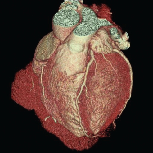
A fully evaluable data set of the coronary arteries was obtained, with no significant motion artefact (Figures 1 and 2). The right coronary artery was dominant. There was no evidence of coronary artery calcification or noncalcified atherosclerotic plaque. Functional analysis was not performed because of the use of prospective gating. The test was interpreted as normal, with no evidence of coronary atherosclerosis.
Figure 1).
A Curved multiplanar reformat of the left anterior descending coronary artery showing excellent image quality without evidence of stenosis or computed tomography-discernible atherosclerosis. B Curved multiplanar reformat of the right coronary artery showing similar findings of crisp image quality and an unremarkable vessel. C Curved multiplanar reformat of the left circumflex coronary artery
Figure 2).
Three-dimensional volume-rendered image of the heart. View a video displaying the coronary circulation with transparent myocardium online at www.canjcardiol.com or www.pulsus.com
DISCUSSION
To our knowledge, we have reported the first CCTA performed in Canada with less than 1 mSv of radiation exposure. We believe that this heralds a new era in the noninvasive evaluation of coronary artery disease.
The most commonly used data acquisition mode has been ‘spiral’ or ‘helical’ computed tomography, during which the table supporting the patient moves continuously while the x-ray tube rotates around the patient. ‘Spiral’ acquisition provides the ability to retrospectively select the phase of the cardiac cycle during which images are reconstructed. ECG-correlated tube current modulation is applied to retrospectively gated helical acquisitions to reduce tube current and radiation exposure during periods of high motion of the cardiac cycle, when cardiac motion precludes successful reconstruction of the coronary arteries. Using ECG modulation, the tube current is at its maximum during mid-diastole (60% to 80% of the R-R interval) and is reduced by approximately 46% to 80% for the remainder of the cardiac cycle. In spite of ECG-gated dose modulation, retrospectively gated studies consistently result in higher patient doses. As a result, we have moved almost exclusively (92% of our most recent 140 cases) to prospective gating to lower the radiation exposure to patients.
Multiple recent studies have raised concerns about the burgeoning radiation exposure from medical imaging tests. The Prospective Multicenter Study On Radiation Dose Estimates Of Cardiac CT Angiography In Daily Practice (PROTECTION) conducted by Hausleiter et al (2) reported an average radiation exposure for CCTA of 12 mSv and was the most recent of a number of papers raising concern regarding the radiation dose of CCTA examinations (3–4). Physicians who practice CCTA must always keep the basic principle of ALARA (as low as reasonably achievable) in mind, and must familiarize themselves with appropriate indications and all the dose reduction strategies available (5). They should individualize protocols based on patient body habitus, clinical indication and the scanner technology available. These techniques include, but are not limited to, prospective axial scanning, 100 kVp scanning for patients with a body mass index of less than 30 kg/m2 and limited scan length.
In the present case, we used ASIR, which is a more efficient image reconstruction algorithm than filtered backprojection (currently the standard for computed tomography imaging) and enables the acquisition of diagnostic-quality images at lower tube current levels. ASIR uses advanced statistical modelling to reduce image noise, which allows for high-quality CCTA imaging with reduced absolute photon numbers and tube current requirements. The combination of low tube current, low voltage, minimized field of view and prospective gating with zero padding results in an extremely low dose. Subsequent to the present case, we have performed a number of diagnostic-quality submillisievert CCTAs, some with radiation doses even lower than was noted in the present case. The median dose used in our 100 most recent consecutive cases was 1.3 mSv.
The ability to perform a diagnostic-quality CCTA with such a low radiation exposure compares favourably with that of coronary artery calcium scoring (which requires 0.6 mSv to 1.5 mSv). The ability to image and quantify both the calcified and noncalcified atherosclerotic plaque may have therapeutic implications for selected patients, including decisions regarding long-term therapy with acetylsalicylic acid and/or lipid-lowering agents.
REFERENCES
- 1.Earls JP, Berman EL, Urban BA, et al. Prospectively gated transverse coronary CT angiography versus retrospectively gated helical technique: Improved image quality and reduced radiation dose. Radiology. 2008;246:742–53. doi: 10.1148/radiol.2463070989. [DOI] [PubMed] [Google Scholar]
- 2.Hausleiter J, Meyer T, Hermann F, et al. Estimated radiation dose associated with cardiac CT angiography. JAMA. 2009;301:500–7. doi: 10.1001/jama.2009.54. [DOI] [PubMed] [Google Scholar]
- 3.Einstein AJ, Henzlova MJ, Rajagopalan S. Estimating risk of cancer associated with radiation exposure from 64-slice computed tomography coronary angiography. JAMA. 2007;298:317–23. doi: 10.1001/jama.298.3.317. [DOI] [PubMed] [Google Scholar]
- 4.Mayo J, Leipsic JA. Radiation dose in cardiac CT. AJR Am J Roentgenol. 2009;192:646–53. doi: 10.2214/AJR.08.2066. [DOI] [PubMed] [Google Scholar]
- 5.Gottlieb I, Lima JA. Screening high-risk patients with computed tomography angiography. Circulation. 2008;117:1318–32. doi: 10.1161/CIRCULATIONAHA.107.670042. [DOI] [PubMed] [Google Scholar]


