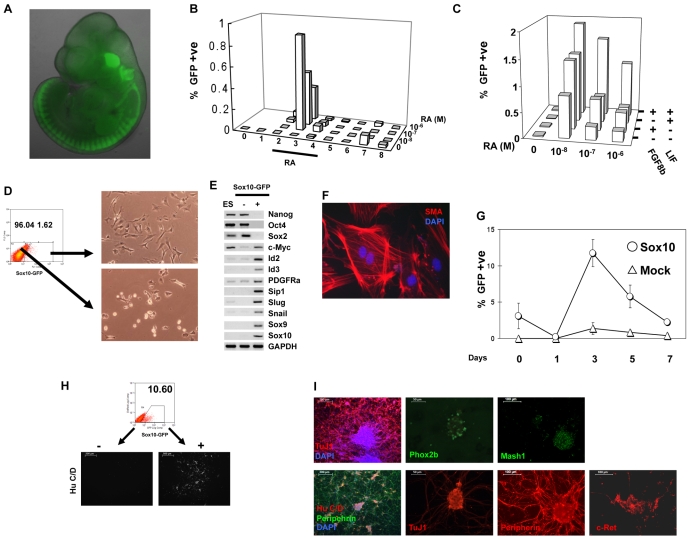
Fig. 1.
Characterisation of Sox10 reporter (S10G) and Sox10 constitutive expression (S10G-S) mouse ES cells. (A) Fluorescence image of E11.5 chimera produced by morula aggregation with S10G cells. (B) Flow cytometry analysis of Sox10-GFP-positive cells from embryoid bodies made with S10G cells. (C) Effect of Lif and Fgf8b on generation of Sox10-GFP-positive cells from embryoid bodies. Bars represent mean value of three independent plates. (D) Flow cytometry scatter plot for sorting Sox10-GFP-positive cells and bright-field images of sorted cells cultured overnight. (E) RT-PCR analysis of pluripotency and neural crest markers in undifferentiated ES cells, Sox10-GFP-negative, and Sox10-GFP-positive populations. (F) Sox10-GFP-positive purified cells cultured for 10 days in the presence of serum and immunostained for smooth muscle actin. (G) GFP-positive cells quantified by flow cytometry during differentiation of S10G-S cells and cells transfected with empty vector (Mock). RA was applied on day 2 at 10–8 M. (H) FACS purification of Sox10-GFP-positive S10G-S cells from day 3 embryoid bodies and subsequent neurogenesis in serum-free N2B27 medium supplemented with Bmp4. Images show immunostaining for HuC/D. (I) Sox10-GFP-positive cells purified as in H differentiate into neurons upon culture in Bmp4 and Gdnf for 4 days. Fixed cells were immunostained for the indicated markers. Scale bars: 500 μm in H; in I, 200 μm left, 50 μm centre, 100 μm right-hand columns.

