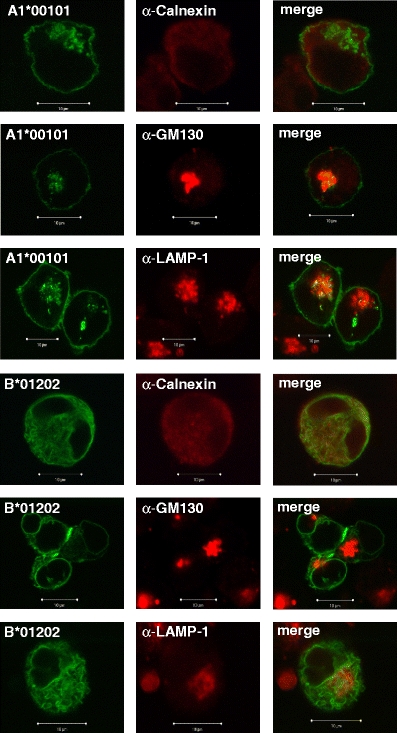
Fig. 3.

Co-localisation of AcGFP-tagged Mamu-A1*00101 and Mamu-B*01202 molecules with organelle-specific markers by confocal microscopy (×63 magnification). Left panels show AcGFP, middle panels show markers for ER (calnexin), Golgi apparatus (GM130), late endosomal/lysosomal compartment (LAMP-1) and right panels show merge. Co-localisation of class I and the marker protein is shown in yellow. Note that co-localisation of B*01202 and calnexin is evident, but the yellow colour is pale
