

Abstract
Previous investigations have shown that magnetic resonance imaging (MRI) can be employed as an efficient non-invasive diagnostic tool in studies on Egyptian mummies. MRI, moreover, because it produces especially clear images of well-hydrated tissue, could be a particularly effective diagnostic option for mummies that still retain humidity within tissues or organs. Therefore, in the present study, we tested MRI on a 17th century mummy, one of the most perfectly preserved ‘hydrated mummies’ ever found in Korea, in order to determine the quality of images that could be obtained. We found that the diagnostic value of an MRI scan of the hydrated mummy was not inferior to that of a computed tomography scan. The T1- and T2-weighted magnetic resonance (MR) signals showed unique patterns not easily obtained by computed tomography, the resultant MR images revealing the organ specificities clearly. Overall, the quality of the MR images from the hydrated mummy was superb and the scientific value of MRI in the study of hydrated mummies should not be underestimated.
Keywords: brain, hydrated mummy, intervertebral disc, Korean mummy, magnetic resonance imaging
Introduction
Whenever invasive investigations into mummified human remains are, for various reasons, not permitted by museum curators or descendants (Kim et al. 2006), computed tomography (CT) has been the first choice of Korean anthropologists (Lee et al. 2007). However, as mummies in most cases are severely dehydrated, even well-trained radiologists have difficulties in interpreting the CT images of mummified remains (Lim et al. 2008). In this regard, recent reports on the successful application of magnetic resonance imaging (MRI) to dehydrated Egyptian mummies (Karlik et al. 2007; Münnemann et al. 2007; Rühli et al. 2007) were very meaningful to us because they showed that the technique can provide radiologists with information that is not easily acquired by CT scan. Moreover, the MRI technique, considering that it is known to produce particularly clear images when applied to relatively well-hydrated tissue, might be a better diagnostic option for so-called ‘hydrated mummies’ (i.e. mummies such as Otzi the Iceman) than for desiccated (e.g. Egyptian) mummies.
Over the past several years, we have studied Korean mummies found in medieval tombs that are well encapsulated by a thick lime soil mixture barrier (Kim et al. 2006; Lim et al. 2008; Lee et al. 2009). These mummies were all in a relatively hydrated condition with, even after several hundreds of years of burial, superbly well-preserved internal organs in comparison with mummies found in other parts of the world. Anticipating that MRI application to a hydrated mummy, the first investigation of its kind, would yield comprehensive and enlightening information for the benefit of future studies, we conducted a study to assess the scientific value of MRI in that context.
Materials and methods
According to historic documents, the mummified individual found within the lime soil mixture barrier tomb had lived in Korea from 1561 to 1622. The skin was discovered to have not completely dried, having retained its elasticity (Data S1A). After CT scanning was completed (Lee et al. 2009), magnetic resonance (MR) images were obtained for the entire body of the mummy using a 1.5-T MR scanner (Signa twinspeed; GE Healthcare, Milwaukee, WI, USA). Pre-contrast T1-weighted spin-echo images [Repetition Time/Echo Time/Number of Excitation (TR/TE/NEX), 550 ms/12 ms/2] and T2-weighted fast spin-echo images (TR/TE/NEX, 4000 ms/126 ms/2) on the axial, sagittal and coronal planes (5 mm section thickness, 1 mm intersection gap, 512 ×512 matrix) were also obtained. The gray colors of the T1- and T2-weighted images of the intervertebral discs (IVDs) were changed to red and green, respectively, using an LSM Image Browser (Carl Zeiss, Germany), after which the two images were merged using Adobe Photoshop CS3 Extended (Adobe Systems Inc., USA). The organs identified by CT or MR images were confirmed by post-factum dissection. Upon incising and opening the body, we observed that the hydrated state of the internal organs had been maintained (Data S1B,C).
Results
Generally, T1-weighted MR images exhibit high signal intensities for soft tissues but only very weak ones for bony parts. Considering the head of our mummy, for instance, the scalp, dura matter and brain showed high signal intensities on both the T1- and T2-weighted images. The T1- and T2-weighted images exhibited mixed signal intensities for the brain, however, whereas the CT scan showed only a uniform radiodensity. The skull showed an intense radiopacity on the CT scan but very weak or no signals on the T2- and T1-weighted MR images, respectively (Fig. 1).
Fig. 1.
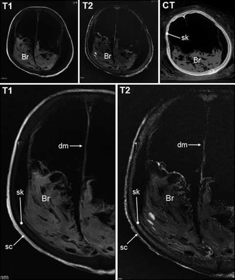
Magnetic resonance imaging on the head of the hydrated mummy. T1- and T2-weighted magnetic resonance (MR) images proved not to be very inferior to the computed tomography (CT) image. Although the skull (sk) is clearly seen on the CT scan, the T1- and T2-weighted MR images showed very weak or no signals, respectively. Magnified MR images exhibited the structures very clearly. The dura mater (dm), brain (Br) and scalp (sc) showed high signal intensities. The skull showed weaker signals on both images, especially on the T1-weighted image. Note the mixed signal intensities for the brain, which might have been due to the retention of the cortex and medulla.
In the chest and abdominal areas, soft tissues such as the skin, tracheal cartilage, bronchus, lung parenchyme, muscle fascia, heart, liver, spleen and intestine exhibited high signal intensities on the T1-weighted images. By contrast, the signal intensities of the T2-weighted images for the same soft-tissue organs were not very different from those of the skeletons (Fig. 2A–C). The MR images of the material within the sigmoid colon and rectum were also very interesting. Whereas the CT images showed radiodensities that were not very different from those of the skin or intestinal wall of the mummy, the MR images showed unique signal patterns; in the case of the T1-weighted images, the signals were much stronger than for any of the other structures within the pelvic cavity, whereas the T2-weighted images showed no signals other than very weak ones representing the contours of the materials (Fig. 2D). When we performed a post-factum dissection on the pelvic cavity, we confirmed that the material was feces (Data S2).
Fig. 2.

Magnetic resonance (MR) images for (A and B) chest, (C) abdominal and (D) pelvic regions. Note that the radiodensities of bone are very high, whereas signals on the T1- and T2-weighted MR images (indicated by white arrows) were not so intense. The lung (indicated by blank arrow) could be seen much more clearly on the T1- than on the T2-weighted image. Whereas the signal intensities of each organ or structure on the T1-weighted image all differed, the T2-weighted image showed much more homogeneous intensities. The tracheal (asterisks) or costal (gray arrows) cartilages were much more clearly seen on the T1- than on the T2-weighted MR image. Ht, heart. In (C), the intestines (indicated by the dotted circle) were very clearly seen on the T1- and T2-weighted MR images. (D) The feces (indicated by asterisks) could be clearly seen on the T1-weighted MR image but not on the T2-weighted image. Whereas the radiopacity of the feces on the computed tomography (CT) image was not very different from those of the other structures in the pelvic cavity, the same materials on the T1-weighted image showed intense signals only in the feces. As pelvic structures such as intestinal walls (indicated by arrowheads) could all be observed on the CT, T1- and T2-weighted MR images, feces showed unique signals on the MR images.
The MRI scan of the vertebral column also provided us with crucial information. Both the T1- and T2-weighted MR images, compared with the CT images of the spine, in which the IVDs were not clearly identified (Data S3), showed high IVD signal intensities (Fig. 3). Moreover, an IVD signal-intensity difference between the T1- and T2-weighted MR images could also be observed (Fig. 3). Thus, simultaneously examining the T1- and T2-weighted MR images of the IVDs clearly differentiated the internal structures of the IVDs. Whereas the cartilaginous end of the vertebral body or annulus fibrosus (intervertebral fibrocartilage) showed high signal intensities on both the T1- and T2-weighted MR images, the nucleus pulposus could be seen only on the T2-weighted images (Fig. 3). Therefore, based on the MR images of the vertebral column, we could even speculate that the mummified individual might have suffered from spinal stenosis caused by diffuse disc bulging, which could not have been easily diagnosed with CT images (Fig. 4).
Fig. 3.
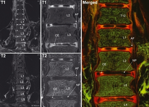
Magnetic resonance imaging axial view of vertebral column. The figures in the second column are magnified images of those in the first column. After we changed the gray colors in the T1-weighted magnetic resonance image to red, and those in the T2-weighted image to green, the two figures were merged into a single plate (merged). The cartilaginous end of the vertebral body (CE) or annulus fibrosus (AF) was seen clearly on the T1-weighted image. Even though the same structures (CE and AF) were also seen on the T2-weighted image, a possible nucleus pulposus (NP) could be identified in that T2-weighted image. In the merged T1- and T2-weighted images, the difference could be much more clearly observed. Only the signals on the T2-weighted image could be seen in the space where the NP should have been positioned, as signaled by the green color there.
Fig. 4.
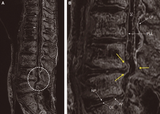
Magnetic resonance imaging sagittal view of vertebral column. (A) High signal intensities could be seen in the vertebral column. (B) Magnified image of the dotted circle area in (A). Spinal canal (SC), ligamentum flavum (LF), posterior longitudinal ligament (PLL), nucleus pulposus (NP), annulus fibrosus (AF) and cartilaginous end of vertebral body (CE) can be seen clearly in (A). Spinal stenosis due to diffuse disc bulging (indicated by yellow arrows) can be observed.
Discussion
As a number of researchers (Andreasen et al. 1991; Reihard, 1996; Spindler et al. 1996; Hess et al. 1998; Shin et al. 2003) have pointed out, different types of mummies, reflecting various mummification processes, have been found in the world. Some were artificially mummified by well-trained embalmers (i.e. Egyptian mummies), whereas others were naturally mummified in dry (i.e. Taklimakan or northern Chile), permafrost (i.e. 15th century Greenland and Otzi the Iceman) or waterlogged (i.e. bog bodies) conditions. Some of these types of mummies, such as Otzi the Iceman or bog bodies, belong to the so-called hydrated mummy (or damp mummy) category, as their tissues have retained humidity (Fleckinger, 2007). Therefore, even if previous studies have shown that MR images could be obtained from dried Egyptian mummies (Karlik et al. 2007; Münnemann et al. 2007; Rühli et al. 2007), MRI diagnosis of hydrated mummies elicits our particular interest because MRI scanning is well known to produce superb images of tissues that remain relatively hydrated. In this regard, we undertook in the present study to examine the internal organs of a Korean mummy under MRI. Indeed, one of the unique findings on medieval Korean mummies is that they are hydrated mummies with exceptionally well-preserved internal organs that, of course, were not removed by embalmers, as was the case in Egypt. Thus, we were able to obtain very comprehensive information on the application of MRI to the internal organs of hydrated mummies.
In our findings, it was clear that MR images of a hydrated mummy are not inferior to corresponding CT images. In fact, whereas CT images are difficult to interpret even for well-trained radiologists, owing to the fact that the organ radiodensities are not very different from each other, in the case of our MRI scan of the hydrated mummy, that pertinent information was easily obtained. Specifically, as the signal distributions of the T1- and T2-weighted MR images were very organ-specific, we concluded that MRI scanning on a hydrated mummy offers the advantage of the precise differentiation of each mummified organ. The MR signals for the mummy's IVD structures are a good example of the remarkable differences between CT and T1- and T2-weighted MR images, i.e. the distributions of the T1- and T2-weighted MR signals for the IVDs differed from each other, especially for the nucleus pulposus, which could be seen only on the T2-weighted MR image. Such clear MR IVD images could even help us to diagnose a vertebral disease, which would not have been easily accomplished by CT scanning alone. Specifically, we found, thanks to clearly differentiated IVD, spinal canal and posterior longitudinal ligament structures on MR images, that the mummified individual had suffered from a spinal stenosis due to diffuse disc bulging.
The confirmation of feces in the sigmoid colon and rectum could be considered to be another achievement of MRI application to a hydrated mummy. In our study, the CT scan showed only the feces’ radiodensity, which was not very different from those of other, adjacent structures, making the differentiation difficult (we originally suspected that the materials might be tumors in the pelvic cavity). By contrast, the feces showed very high intensities in the T1-weighted images, whereas no signals were detected in the T2-weighted MR images. Considering that the images of the intestinal wall on the CT, T1- and T2-weighted MRI scans looked similar, the difference between the CT and MR images of the feces was an exception rather than the rule. MRI could become a very useful tool for the differentiation of feces from other structures within body cavities; further findings from our forthcoming studies will hopefully contribute to this and other, related goals.
Finally, we should point out that our MRI study has value in its comparability with previous MRI studies on desiccated Egyptian mummies. To obtain MR images from those mummies, researchers had to rehydrate their tissue specimens prior to scanning (Piepenbrink et al. 1986) or use the newly developed Ultra-short-Echo Time sequence technique (Rühli et al. 2007). However, in the present study, we were able to obtain clear MR images from a Korean hydrated mummy without having to employ any rehydration method or the UTE sequence, thanks to the water remaining within the tissues. This means that MRI would be a convenient and powerful, non-invasive tool for the investigation of hydrated mummies (examined before becoming completely dehydrated) around the world.
Conclusion
Taken together, MRI applied to a Korean hydrated mummy could provide us with information that is not inferior in quality to that obtained from CT scans. Considering that we could clearly delineate our mummy's organ specificities from the T1- and T2-weighted MR signals, the qualities of the resultant MR images being superb even in comparison with CT images, the scientific value of MRI as applied to a hydrated mummy should not be underestimated. Moreover, as the MR images could be obtained without any pre-scan rehydration or other newly developed techniques, MRI promises to be a very powerful non-invasive tool for estimating the preservation status of or making diagnoses of pathological changes in mummified organs within hydrated mummies.
Acknowledgments
This work was supported by the Korea Research Foundation Grant funded by the Korean government (MOEHRD) (KRF-2007-313-E00019).
Supporting Information
Additional Supporting Information may be found in the online version of this article.
Data S1. (A) The face of the mummified person. The skin was very elastic even after several hundred years of burial. Organs seen during dissection performed after computed tomography or magnetic resonance imaging scans: (B) intestine and (C) lung. Both organs showed high signal intensities on magnetic resonance images. Some parts of the organ surfaces were not yet dehydrated completely, which seems to improve magnetic resonance images on those organs.
Data S2. (Left) Feces (arrow) filled in the sigmoid colon or rectum, which could be identified during post-factum dissection after computed tomography and magnetic resonance imaging scans. (Right) The feces are still well hydrated even after sampling.
Data S3. Computed tomography scan on the vertebral column. Radiopacities could be seen in the intervertebral space, possibly intervertebral disc.
As a service to our authors and readers, this journal provides supporting information supplied by the authors. Such materials are peer-reviewed and may be reorganized for online delivery but are not copy-edited or typeset. Technical support issues arising from supporting information (other than missing files) should be addressed to the authors.
References
- Andreasen C, Gullov HC, Hart-Hansen JP, et al. The Greenland Mummies. London: The Trustees of the British Museum; 1991. [Google Scholar]
- Fleckinger A. Ötzi, the Iceman. The Full Facts at a Glance. Vienna/Bolzano: Folio, and the South Tyrol Museum of Archaeology; 2007. [Google Scholar]
- Hess MW, Klima G, Pfaller K, et al. Histological investigations on the Tyrolean Ice Man. Am J Phys Anthropol. 1998;106:521–532. doi: 10.1002/(SICI)1096-8644(199808)106:4<521::AID-AJPA7>3.0.CO;2-L. [DOI] [PubMed] [Google Scholar]
- Karlik SJ, Bartha R, Kennedy K, et al. MRI and multinuclear MR spectroscopy of 3,200-year-old Egyptian mummy brain. AJR Am J Roentgenol. 2007;189:105–110. doi: 10.2214/AJR.07.2087. [DOI] [PubMed] [Google Scholar]
- Kim SB, Shin JE, Park SS, et al. Endoscopic investigation of the internal organs of a 15th-century child mummy from Yangju, Korea. J Anat. 2006;209:681–688. doi: 10.1111/j.1469-7580.2006.00637.x. [DOI] [PMC free article] [PubMed] [Google Scholar]
- Lee IS, Kim MJ, Yoo DS, et al. Three-dimensional reconstruction of medieval child mummy in Yangju, Korea, using multi-detector computed tomography. Ann Anat. 2007;189:558–568. doi: 10.1016/j.aanat.2007.03.005. [DOI] [PubMed] [Google Scholar]
- Lee IS, Lee EJ, Park JB, et al. Acute traumatic death of a 17th century general based on examination of mummified remains found in Korea. Ann Anat. 2009;191:309–320. doi: 10.1016/j.aanat.2009.02.006. [DOI] [PubMed] [Google Scholar]
- Lim DS, Lee IS, Choi KJ, et al. The potential for non-invasive study of mummies: validation of the use of computerized tomography by post factum dissection and histological examination of a 17th century female Korean mummy. J Anat. 2008;213:482–495. doi: 10.1111/j.1469-7580.2008.00955.x. [DOI] [PMC free article] [PubMed] [Google Scholar]
- Münnemann K, Böni T, Colacicco G, et al. Noninvasive (1)H and (23)Na nuclear magnetic resonance imaging of ancient Egyptian human mummified tissue. Magn Reson Imaging. 2007;25:1341–1345. doi: 10.1016/j.mri.2007.03.023. [DOI] [PubMed] [Google Scholar]
- Piepenbrink H, Frahm J, Haase A, et al. Nuclear magnetic resonance imaging of mummified corpses. Am J Phys Anthropol. 1986;70:27–28. doi: 10.1002/ajpa.1330700107. [DOI] [PubMed] [Google Scholar]
- Reihard J. Peru's ice maidens. Natl Geogr. 1996;18:62–81. [Google Scholar]
- Rühli FJ, von Waldburg H, Nielles-Vallespin S, et al. Clinical magnetic resonance imaging of ancient dry human mummies without rehydration. JAMA. 2007;298:2618–2620. doi: 10.1001/jama.298.22.2618-b. [DOI] [PubMed] [Google Scholar]
- Shin DH, Youn M, Chang BS. Histological analysis on the medieval mummy in Korea. Forensic Sci Int. 2003;137:172–182. doi: 10.1016/s0379-0738(03)00335-9. [DOI] [PubMed] [Google Scholar]
- Spindler K, Wilfing H, Rastbichler-Zissernig E, et al. The Man in the Ice. New York: Springer Wien; 1996. [Google Scholar]
Associated Data
This section collects any data citations, data availability statements, or supplementary materials included in this article.