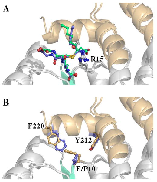
FIGURE 5.

Position of residues 10 and 220 within human A-class GST active sites. (A) Structural superposition of 3S,4R-GSDHN-bound GSTA4-4 (3IK7, blue ligand) and 3S,4R-GSDHN-bound GSTA1-1 GIMFhelix (3IK9, green ligand) (B) Structural superposition contrasting the side chain of F/P10 and F220. The ball and stick representation of residues from GSTA4-4 and GSTA1-1 GIMFhelix are colored blue and orange, respectively. The ligands are not shown for clarity in (B).
