Abstract
Background
Hypereosinophilic syndromes (HES) are a heterogeneous group of rare disorders defined by persistent blood eosinophilia ≥1.5 × 10⁹/L, absence of a secondary cause, and evidence of eosinophil-associated pathology. With the exception of a recent multicenter trial of mepolizumab (anti-IL-5 monoclonal antibody), published therapeutic experience has been restricted to case reports and small case series.
Objective
The purpose of the study was to collect and summarize baseline demographic, clinical and laboratory characteristics in a large, diverse cohort of patients with HES and to review responses to treatment with conventional and novel therapies.
Methods
Clinical and laboratory data from 188 patients with HES, seen between January 2001 and December 2006 at eleven institutions in the United States and Europe, were collected retrospectively by chart review.
Results
Eighteen of 161 patients (11%) tested were FIP1L1-PDGFRA mutation-positive and 29/168 patients tested (17%) had a demonstrable aberrant or clonal T cell population. Corticosteroid monotherapy induced complete or partial responses at 1 month in 85% (120/141) of patients with most remaining on maintenance doses (median 10 mg prednisone equivalent daily for 2 months-20 years). Hydroxyurea and interferon-alpha (used in 64 and 46 patients, respectively) were also effective, but their use was limited by toxicity. Imatinib (used in 68 patients) was more effective in patients with the FIP1L1-PDGFRA mutation (88%) than in those without (23%; p<0.001).
Conclusion
This study, the largest clinical analysis of patients with HES to date, not only provides useful information for clinicians but should stimulate prospective trials to optimize treatment of HES.
Keywords: eosinophil, hypereosinophilic syndrome, FIP1L1-PDGFRA
Introduction
The hypereosinophilic syndromes (HES) are a diverse group of rare disorders defined by the presence of persistent peripheral blood eosinophilia ≥1.5 × 10⁹/L, the absence of a secondary cause of eosinophilia, and evidence of eosinophil-associated end organ damage1. The clinical heterogeneity of HES has long been recognized 2; however, it is only recently that the techniques have become available to identify subtypes of HES with different underlying etiologies. The two best described of these subtypes are lymphocytic variant HES (L-HES) 3, in which the underlying cause of the eosinophilia is secretion of eosinophilopoietic cytokines by T lymphocytes, and myeloproliferative HES/chronic eosinophilic leukemia (CEL) 4, 5, most commonly due to an interstitial deletion in chromosome 4. These subtypes are associated with dramatic differences in clinical presentation, prognosis and responses to therapy 6. The availability of new targeted therapies, including tyrosine kinase inhibitors and humanized monoclonal antibodies, have only increased the importance of developing a better understanding of the etiologies and pathogenesis of HES, as these may be predictors of treatment response.
Despite the fact that a wide variety of agents have been used for the treatment of HES, published therapeutic experience has been largely restricted to case reports and small case series, the exception being a recently published multicenter trial of the monoclonal anti-IL-5 antibody, mepolizumab, as a steroid-sparing agent in HES 7. The lack of published data is due, in large part, to the paucity of affected patients as well as to the heterogeneity of presenting symptoms. This retrospective, multicenter analysis sought to collect and summarize the baseline demographic, clinical and laboratory characteristics in a large, diverse cohort of patients with HES and to review responses to treatment with both conventional and novel therapies.
Methods
Patients meeting diagnostic criteria for HES6, evaluated between January 2001 and December 2006, at eleven participating institutions with expertise in the evaluation of eosinophilic disorders, were included in the study. Inclusion criteria were: documentation of a peripheral eosinophil count of ≥1.5 × 10⁹/L and signs or symptoms of end organ involvement for which another etiology could not be found. Patients with neoplasms (other than FIP1L1/PDGFRA (FP)-positive CEL), biopsy-positive Churg-Strauss vasculitis, single organ eosinophilic diseases without blood eosinophilia, and overlap syndromes 6 were excluded from the study. Patients were enrolled consecutively at each site beginning with patients seen at initial or follow-up visit in December 2006 and proceeding backwards in time until 50 patients were included from a given site or the year 2001 was reached.
Clinical and laboratory data pertaining to baseline characteristics and treatment responses were collected following chart review, entered without identifiers into a database and compiled for analysis (see Online Repository). Potential duplicates were removed on the basis of a combination of factors, including birth year, gender, and clinical features. IRB approval and informed consent were obtained as required by each institution.
All 188 patients underwent detailed evaluation at the contributing clinical sites, including a complete history, physical exam, and laboratory evaluation (Table 1). Parameters previously shown or suspected to have prognostic significance in HES, e.g. peak eosinophil count, serum IL-5, total IgE, vitamin B12, tryptase, and TARC (thymus and activation-related chemokine (CCL17)) levels, were assessed. Results of FP mutation analysis (by nested polymerase chain reaction (PCR) or fluorescence in situ hybridization FISH)) and assessment of T cell clonality and phenotype by peripheral blood flow-cytometry and/or T cell receptor rearrangement PCR (TCR) were also included in the analysis. Although whole blood flow cytometry techniques and antibody panels varied among the sites, CD3, CD4 and CD8 antibodies, which detect the most common aberrant phenotypes in L-HES (CD3−CD4+ and CD3+CD4−CD8−)8, were included in all panels. Serum TARC levels were determined using commercially available ELISA kits from R&D Systems Europe (BRU, GER, LEIC; detection limit 7 pg/ml) or by ELISA from Thermo Fisher Scientific (BETH; detection limit 0.8 pg/ml). Serum IL-5 levels were determined using commercially available ELISA kits from BD Biosciences Pharmingen or R&D Systems (CIN, BRU, LEIC, SUR, MINN, BOS, SLC; detection limit 7.8 pg/ml) or by ELISA by Thermo Fisher Scientific (BETH; detection limit 0.8 pg/ml). Laboratory information, including results of FP mutation testing, T cell studies and serum TARC levels, was requested on all patients; however, due to the retrospective nature of the study, all results were not available in all cases. Normal values were defined as follows: IgE<100 IU/ml, serum IL-5 <14.1 pg/ml, tryptase <11.5 ng/ml, vitamin B12 <950 pg/ml, and TARC < 500 pg/ml.
Table 1.
Baseline characteristics of patients
| Center* | Number of patients |
Gender (male/female) |
Median age at diagnosis (range) |
GM peak eosinophil count ×109/L (range) |
|---|---|---|---|---|
| BRU | 22 | 12/10 | 48 (15–81) |
6.29 (2.0–16.7) |
| MUN | 8 | 5/3 | 43 (12–65) |
3.37 (1.5–8.32) |
| LEIC | 14 | 10/4 | 62 (30–78) |
10.52 (2.12–80.90) |
| BALT | 15 | 8/7 | 46 (23–85) |
5.86 (2.0–59.75) |
| CIN | 21 | 10/11 | 37 (15–56) |
5.45 (1.5–42.0) |
| MINN | 16 | 7/9 | 54 (9–69) |
5.91 (1.89–29.02) |
| BOS | 7 | 4/3 | 45 (23–59) |
7.30 (3.5–14.39) |
| SLC | 13 | 8/5 | 30 (6–74) |
11.34 (2.4–85.0) |
| BETH | 39 | 12/14 | 42 (16–69) |
8.98 (2.2–400) |
| BER | 14 | 6/8 | 54 (8–69) |
5.18 (1.5–340) |
| SUR | 19 | 9/10 | 43 (14–65) |
5.61 (1.7–31.60) |
| Total | 188 | 105/82 | 45 (6–85) |
6.602 (1.5–400) |
Gender distribution, median age at diagnosis, and geometric mean peak eosinophil count are provided for each of the participating centers: BRU=Erasme Hospital, Brussels, Belgium; MUN=Technical University of Munich, Munich, Germany; LEIC=University of Leicester, Leicester, UK ; BALT=Johns Hopkins Hospital, Baltimore, USA; CIN= Cincinnati Children’s Hospital Medical Center, Cincinnati, USA; MINN= Mayo Clinic, Rochester, USA; BOS=Harvard Medical School, Boston, USA; SLC= University of Utah, Salt Lake City, USA; BETH= Laboratory of Parasitic Diseases, National Institutes of Health, Bethesda, USA; BER=University of Bern, Bern, Switzerland; SUR=Hopital Foch, Suresnes, France
Clinical responses at 1 month of treatment (full, partial or no response) were recorded, as well as the maintenance dose, the maximal dose (for prednisone only), and whether each drug was given as monotherapy. Complete responses were defined as decrease of the eosinophil count to the normal range (0–0.5 × 10⁹/L) and symptomatic improvement after one month of treatment. Partial responses were defined as decrease of the eosinophil count, but not to the normal range, and/or symptomatic improvement after one month of treatment. No response was defined as a stable or increasing eosinophil count and no symptomatic improvement after one month of treatment.
Statistical Analysis
Nonparametric comparisons of group means were made using the Mann-Whitney U test. Proportions were compared using Fisher exact test. A p-value of less than 0.05 was considered significant for all tests.
Results
Baseline characteristics
Of the 188 patients evaluated, 104 were male (55%) and 84 were female (45%). The median age at diagnosis was 45 years of age (range 6–85 years). The peak recorded absolute eosinophil counts ranged from 1.5–400 × 109/L with a geometric mean (GM) peak eosinophil count of 6.6 × 10⁹/L (Table 1). Of the 161 patients tested for the FP mutation, 18 (11%), all of whom were male, were positive. Of the 168 patients who were evaluated for clonal or aberrant populations of T cells, 29 (17%; 15 male and 14 female) were positive by PCR (n=14), flow cytometry (n=3), or both (n=12). These patients were classified as lymphocytic variant HES (L-HES). There were 4 patient deaths reported during the study period, 2 of which were thought to be secondary to HES (due to eosinophilic heart disease).
Serum tryptase levels were reported for 123 patients (66%) and ranged from 1– 131 ng/ml (GM 7.6 ng/ml). Elevated serum tryptase levels were significantly more common in patients with the FP mutation (9/11 (82%)) than in patients who tested negative for the mutation (21/105 (20%); p=.001). Furthermore, among the 30 patients with elevated levels, the GM tryptase level was greater in the FP-positive patients (31 vs. 19 ng/ml, p=0.03). Serum vitamin B12 levels were reported for 120 patients and ranged from 156–10995 pg/ml (GM 632 pg/ml). Elevated serum vitamin B12 levels were also more common in the FP-positive patients (range 401–10,955 pg/ml and present in 13/14 (93%) vs. FP-negative patients (range 156–2000 pg/ml and present in 20/106 (18%); p<0.0001). Elevated serum tryptase and/or B12 levels were uncommon in patients with L-HES, occurring in only 3/25 and 1/22 patients tested, respectively,
Serum TARC levels were reported for 82 patients from 4 centers (see methods) and ranged from 22–150100 pg/ml (GM 769 pg/ml). Serum TARC levels were elevated in 75% (12/16) of patients with L-HES as compared to 36% (24/66) of patients without a clonal or aberrant T cell population (p=.0017). Furthermore, GM serum TARC levels in the subset of patients with elevated levels were significantly increased in patients with L-HES as compared to those without (GM 12,979 vs. 3,406 pg/ml, p=0.02). Serum IgE levels were elevated in 83/150 patients tested (55%). In the 135 patients who were also evaluated for clonal or aberrant T cell populations, serum IgE was elevated in 19/28 (68%) patients with L-HES and 56/107 (52%) in whom a clonal or aberrant population was sought but not found (p<0.0001). Only one patient in the FP group had an elevated IgE level, and none had elevated serum TARC levels.
Serum IL-5 levels were assessed in 107 patients and ranged from undetectable to 377 pg/ml. IL-5 levels were elevated in 28/107 (26%) patients: 6 patients with L-HES, 1 patient with the FP mutation, and 21 patients with HES in whom FP mutation analysis and at least one test for T cell clonality/phenotype had been performed and were negative.
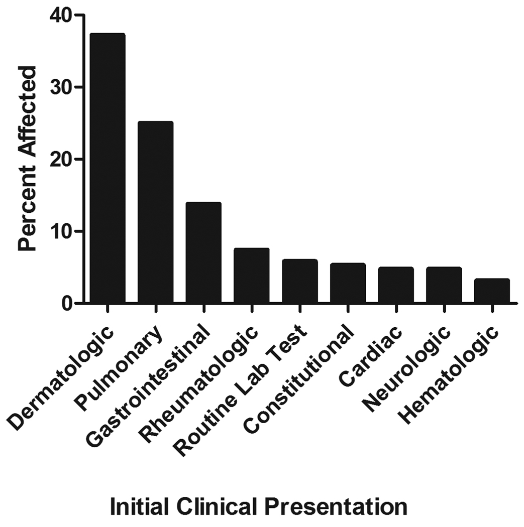
Data pertaining to initial clinical presentation was collected for all patients (Figure 1a and Online Repository Table E1). The most common presenting manifestations of HES were dermatologic (70/188, 37%), followed by pulmonary (47/188, 25%), and gastrointestinal (25/188, 14%). Less than 5% (9/188) of patients had cardiac manifestations at the time of diagnosis. Of note, 6% (11/188) of patients presented with clinically asymptomatic eosinophilia identified on laboratory testing performed for unrelated reasons.
Figure 1.
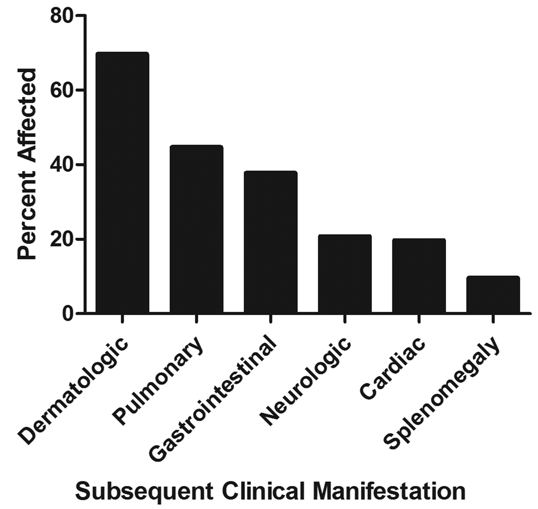
Clinical manifestations of HES. The clinical manifestations at initial presentation (A) and at the time of the retrospective analysis (B) are shown as the percent of patients with evidence of organ involvement referable to a given category. Reported manifestations in each of the categories are listed in Table E1 of the Online Repository.
Dermatologic involvement was also the most common subsequent clinical manifestation of HES and was reported in 69% (130/188) of patients. This was followed in frequency by pulmonary (44%) and gastrointestinal (38 %) manifestations (Figure 1b). Cardiac disease unrelated to hypertension, atherosclerosis or rheumatic disease was reported in 20% (37/188) of the patients. The frequency of cardiac disease was similar in patients with and without the FP mutation (4/18 (22%) vs. 33/170 (19%), respectively).
Treatment
Corticosteroids
Corticosteroids have long been the mainstay of therapy for HES, although the dosing has not been standardized 6, 9. In this series, 179 (95%) patients were treated with corticosteroids, most of whom (163/188; 81%) received corticosteroids as initial therapy. The median maximal daily dose of prednisone (or prednisone equivalent) was 40 mg (range 5–625 mg). Most patients (130/179; 72%) were maintained on corticosteroids for some period of time, ranging from 2 months-20 years, with a median maintenance dose of 10 mg daily (range 1–40 mg/day).
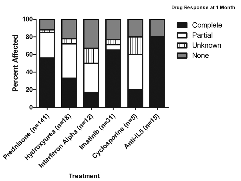
Of 141/188 (75%) patients who received corticosteroid monotherapy, 120 (85%) experienced a complete or partial response after one month of treatment (Figure 2a). When corticosteroids were used in combination therapy (most often due to failure or corticosteroid toxicity), the most frequent second agents added were hydroxyurea (36 patients) and interferon alpha (IFN, 24 patients). Responses (complete or partial) were achieved in 69% (25/36) of patients receiving hydroxyurea and corticosteroids and 75% (18/24) of patients treated with IFN and corticosteroids.
Figure 2.
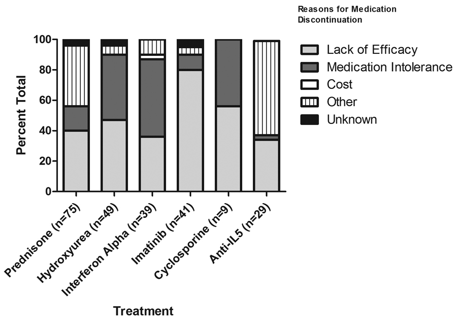
Response to Treatment. The bars represent response rates after one month of therapy (A) and reasons for drug discontinuation (B). Responses were defined as complete (normalization of absolute eosinophil count (AEC) and clinical symptom improvement), partial (reduction of AEC, but not to normal levels, and/or improvement in symptoms) or no response (neither reduction of AEC nor improvement in symptoms).
Prednisone was discontinued in 42% (75/179) of patients. The most frequently cited reasons for discontinuation were lack of efficacy (40%) and “other” (40%) (Figure 2b). The most common reasons for the response “other” were efficacy of another drug, most frequently imatinib or anti-IL-5 antibody (n= 12), and disease remission (n= 12).
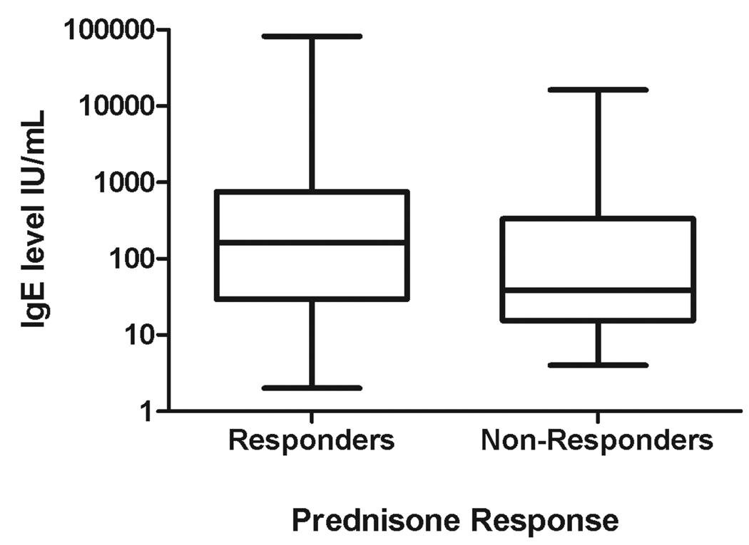
Elevated serum IgE and TARC levels have been associated with corticosteroid responsiveness in patients with HES 10, 11. Serum IgE levels were elevated in 77/142 (54%) patients who received corticosteroid therapy and for whom serum IgE levels were available. The percentage of patients with elevated IgE was comparable in patients who responded to steroids and those who did not (73/129 (57%) vs. 4/13 (31%); p=0.09, Fisher’s exact test). Similarly, GM serum IgE levels were similar in patients with a complete (125 IU/ml), partial (222 IU/ml) or lack of response (70 IU/ml) to corticosteroid therapy (Figure 3a).
Figure 3.
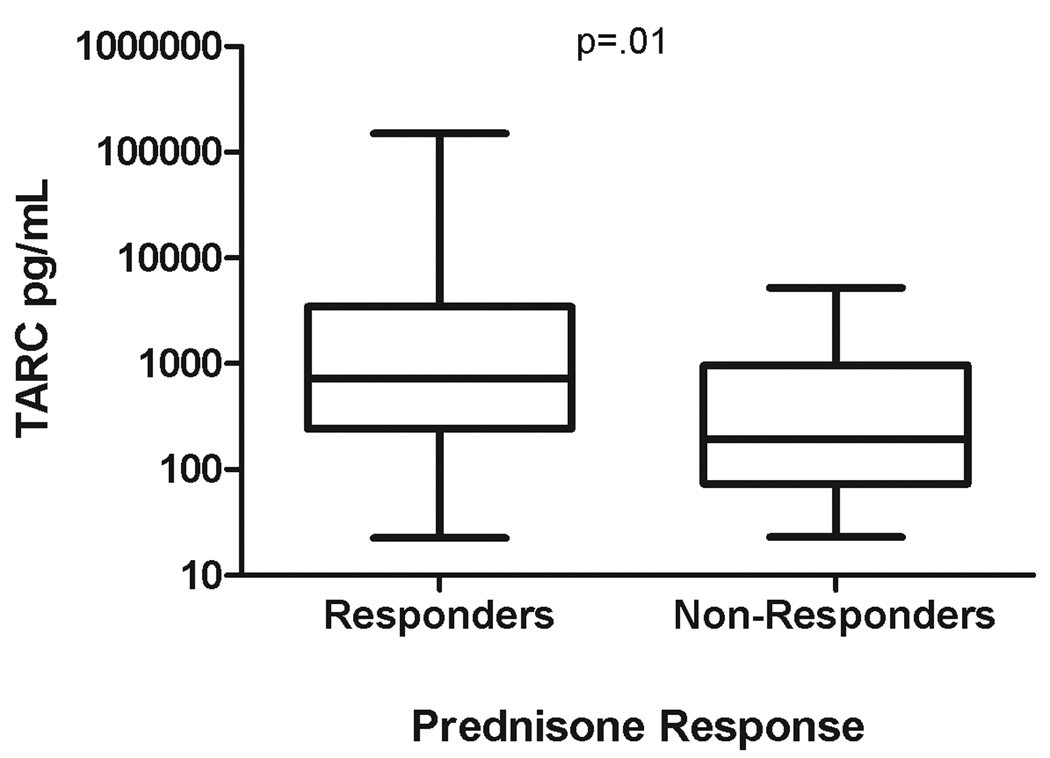
Association of serum TARC, but not serum IgE levels with prednisone responsiveness. Serum IgE (A) and TARC (B) levels for prednisone responders (n=129 and 75, respectively) and non-responders (n=13 and 8, respectively) are shown using box and whiskers plots. The whiskers represent the minimum and maximum values and the horizontal lines represent the lower quartile, median, and upper quartile.
Serum TARC levels were elevated in 41/82 (50%) patients who received corticosteroid therapy and for whom levels were available. Not only were GM TARC levels significantly elevated in patients who responded to corticosteroids as compared to non-responders (979 vs. 242; p=.01), but serum TARC levels >10,000 pg/ml (n=11) were reported only among prednisone responders (Figure 3b).
Hydroxyurea
Thirty-four percent (64/188) of patients were treated with the oral cytotoxic agent hydroxyurea, with a median maximum daily dose of 1000 mg (range 500–2000 mg). Eighteen patients received hydroxyurea as monotherapy, of whom 6 (33%) achieved a complete response and 7 (39%) a partial response (Figure 2a). Hydroxyurea was discontinued in the majority of patients (49/64; 77%), primarily due to lack of efficacy (23/49; 47%) and medication intolerance secondary to treatment-related side effects (21/49; 43%) (Figure 2b).
Interferon alpha (IFN)
Approximately one quarter of the patients (46/188) were treated with IFN, with a median maximal dose of 14 million units per week (range 3–40 million units per week). Only 12 patients received IFN as monotherapy, of whom 2 (17%) achieved a complete response and 4 (33%) a partial response at 1 month (Figure 2a). IFN was discontinued in all but 6 patients (87%). The most common reasons for drug discontinuation were medication intolerance (20/40; 50%), lack of efficacy (14/40; 36%) and cost (1/40; 3%) (Figure 2b).
Cyclosporine
11/188 (6%) patients were treated with cyclosporine (median maximal daily dose of 200 mg; range 150–500 mg). Of the 5 patients who received cyclosporine monotherapy, one patient achieved a complete response (20%), and 2 patients (40%) achieved partial responses (Figure 2a). The medication was discontinued in 9/11 patients (82%), including the patient who responded completely for reasons of medication intolerance (Figure 2b).
Imatinib
The tyrosine-kinase inhibitor, imatinib, was initiated in 68/188 patients (36%), of whom 17 were known to be positive for the FP mutation. The median maximal dose used was 400 mg daily (range 100 mg twice weekly to 600 mg daily). Of the 31 patients who received imatinib monotherapy, 20/31 (65 %) achieved a complete response and 2/31 (6%) a partial response (Figure 2a). Of the FP-positive patients, 15/17 (88%) responded completely and two (12%) had no response12, 13. In contrast, only 10/43 (23%) FP-negative patients who received imatinib experienced a complete (n=6) or partial (n=4) response (p<.0001 compared to the FP-positive patients).
Imatinib was discontinued in 41/68 patients, mostly due to lack of efficacy (33/41; 80%) (Figure 2b). The remaining 8/41 patients discontinued imatinib therapy because of medication intolerance (4 patients), unspecified reasons (2 patients), the initiation of an anti-IL-5 clinical trial (1 patient), or the desire to have children (1 patient).
Anti-IL-5 antibody
A total of 62/188 (33%) patients received at least one dose of anti-IL-5 monoclonal antibody therapy with mepolizumab (GlaxoSmithKline; 750 mg/month; n=59) or reslizumab (Ception; 1–3 mg/kg/month; n=2), or both (n=1). Of the 15 patients who received mepolizumab monotherapy, 12/15 (80%) achieved a complete response one month after the initiation of therapy (Figure 2a). As has been previously reported17, two patients received reslizumab monotherapy, both of whom responded after 1 month of therapy. The remaining 45 patients were treated concomitantly with mepolizumab and corticosteroids. Thirty-four of 45 (76%) responded completely after 1 month of mepolizumab and an additional 5 (11%) had a partial response.
Although anti-IL-5 therapy was discontinued in 29/62 (47%) patients the most frequent cause for discontinuation (18 patients; 62%) was the completion of a clinical trial (listed as “other”) (Figure 2b). An additional 10 patients (34%) discontinued due to lack of efficacy. Only 1 patient discontinued anti-IL-5 treatment due to medication intolerance.
Other agents
Several other medications were used for treatment of HES (see Table E2 in the Online Repository); however too few patients were treated to draw conclusions for individual agents.
Discussion
One of the major problems with previously reported series of HES patients has been referral bias, with single centers more likely to see patients with end organ manifestations that fall within the area of expertise of a particular subspecialty interested in HES. This has been compounded by the tendency to publish cases of the most severely affected patients, a disproportionate number of whom, in retrospect, were likely FP-positive. Patients in the current series were referred to groups with international recognition in the diagnosis and treatment of HES, composed of a wide variety of subspecialty physicians. Consequently, the data compiled are more likely to represent the true spectrum of HES. The major limitation of this study was the retrospective design and resultant lack of standardization of laboratory studies between the different sites. Despite these limitations, however, a number of interesting conclusions can be drawn.
In contrast to the marked (9:1) male predominance of HES reported in the literature, the ratio of males to females in the current study was 11:8. This likely reflects the fact that FP-positive patients, who are almost exclusively male, were overrepresented in previous series and confirms data from recent studies in which FP-positive patients were excluded7. Similarly, early assessments of the relative prevalence of end organ manifestations were gleaned from case reports and small series and tended to overestimate more serious consequences of HES, including cardiac and neurologic involvement 2. In the current series, the most common presenting manifestations of HES were dermatologic, pulmonary and gastrointestinal. Cardiac and neurologic complications did occur, but were relatively uncommon at the initial presentation. Earlier diagnosis and the availability of better therapies likely contributed to the improved HES patient outcomes in the current series. It is important to note that 11 of the 188 patients presented with asymptomatic eosinophilia found on routine lab testing. Of these, two were found to have FP-mutation positive CEL, a condition associated with high morbidity and mortality (up to 50% at 5 years) in the absence of imatinib therapy 5. These findings highlight the importance of a comprehensive evaluation and close follow-up of patients presenting with unexplained hypereosinophilia.
There has been considerable controversy regarding the prevalence of L-HES and the FP-mutation among patients meeting criteria for HES. Early studies likely overestimated the prevalence of these two entities due to selection bias 3, 4. More recently, the prevalence of the FP mutation has been estimated at 14% in a retrospective analysis of 81 patients with primary eosinophilia ≥1.5 × 10⁹/L who underwent bone marrow examination 14. The prevalence of FP-positive patients was slightly lower in our study (10%), possibly due to the inclusion of patients in whom a bone marrow examination was not deemed necessary (i.e. less likely to have myeloproliferative disease). The association between elevated serum vitamin B12 and tryptase levels and the presence of the FP mutation was confirmed in the present study. Of note, extremely high levels of serum vitamin B12 (>2000 pg/ml) were observed only in the FP-positive group, suggesting that this may be a more discriminating biomarker of myeloproliferative HES.
Confirmation of L-HES is complex, ideally involving both lymphocyte phenotyping and TCR analysis. Consequently, differences in technique as well as the characteristics of the population studied likely account for the variability in the reported prevalence of L-HES, ranging from 14–31% 3, 15, 16. In the present study, 17% of patients who were tested had clonal/abnormal populations of T cells detected by PCR or flow cytometry. Although it is debatable whether L-HES can be diagnosed on the basis of a clonal population identified by PCR in the absence of a detectable aberrant phenotype by flow cytometry (8.3% of the patients in the current study), the fact that some patients with clonal TCR rearrangement patterns have strikingly elevated serum TARC and/or IgE levels 11 suggests that aberrant T cell populations not identified using current antibody panels may be responsible for the eosinophilia in these patients. Nevertheless, a prospective trial using standardized techniques is clearly necessary to obtain an accurate assessment of the prevalence of the lymphocytic variant of HES.
Early efficacy studies and extensive clinical experience have proven corticosteroids to be the first line agent for the treatment of FP-negative HES 6. Although the previously reported association between prednisone responsiveness and elevated serum IgE levels 10 was not confirmed in this study, TARC levels were significantly higher in patients who responded to prednisone. There was no difference between the serum IL-5 levels in patients who responded to prednisone compared to those who failed to respond (data not shown). Most patients were maintained on low to moderate dose prednisone therapy (5–20 mg daily). Although this would seem to indicate that the need for alternative therapies is small, the study questionnaire was not designed to detect steroid toxicities that did not lead to drug discontinuation. Furthermore, the majority of corticosteroid-responsive patients went on to receive a second-line agent, consistent with a higher rate of steroid toxicity and/or resistance than is evident from the collected data.
In the past 5–10 years, HES subtypes have been defined and a number of new, targeted agents have been developed, including imatinib mesylate and humanized monoclonal antibodies to IL-5. The restriction of this retrospective study to patients seen after 2001 was intended to maximize inclusion of data on these newer agents. As expected, imatinib was extremely effective in FP-positive patients with resistance described in only 2 patients. The 23% response rate in FP-negative patients is more difficult to interpret as the database did not include sufficient information to distinguish patients with myeloproliferative features who may be more likely to respond to imatinib. Although anti-IL-5 therapy was well-tolerated with an overall response rate of 80%, all patients received this agent in clinical trials, the majority of which required patients to be steroid-responsive 7, 13, or were limited to a small number of doses 17. Consequently, conclusions regarding the long-term efficacy of anti-IL-5 therapy and the most appropriate patients for treatment await further study.
In summary, HES is a rare group of disorders for which unbiased information on demographics, clinical manifestations and treatment responses is scarce. Although the current study has limitations due to its retrospective design, it represents the largest multicenter study of unselected patients with HES to date. As such, it not only provides useful information for clinicians involved in the care of HES patients, but will hopefully stimulate carefully designed prospective trials for the treatment of this disorder.
Key Messages
Classically defined hypereosinophilic syndrome is a heterogeneous group of varied disorders, the majority of which remain idiopathic.
Corticosteroids are extremely effective in the treatment of FIP1L1/PDGFRA-negative HES, but their use may be limited by side effects.
Carefully designed prospective trials are required to advance the diagnosis and treatment of HES.
Supplementary Material
Acknowledgments
The authors would like to thank Dr. Amal Assa’ad, Bridget Buckmeier, Cheryl Talar-Williams, Melissa Law and Marilyn Hartsell for their participation in the clinical care of the patients.
Funding/Support: This study was supported by the Division of Intramural Research of the NIAID/NIH (Drs. Klion, Ogbogu and Nutman), grants AI41472 and AI72265 from the NIH (Dr. Bochner), grant AI061097 from the NIH (Dr. Gleich), the Human Immunology grant program of the Dana Foundation (Dr. Bochner), the Swiss National Science Foundation (Drs. Simon), the Belgian National Fund for Scientific Research (Dr. Roufosse) and the Campaign Urging Research for Eosinophilic Disorders (Dr. Rothenberg). Dr. Bochner is a Cosner Scholar in Translational Research from Johns Hopkins University. The funding organizations had no role in the design and conduct of the study; collection, management, analysis, and interpretation of the data; and preparation, or review of the manuscript. The manuscript was approved by the Division of Intramural Research, NIAID/NIH.
Abbreviations
- FIP1L1
Fip1-like 1
- FP
FIP1L1-PDGFRA
- HES
Hypereosinophilic syndrome
- L-HES
Lymphocytic variant hypereosinophilic syndrome
- M-HES
Myeloproliferative variant hypereosinophilic syndrome
- PDGFRA
Platelet derived growth factor receptor alpha
- CEL
Chronic eosinophilic leukemia
- TARC
Thymus and activation-related chemokine
- GM
Geometric mean
Footnotes
Publisher's Disclaimer: This is a PDF file of an unedited manuscript that has been accepted for publication. As a service to our customers we are providing this early version of the manuscript. The manuscript will undergo copyediting, typesetting, and review of the resulting proof before it is published in its final citable form. Please note that during the production process errors may be discovered which could affect the content, and all legal disclaimers that apply to the journal pertain.
Author Contributions.
P.O. collected and analyzed the data and wrote the paper; B.S.B,, J.H.B., G.J.G., J.H., J.E.K., J.R., M.E.R., F.R., H.U.S., A.W., and P.F.W helped design the research, collect and analyze the data and edit the paper, J.H.B. helped design the research, collect and analyze the data and edit the paper, K.M.L., F.P., M.S., J.S., D.S., and M.L.S. helped collect and analyze the data and edit the paper, T.B.N. helped design the research, analyze the data and edit the paper, and A.D.K. helped design the study, collect and analyze the data and write the paper.
Financial disclosures: Dr. Sheikh has served as a consultant, speaker’s bureau and received research support from GlaxoSmithKline and has been on speaker’s bureau for Novartis. Drs. Weller and Kahn have served as consultants to GlaxoSmithKline. Drs. Wardlaw and Kahn have received funding for research from and have served on the advisory board of GlaxoSmithKline.
References
- 1.Chusid MJ, Dale DC, West BC, Wolff SM. The hypereosinophilic syndrome: analysis of fourteen cases with review of the literature. Medicine (Baltimore) 1975;54:1–27. [PubMed] [Google Scholar]
- 2.Weller PF, Bubley GJ. The idiopathic hypereosinophilic syndrome. Blood. 1994;83:2759–2779. [PubMed] [Google Scholar]
- 3.Simon HU, Plotz SG, Dummer R, Blaser K. Abnormal clones of T cells producing interleukin-5 in idiopathic eosinophilia. N Engl J Med. 1999;341:1112–1120. doi: 10.1056/NEJM199910073411503. [DOI] [PubMed] [Google Scholar]
- 4.Cools J, DeAngelo DJ, Gotlib J, Stover EH, Legare RD, Cortes J, et al. A novel tyrosine kinase created by the fusion of the PDGFRA and FIP1L1 genes is a therapeutic target of imatinib in idiopathic hypereosinophilic syndrome. N Engl J Med. 2003;348:1201–1214. doi: 10.1056/NEJMoa025217. [DOI] [PubMed] [Google Scholar]
- 5.Klion AD, Noel P, Akin C, Law MA, Gilliland DG, Cools J, et al. Elevated serum tryptase levels identify a subset of patients with a myeloproliferative variant of idiopathic hypereosinophilic syndrome associated with tissue fibrosis, poor prognosis and imatinib-responsiveness. Blood. 2003;101:4660–4666. doi: 10.1182/blood-2003-01-0006. [DOI] [PubMed] [Google Scholar]
- 6.Klion AD, Bochner BS, Gleich GJ, Nutman TB, Rothenberg ME, Simon HU, et al. Approaches to the treatment of hypereosinophilic syndromes: a workshop summary report. J Allergy Clin Immunol. 2006;117:1292–1302. doi: 10.1016/j.jaci.2006.02.042. [DOI] [PubMed] [Google Scholar]
- 7.Rothenberg ME, Klion AD, Roufosse FE, Kahn JE, Weller PF, Simon HU, et al. Treatment of patients with the hypereosinophilc syndrome with mepolizumab. N Engl J Med. 2008;358:1215–1228. doi: 10.1056/NEJMoa070812. [DOI] [PubMed] [Google Scholar]
- 8.Roufosse F, Cogan E, Goldman M. Lymphocytic variant hypereosinophilic syndromes. Immunol Allergy Clin N Am. 2007;27:389–413. doi: 10.1016/j.iac.2007.07.002. [DOI] [PubMed] [Google Scholar]
- 9.Simon HU, Cools J. Novel approaches to therapy of hypereosinophilic syndromes. Immunol Allergy Clin North Am. 2007;27:519–527. doi: 10.1016/j.iac.2007.07.003. [DOI] [PubMed] [Google Scholar]
- 10.Bush RK, Geller M, Busse WW, Flaherty DK, Dickie HA. Response to corticosteroids in the hypereosinophilic syndrome. Association with increased serum IgE levels. Arch Intern Med. 1978;138:1244–1246. [PubMed] [Google Scholar]
- 11.de Lavareille A, Roufosse F, Schmid-Grendelmeier P, Roumier AS, Schandene L, Cogan E, et al. High serum thymus and activation-regulated chemokine levels in the lymphocytic variant of the hypereosinophilic syndrome. J Allergy Clin Immunol. 2002;110:476–479. doi: 10.1067/mai.2002.127003. [DOI] [PubMed] [Google Scholar]
- 12.Simon D, Salemi S, You sefi S, Simon HU. Primary resistance to imatinib in Fip1-like 1-platelet-derived growth factor receptor alpha-positive eosinophilic leukemia. J Allergy Clin Immunol. 2008;121:1054–1056. doi: 10.1016/j.jaci.2007.11.027. [DOI] [PubMed] [Google Scholar]
- 13.Stein ML, Villanueva JM, Buckmeier BK, Yamada Y, Filipovich AH, Assa'ad AH, et al. Anti-IL-5 (mepolizumab) therapy reduces eosinophil activation ex vivo and increases IL-5 and IL-5 receptor levels. J Allergy Clin Immunol. 2008;121:1473–1483. doi: 10.1016/j.jaci.2008.02.033. [DOI] [PMC free article] [PubMed] [Google Scholar]
- 14.Pardanani A, Brockman SR, Paternoster SF, Flynn HC, Ketterling RP, Lasho TL, et al. FIP1L1/PDGFRA fusion: prevalence and clinicopathologic correlates in 89 patients with moderate to severe eosinophilia. Blood. 2004;104:3038–3045. doi: 10.1182/blood-2004-03-0787. [DOI] [PubMed] [Google Scholar]
- 15.Vaklavas C, Tefferi A, Butterfield J, Ketterling RP, Vertovsek S, Kantarjian H, et al. 'Idiopathic' eosinophilia with an Occult T-cell clone: prevalence and clinical course. Leuk Res. 2007;31:691–694. doi: 10.1016/j.leukres.2006.10.005. [DOI] [PubMed] [Google Scholar]
- 16.Roche-Lestienne C, Lepers S, Soenen-Cornu V, Kahn JE, Lai JL, Hachulla E, et al. Molecular characterization of the idiopathic hypereosinophilic syndrome (HES) in 35 French patients with normal conventional cytogenetics. Leukemia. 2005;19:792–798. doi: 10.1038/sj.leu.2403722. [DOI] [PubMed] [Google Scholar]
- 17.Klion AD, Law MA, Noel P, Haverty TP, Nutman TB. Safety and efficacy of the monoclonal anti-interleukin 5 antibody, SCH55700, in the treatment of patients with the hypereosinophilic syndrome. Blood. 2004 doi: 10.1182/blood-2003-10-3620. [DOI] [PubMed] [Google Scholar]
Associated Data
This section collects any data citations, data availability statements, or supplementary materials included in this article.


