Abstract
WEB SITE FEATURE
A 72-year-old woman was admitted to our hospital with mild chest discomfort. She had no respiratory symptoms. Clinical examination and cardiac auscultation revealed nothing unusual. A 12-lead electrocardiogram showed sinus rhythm of 64 beats/min and occasional premature ventricular complexes. A chest radiograph showed a cardiothoracic ratio of 0.5 and no evidence of increased pulmonary blood flow. A transthoracic echocardiogram revealed normal-sized cardiac chambers and normal left ventricular systolic function.
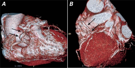
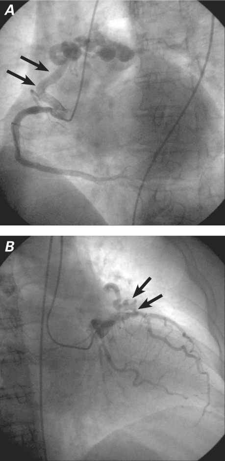
Electrocardiographically-gated, 64-slice multidetector-row computed tomography (MDCT) showed 2 large, tortuous, abnormal vessels that arose from the conal branch of the proximal right coronary artery and the left anterior descending coronary artery and drained into the main pulmonary artery (Fig. 1). The abnormal vessels had multiple aneurysmal dilations along their course. Bilateral coronary artery-to-main pulmonary artery fistulae were suspected, and coronary angiography confirmed this diagnosis (Fig. 2). No significant stenosis was detected in any coronary vessel. The patient responded well to a medical regimen of nitrates, aspirin, and β-blockers.
Fig. 1 Three-dimensional, volume-rendered multidetector-row computed tomographic images show bilateral coronary artery fistulae with multiple aneurysmal dilations. The fistulae arise from A) the conal branch of the proximal right coronary artery (arrows) and B) the proximal left anterior descending coronary artery (arrows).
Fig. 2 Coronary angiography confirms that bilateral coronary artery fistulae arise from A) the conal branch of the proximal right coronary artery (arrows) and B) the proximal left anterior descending coronary artery (arrows), with drainage into the main pulmonary artery. Real-time motion images are available at www.texasheart.org/journal.
Comment
Coronary artery fistulae communicate directly between a coronary artery and a cardiac chamber or a vessel around the heart. They are rare abnormalities (whether congenital or acquired), with an estimated incidence of 0.002% in the general population; however, they are found in 0.05% to 0.25% of patients who undergo coronary angiography.1,2 The most common sites of drainage are the right ventricle (41%), the right atrium (26%), and the pulmonary artery (17%).3 Bilateral fistulae that originate from the coronary system account for 5% of all coronary artery fistulae. Bilateral fistulae terminate more often into the pulmonary artery (56%) than do unilateral fistulae (17%).3,4 Fistulae are usually discovered incidentally upon coronary angiography. Although advances in MDCT technology and its use continue, the method remains inferior to coronary angiography in the detection of arrhythmias, in temporal resolution, and as a tool for use in performing intervention. Regardless, MDCT is an emerging option for noninvasively evaluating the anatomy of coronary artery fistulae, and it provides good noninvasive anatomic correlation with coronary angiography and surgery.5
Supplementary Material
Footnotes
Address for reprints: Se Hwan Kwon, MD, Department of Radiology, Kyung Hee University Medical Center, Hoeki-dong 1, Dongdaemun-gu, Seoul 130-702, ROK
E-mail: radkwon@dreamwiz.com
References
- 1.Burch GH, Sahn DJ. Congenital coronary artery anomalies: the pediatric perspective. Coron Artery Dis 2001;12(8):605–16. [DOI] [PubMed]
- 2.Okwuosa TM, Gundeck EL, Ward RP. Coronary to pulmonary artery fistula–diagnosis by transesophageal echocardiography. Echocardiography 2006;23(1):62–4. [DOI] [PubMed]
- 3.Levin DC, Fellows KE, Abrams HL. Hemodynamically significant primary anomalies of the coronary arteries. Angiographic aspects. Circulation 1978;58(1):25–34. [DOI] [PubMed]
- 4.Baim DS, Kline H, Silverman JF. Bilateral coronary artery–pulmonary artery fistulas. Report of five cases and review of the literature. Circulation 1982;65(4):810–5. [DOI] [PubMed]
- 5.Huang YK, Lei MH, Lu MS, Tseng CN, Chang JP, Chu JJ. Bilateral coronary-to-pulmonary artery fistulas. Ann Thorac Surg 2006;82(5):1886–8. [DOI] [PubMed]
Associated Data
This section collects any data citations, data availability statements, or supplementary materials included in this article.


