Abstract
Soft X-rays (< 1Kev) are of medical interest both for imaging and microdosimetry applications. X-ray sources at this low energy present a technological challenge. Synchrotrons, while very powerful and flexible, are enormously expensive national research facilities. Conventional X-ray sources based on electron bombardment can be compact and inexpensive, but low x-ray production efficiencies at low electron energies restrict this approach to very low power applications. Laser-based sources tend to be expensive and unreliable. Energetiq Technology, Inc. (Woburn, MA, USA) markets a 92 eV, 10W(2pi sr) electrode-less Z-pinch source developed for advanced semiconductor lithography. A modified version of this commercial product has produced 400 mW at 430 eV (2pi sr), appropriate for water window soft X-ray microscopy. The US NIH has funded Energetiq to design and construct a demonstration microscope using this source, coupled to a condenser optic, as the illumination system. The design of the condenser optic matches the unique characteristics of the source to the illumination requirements of the microscope, which is otherwise a conventional design. A separate program is underway to develop a microbeam system, in conjunction with the RARAF facility at Columbia University, NY, USA. The objective is to develop a focused, sub-micron beam capable of delivering > 1 Gy/second to the nucleus of a living cell. While most facilities of this type are coupled to a large and expensive particle accelerator, the Z-pinch X-ray source enables a compact, stand-alone design suitable to a small laboratory. The major technical issues in this system involve development of suitable focusing X-ray optics. Current status of these programs will be reported.
1. Introduction
Soft x-ray microscopy shows enormous promise for imaging cellular structures at resolutions beyond what can be achieved in optical microscopy, and with much simpler sample preparation than is required for electron microscopy[1]. In addition, the lower radiation dose required (compared to electron microscopy) allows tomographic investigation of subcellular structures in three dimensions. Existing synchrotron based microscopes have shown the potential of the soft x-ray microscope as a research tool, but they have the disadvantage of being tied to a massive light source at only a few national laboratories. A suitably bright, compact, low cost light source is the enabling innovation needed to realize a commercial biological soft x-ray microscope.
Energetiq has developed a unique light source based on an electrodeless, inductively coupled plasma Z-pinch. A commercial version optimized for EUV semiconductor lithography[2] has been modified to produce light in the soft x-ray range, and a program is under way to develop a demonstration microscope based on use of this source.
The overall design of the microscope is relatively conventional [3,4]. An illumination system composed of the source and condenser optics generates a hollow cone of light with a focus at the sample. An objective lens with short focal length produces a magnified image. As in other microscopes of this type, we use a zone plate objective, and match the illuminator numerical aperture to that of the zone plate. A cooled, back-thinned CCD camera detects the image.
The chromatic aberration inherent in a diffractive optic imposes spectral purity requirements on the illumination system. If the illumination is not nearly monochromatic, resolution will suffer. This requirement leads one to consider rather simple radiating systems. Helium-like nitrogen (N VI) is nearly ideal, with the 1S2-1S2P transition at about 430 eV.
2. Source Performance
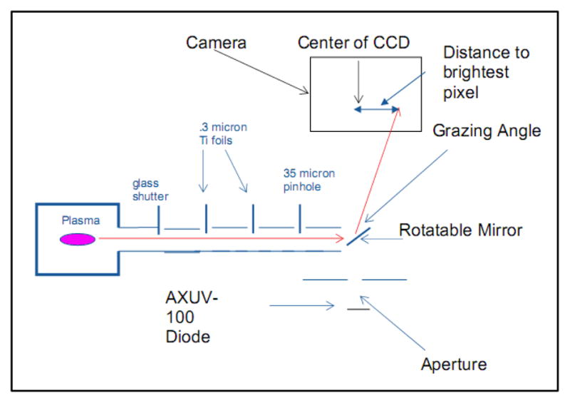
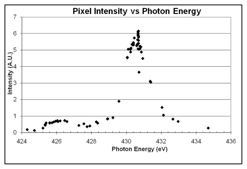
The spectral performance of the source was measured using a graded multi-layer mirror as a spectrometer, as shown in Figure 1. The mirror is treated as a Bragg diffractor. The grazing angle is varied by rotating the mirror; given the grazing angle calculated to the brightest pixel in the CCD, the photon energy can be calculated. The spectrum produced in this manner is shown in Figure 2.
Figure 1.

Spectral and Power Diagnostic
Figure 2.

Measured Spectrum
We plot versus photon energy. The peaks at 430 and 426 eV are the primary resonance lines of helium-like nitrogen. The widths of the peaks are entirely instrumental. We estimate the power at 426 ev to be down by a factor of roughly 10 from that at 430 eV; we expect negligible chromatic aberration at the image due to this component.
By rotating the mirror to throw the beam onto the X-ray diode, in-band power can be measured as well. We estimate the power radiated into 2π to be about 0.2 – 0.4 W, depending on source operating conditions.
3. Condenser Optic
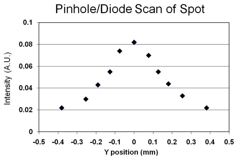
The condenser optic is an elliptical grazing incidence mirror. The focal points are 1 M apart, and the output numerical aperture is matched to that of the objective zone plate. A copper-nickel alloy was selected both for its resistance to oxidation and corrosion, and for good machining properties. A surface finish of 4 nm RMS was achieved using conventional diamond turning. Testing of the condenser is still under way; figure 3 shows a scan with a 25 micron pinhole/diode across the focal spot. The design demagnification of the prototype is about 1/4, which, with an 800 micron FWHM plasma should yield a spot size of 200 microns FWHM. In fact, the observed FWHM is about twice the ideal size. We believe this defect is due primarily to imperfections in both the figure and mounting geometry of the condenser, and will be corrected.
Figure 3.

Profile of illumination field
4. Objective lens and camera beamline
The objective portion of the microscope was designed to use zone plates of 120 microns diameter, in conjunction with a standard 1K × 1K, 13 micron pixel size back-thinned CCD X-ray camera.
We have acquired zone plates with outer zone widths (dr) of 40, 30, and 25 nm; the condenser numerical aperture was designed to match the .057 NA of the 25 nm zone plate. We have simulated the optical behaviour of the hollow-cone illumination (simulations courtesy of Patrick Naulleau, LBNL). The basic issue is that for illumination within the NA of the objective, the sample is observed in transmission. However, if the NA of the objective is overfilled, photons are diffracted into the NA of the objective by the smaller sample features – increasing (theoretical) resolution, but at the cost of potentially introducing halos and artefacts into the image. The simulation results imply that we will have acceptable contrast, without significant diffraction-produced artefacts.
In parallel with the microscope program, we are also exploiting many of the same technologies to produce a microbeam system, in conjunction with Columbia University (RARAF). The goal is to produce a sub-micron spot capable of delivering ~ 1 Gy/second to a cell nucleus. Motivated by x-ray proximity printing technology, we plan to use an optic similar to the condenser described above, in combination with an ion-milled aperture. The optics are currently in design.
Acknowledgments
(Supported by NIH grants 5R44RR022488-03 and 5R44RR023753-03)
References
- 1.Sayre D, Kirz J, Feder R, Kim DM, Spiller E. Science. 1977;196:1339. doi: 10.1126/science.867033. [DOI] [PubMed] [Google Scholar]
- 2.Blackborow P, Gustafson D, Smith DK, Besen M, Horne S, D’Agostino R, Minami Y, Denbeaux G. Application of the Energetiq EQ-10 electrodeless Z-Pinch EUV light source in outgassing and exposure of EUV photoresist. In: Lercel MJ, editor. Emerging Lithographic Technologies XI.: Proc SPIE; San Jose, USA. Feb 25-Mar 2; 2007. p. 65171. [Google Scholar]
- 3.Attwood D. Soft X-rays and Extreme Ultraviolet Radiation:Principles and Applications. Cambridge University Press; 2000. [Google Scholar]
- 4.Kim KW, et al. Phys Med Biol. 2006;51:N99. doi: 10.1088/0031-9155/51/6/N01. [DOI] [PubMed] [Google Scholar]


