Abstract
A new semi-automatic method for segmenting the spinal cord from MR images is presented. The method is based on an active surface (AS) model of the cord surface, with intrinsic smoothness constraints. The model is initialized by the user marking the approximate cord center-line on a few representative slices, and the compact surface parametrization results in a rapid segmentation, taking on the order of one minute. Using 3-D acquired T1-weighted images of the cervical spine from human controls and patients with multiple sclerosis, the intra- and inter-observer reproducibilities were evaluated, and compared favorably with an existing cord segmentation method. While the AS method overestimated the cord area by approximately 14% compared to manual outlining, correlations between cord cross-sectional area and clinical disability scores confirmed the relevance of the new method in measuring cord atrophy in multiple sclerosis. Segmentation of the cord from 2-D multi-slice T2-weighted images is also demonstrated over the cervical and thoracic region. Since the cord center-line is an intrinsic parameter extracted as part of the segmentation process, the image can be resampled such that the center-line forms one coordinate axis of a new image, allowing simple visualization of the cord structure and pathology; this could find wider application in standard radiological practice.
INTRODUCTION
In multiple sclerosis (MS), both focal and diffuse damage occurs in brain and spinal cord tissues, and magnetic resonance imaging (MRI) is the most sensitive technique for detecting changes in the integrity of tissue over time (Bakshi et al., 2009; Filippi et al., 2006). While quantitative measures have concentrated mainly on the brain, the relationship between spinal cord atrophy and development of disability has been the focus of growing interest (Kidd et al., 1993; Filippi et al., 1996; Lin et al., 2004) and a correlation between cord atrophy and disability has been shown (Losseff et al., 1996; Lin et al., 2004). For assessing atrophy, high-resolution T1- or T2-weighted MR images of the cord can be acquired either as multi-slice axial 2-D images, or with 3-D pulse sequences, although it is most common to use 3-D T1-weighted images. Spinal cord atrophy is a putative outcome measure that assesses the effects of emerging neuroprotective therapies (Kalkers et al., 2002), which represent a major unmet need in the current approach to managing MS. However, there is still a requirement for a fast, reliable post processing method to assess spinal cord atrophy from such scans, since the rate of atrophy is only on the order of 1% per year in the relapsing-remitting form of MS (Rashid et al., 2006).
The most common method of assessing atrophy is to measure the cross-sectional area at specific anatomical levels, typically in the cervical region in the area from C2 to C5 (Filippi et al., 1996; Losseff et al., 1996; Tench et al., 2005). Normally, the images must be acquired perpendicular to the cord axis at these levels, or else the cord images must be reformatted to the correct plane orientation. The cord surface is then identified semi-automatically using a combination of intensity and gradient information to determine the location of the cord edge. The general method has proved reliable for assessing cross-sectional area (CSA) over a limited cord region. Since the cord curves in three dimensions, the method is not suitable for measuring the cord over an extended region. Other methods have been developed to segment the cord over an extended region, covering more of the cord length (Coulon et al., 2002; McIntosh et al., 2006). Active surface (AS) models are an extension of the active contour (Kass et al., 1987) methodology to three dimensional images and, while they can give reproducible results, methods based on AS models can require extensive computer analysis time, on the order of several hours (Coulon et al., 2002). We report here on a new AS method that requires much less computation time, on the order of a minute. It is based on a compact parametrization of the cord surface, and uses the notion that the cord has a smooth surface with a cross-sectional shape that varies only slowly along the cord axis. All processing steps are implemented using locally-produced software.
We demonstrate that the method provides reproducible measures of cord cross-sectional area from the cervical region, and that it can be used to segment T1-weighted images of the cord from the foramen magnum to C5. The measures of cord atrophy are shown to have clinical relevance, with strong correlations between cord cross-sectional areas and clinical disability scores in MS patients. Furthermore, we illustrate that the method should also be applicable to 2-D multi-slice T2-weighted images, and that it may be possible to segment the whole spinal cord reliably, from the upper cervical origin to its inferior terminus in the lumbar region.
MATERIALS AND METHODS
Model and Initialization
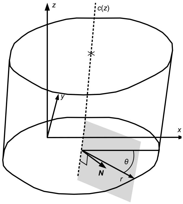
The cord topology is that of a cylinder, and a convenient and compact representation of the surface can be achieved by specifying the center-line c of the cord, and a radius generator (Fig. 1). The center-line is parametrized by the through-plane distance, z:
| [1] |
Figure 1.
Parametrization of the cord surface. The center-line c is parameterised in z and implemented as cubic spline interpolators of the x- and y- coordinates of the center-line curve. The radius r is parameterised in z and θ, the angle between the radius vector and the x-axis. The surface normal vector N is approximated as the vector that is both normal to the center-line tangent vector, and lies in the plane containing both the center-line tangent and the radius vectors.
The functions cx(z) and cy(z) are implemented as cubic spline interpolators of the location of the cord at the slice centers, so that the unit vector tangential to the cord center-line curve (the center-line tangent vector) can easily be found (do Carmo, 1976) as
| [2] |
since the derivatives are straightforward to compute from the cubic spline interpolator coefficients. The cord radius is then a function of z and θ, the angle subtended to the positive x-axis.
Initialization of the center-line is the only interactive part of the segmentation process and requires the operator to provide a set of landmarks that define the initial center-line of the cord. First, the anatomical range of the cord over which it is desired to assess atrophy is decided. Through the user interface, the operator marks the approximate location of the center of the cord on the top and bottom slices of that range, and at regular intervals between them. With human images, we found that marking every 10 mm was sufficient to provide a good initialization, and required minimal operator time, always less than 5 minutes. Using cubic spline interpolators for the x- and y- location of these landmarks, a new set of landmarks is generated on every slice. The radius generator is automatically initialized to a fixed nominal radius value, rNOM; this value is not critical to the final segmented cord, but will generate an initial surface that lies close to the true cord surface for much of the length. For this study of human spinal cord, rNOM was chosen to be 4 mm.
The assumptions underlying the segmentation process are that a) the cord center-line follows a sinuous but smooth path in the superior-inferior direction, close to that initially estimated by the user; b) at the cord surface, the image intensity gradient vector is coincident with the normal to the surface; c) the cross-sectional shape of the cord varies only slowly in the superior-inferior direction; and d) in cross-section, the radius is a smoothly-varying function of θ. The segmentation process involves a two-stage iterative update. The first uses intensity gradient information to update the radii; the second computes the center of area of the cross-section at each slice, and redefines the center-line to pass through the centers of area, but constrains the center-line to pass close to the initial user estimate. Furthermore, to improve the reliability of convergence, the segmentation uses a multi-scale approach whereby the cord radius generator is given a steadily increasing degree of complexity, allowing the surface to be defined in finer detail as the segmentation progresses.
Radius Update
Cord radii are computed for each slice, and for sixty-four values of θ. Thus, at every slice location, the cord cross-section can be considered as a polygonal shape, with its center of area being at the current center-line location. At each iteration, the radii are updated according to the local intensity gradient in the region of the vertices of the polygon, along the radial direction. Five locations along the radius vector are selected at intervals of 10% of the nominal cord radius, with the central location being at the current vertex position. At each location, the intensity gradient vector is evaluated, and an intensity gradient-related radial update “force” is calculated as:
| [3] |
where is the normalized gradient vector at location j, N is the current estimate of the cord surface normal, and sgn is the sign function:
| [4] |
Of course, the gradient at the vertex does not contribute to the update function since sgn(0)=0, and this term in the sum is not evaluated. The normalized gradient vector Ḡ is defined by:
| [5] |
The gradient vector, G, is evaluated using 3-D Sobel operators (Duda and Hart, 1973) on the original image, with kernel coefficients scaled to take account of the possibly different sample spacing of the pixels in physical space. The Sobel operators form three separate gradient images, and to estimate the intensity gradient vector at any location, the gradients at the pixel centers are interpolated using tri-linear interpolation. |G|max is the maximum gradient magnitude, and |G|min is the minimum gradient magnitude evaluated over the whole image domain. The scaling by ١/(|G|max–|G|min) is done in order to provide an update function that is independent of the absolute image intensities (Coulon et al., 2002).
The surface normal vector, N, is defined to lie in the plane in which both the center-line tangent vector and the vector from the centroid to the vertex also lie. N is perpendicular to the current estimated center-line, and the scalar product between N and the radial vector is positive (see Fig. 1). The “±” sign in Eq. [3] indicates that the direction of the radial update force depends on the brightness of the cord relative to the background intensity: at the surface of the cord, the intensity gradient should be positive in the outward radial direction for a hypointense cord on T2-weighted images, and negative for a hyperintense cord on T1-weighted images. The user indicates to the program the type of image being analyzed. The form of Eq. [3] is such that gradients of the correct orientation will “pull” the cord surface towards them, and gradients of the opposite orientation will “push” the surface away. If the intensity gradient is not normal to the surface, any force will be weakened according the scalar product of the gradient and normal vectors. Each radius is increased by a distance (in mm) equal to the radial update force scaled by an update multiplier. An update multiplier of 0.8 was found experimentally to promote rapid convergence.
The cord radii are then constrained to vary smoothly, both around the cord and along the cord. At each slice location, the radii are considered as a periodically-varying function of θ. These functions are then Fourier transformed and the Fourier coefficients associated with the highest frequencies are discarded (set to zero). The remaining coefficients are then smoothed by fitting a polynomial function separately to each coefficient, to model the variation of these coefficients with z. The polynomial for each coefficient is interpolated to give a smoothed set of coefficients at each slice location, before these are inverse Fourier transformed to give a smoothed set of cord radii. The maxima of the number of Fourier coefficients that is retained and the order of the polynomial variation of the coefficients with z are user-configurable, but in this work they were fixed at 32 coefficients and a polynomial order of 10. When fitting the polynomial model for the coefficients, each point along the z-axis is weighted with a sigmoidal weighting function that varies according to the strength and orientation of the intensity gradients at the current estimated edge location at that slice location:
| [6] |
Nθ is the number of radii defining the cord outline, and again the “±” depends on whether the cord is hyperintense or hypointense. Thus, more weight is given to cord cross-sections which are close to a well-defined edge of the correct orientation, making the smoothing more robust to areas where the image suffers from poor contrast, or is corrupted by artifacts such as occurs when there is CSF flow, and cord and general patient motion.
Center-Line Update
After the radii are updated, the centers of area of the cord cross-sections are computed and the center-line redefined to pass through the centers of area. Using a cubic spline interpolator, the radii are re-sampled at the regular values of θ relative to the new center-line. Next, the cord center-line is adjusted to ensure that the path followed by the cord does not deviate substantially from that initially estimated by the user. A “shear” force acts on the center-line, and acts to restore the center-line towards the initial estimate of the cord center-line provided the user. If the user-set center-line location is cu(z), and the current estimate of the center-line is c(z), the force vector acting to restore the center-line is:
| [7] |
This gives a unit force when the distance between the user-set center-line and the current center-line is half the nominal radius. Implicit in this is the expectation that the user can estimate the cord center of area to an accuracy well within the nominal cord radius. The center-line positions are shifted by a distance (in mm) equal to fC times the update multiplier.
Thus, at each iteration, the vertex locations are driven by the intensity-related forces, but are constrained to vary smoothly both around the cord circumference, and along its length. If there are regions of the image where the cord is poorly defined, image artifacts that obscure the cord, or adjacent other structures such as vertebrae, the segmentation can still proceed successfully because of these smoothness constraints. The cord is surrounded by other structures which also create intensity gradients; the segmentation is prevented from converging on non-cord structures by the constraint on the path of the center line.
Convergence and Termination
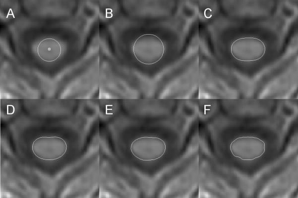
Convergence is made more rapid and reliable using a multi-scale approach. At the coarsest scale, the number of Fourier coefficients is set to one, and the polynomial order is set to zero for the smoothing step. Thus, during this initial stage, the cord is modeled with a circular cross-section of constant radius. After convergence at each scale, the model is refined by incrementing both the number of Fourier coefficients and the polynomial order, until the desired model complexity has been reached. If either the number of coefficients or the polynomial order have reached the maxima set by the user, then that parameter is not incremented further. Fig. 2 shows the successive refinement of the cord cross-section at one z location.
Figure 2.
For the AS method, evolution of the cord active surface at one z location (at the C2/C3 level) from a 3-D T1-weighted dataset. The image is displayed using bi-linear interpolation and thus appears less pixelated than the underlying image data. The cord segmentation algorithm uses a multi-resolution approach with steadily increasing refinement of the surface model by increasing the number of Fourier coefficients and order of polynomial fit to those coefficients that define the radius generator. Not all steps are shown here. A) shows the user-defined initial marker of the cord center-line and the initial surface of constant radius; B) shows the surface at equilibrium with 3 coefficients and a polynomial of order 2 (C3O2); C) shows C5O4; D) shows C7O6; E) shows C11O10; and F) shows C32O11.
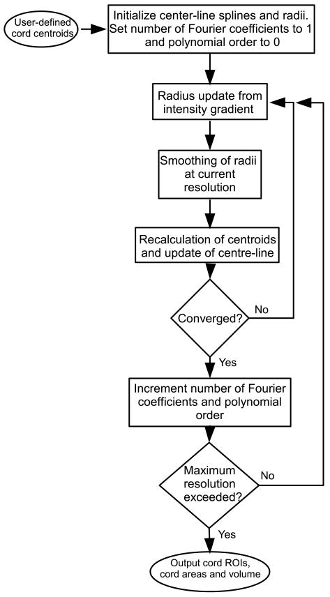
At each iteration, convergence is assessed by evaluating the maximum distance moved by any of the points defining the cord surface. If, during an iteration, none of the points moves by a distance more than a fixed maximum, equal to 0.1% of rNOM, then convergence is deemed to have occurred at that scale. Making the convergence criterion any stricter than this made negligible difference to the final segmentation. Computation time for cord segmentation over 60–70 image slices is between 1 and 2 minutes using a single-core 32-bit 2GHz CPU. After convergence with the maximal number of Fourier coefficients and polynomial order, the program outputs the cord cross-sectional shape as a polygonal region-of-interest (ROI) at each slice location. The user can choose to modify the ROIs at this stage to correct any errors in the cord outline, but for the work presented here no modifications were performed, and subsequent processing was fully automated. A flow diagram for the whole program algorithm is shown in Fig. 3.
Figure 3.
Flow diagram for spinal cord segmentation. The algorithm follows a multi-resolution approach, with the level of detail of the cord outline being controlled at each level by the number of Fourier coefficients used in smoothing the radius generator function, and the polynomial order of the variation of these coefficients with z-coordinate. The inner loop shows a two-stage update, first of the radius generator then of the center-line. The convergence criterion for this inner loop is that none of the vertices of the current cord outline changes position during one iteration by more than a small fraction of the nominal cord radius.
If the cord axis is approximately normal to the image plane, then the areas of these ROIs gives the cord cross-sectional area. However, since over an extended length the cord curves, and therefore does not remain perpendicular along its whole length, a more natural measure of cord area is described below.
Measurements
Since the center-line is an explicit part of the cord model, this allows a natural measure of cord cross-sectional area as the area of the cord outline in a plane to which the center-line tangent vector is normal (Coulon et al., 2002). As long as the cross-section shape changes only slowly with z, and the angle between the center-line tangent vector and z-direction is fairly shallow, the area can be closely approximated by taking the area of the cross-section in the x-y plane, and multiplying it by the z-component of the center-line unit tangent vector. This cross-sectional area can be plotted against the cord arc length, L, which is given by:
| [8] |
where z0 and z1 are the lower and upper range of z over which the cord length is to be measured (do Carmo 1976). The cord volume can also be approximated by:
| [9] |
where l is the local cord arc length, and A is the area perpendicular to the center-line. In practice, the cord volume is calculated using the trapezium rule from the cross-sectional area at the slice locations, and the arc length between slices. The average cord area can then be calculated as the cord volume divided by the cord length.
Since the spinal cord follows a sinuous path, with a complex curvature in three dimensions, it is sometimes difficult to visualize pathology within the cord since, when viewed in the sagittal plane, the regions of damaged cord can curve in and out of the image plane. The definition of the cord center line above allows the cord to be artificially straightened in a further automatic postprocessing step. At regular steps along the center-line of the cord, the original image is re-sampled in planes to which the center-line forms the normal vector. A 3-D image is built up in this way, forming a new image in which the center-line remains at a fixed position in the image planes, allowing the cord to visualized as a straight cylinder-like structure, with any pathology being more readily apparent; we term this the ‘unwrapped cord image’. This type of visualization was first shown by Stroman et al. (Stroman et al., 2005), where the anterior edge of the cord was manually delineated in a sagittal view, and this reference line was used to reslice the image in planes transverse to the spinal cord.
Normalization Procedure
There is considerable natural variation in spinal cord cross-sectional area related to the size of the subjects, and for cross-sectional studies comparing different patients it is important to take account of this variation. In normal controls, the cranial size is significantly correlated with cord area in healthy controls (Vaithianathar et al., 2003; Rashid et al., 2006b), and therefore the cranial cross-sectional area was used a normalization factor, in a way similar to that done by Lin et al. (Lin et al., 2003). In a separate analysis procedure, intra-cranial cross-sectional area (ICCSA) was measured at the level the inferior margins of the corpus callosum on an axial slice of a proton density-weighted image using a semi-automated contouring method. Using the computer mouse, the user indicated the outer contour of the brain and CSF. An edge detection algorithm then identified the position of the border as the point of maximum intensity gradient in the vicinity of the mouse. The brain and CSF space was then automatically outlined as a closed iso-intense contour, with the intensity being that at the border location.
For the control subjects, cord CSA was plotted against ICCSA, and the following form was assumed:
| [10] |
where CSAmean is the average cord CSA and ICCSAmean is the average ICCSA for the control subjects. The term α (ICCSA-ICCSAmean) can therefore be used as a correction factor for the natural variation in cord area between subjects associated with cranial size, by subtracting it from the measured cord CSA. The cord CSA was therefore normalized to the ICCSA using the equation:
| [11] |
where CSAn is the subject’s normalized cord CSA.
Comparison with Existing Method
We compared the results of AS cord segmentation technique with the method published by Losseff et al. (Losseff et al., 1996), which is used to assess the cord cross-sectional area at the C2/C3 intervertebral disk on 3-D T1-weighted scans. In brief, the procedure was to reformat the scans axially, with the image plane perpendicular to the long axis of the cord at the C2/C3 disk level. At the same time, the images were re-sampled to a 3 mm slice thickness. Five contiguous image slices were selected with the most inferior slice passing centrally through the C2/C3 disk. On each of these five slices, two regions of interest (ROIs) were created: the first surrounded the cord but contained only cord parenchyma, while the second surrounded both the cord and the annulus of CSF space. From the mean intensities within these ROIs, and their areas, an intensity threshold was calculated that separated the cord from the CSF. A contour with this threshold intensity was then automatically drawn around the cord on each of the five slices, with an average of the area enclosed by the contour representing the mean cross-sectional area of the cord.
Patients and Controls
For the main study, from a database of MRI scans performed in Milan, we selected sixty subjects at random from our database of images for spinal cord analysis. There were twenty normal control subjects, twenty patients with relapsing-remitting (RR) multiple sclerosis and twenty patients with the secondary progressing (SP) form of the disease (Lublin and Reingold, 1996). None of the patients had any clinical relapses or undertook steroid treatment during the period of at least six months prior to the scans being performed. Demographic details are given in Table 1. There were no significant differences between the ages of the control group and either the RR group (p = 0.09) or the SP group (p = 0.24). However, there was a significant difference between the RR and SP groups (p < 0.001). At the time of the MRI scan, all subjects underwent a standard neurological examination. For the control subjects, the possibility of neurological conditions was excluded, whilst the patients also underwent rating on the expanded disability status scale (EDSS) (Kurtzke, 1983) and ambulation index (AI) (Hauser et al., 1983). The original studies for which the MRI scans were collected were all approved by the local ethical committees, and all subjects provided written informed content prior to inclusion.
Table 1.
Patient and control demographics.
| Controls | Relapsing-remitting | Secondary-progressive | |
|---|---|---|---|
| Number (Male/Female) | 20 (6/14) | 20 (7/13) | 20 (6/14) |
| Mean age (range) [years] | 38.9 (24–72) | 33.0 (25–51) | 43.5 (31–63) |
| Mean disease duration (range) [years] | - | 6.7 (1–13) | 15.9 (8–37) |
| Median EDSS score (range) | - | 2.0 (1–4) | 6.0 (3.5–7.5) |
| Median ambulation index (range) | - | 0 (0–4) | 5.0 (0–8) |
Image Acquisition
To assess the reproducibility and robustness of the technique, we used images of the cord acquired at 1.5 T. The main study was performed on historical data collected during the period 2002–2005 in Milan on a Siemens Symphony scanner (Siemens Medical, Erlangen, Germany), equipped with a tailored phased array coil for signal reception in the cervical cord region. The built-in body coil was used for R.F. transmission. A 3-D MP-RAGE (magnetization-prepared rapid acquisition gradient echo) sequence was applied in the sagittal plane with phase-encoding in the anterior-posterior and left-right directions, a slab thickness of 160 mm, FoV of 280×280 mm, 256×256×128 acquisition matrix, zero filled in the slab direction to give a digital spatial resolution of 1.09×1.09×1.0 mm. TR = 9.7 ms; TE = 4.0 ms; TI = 300 ms to the first line of k-space; receiver bandwidth = 49.92 kHz. The contrast-to-noise ratio between the cord and the surrounding CSF was approximately 15:1, where the noise magnitude was measured in a region of image background containing no signal (Henkelman, 1984).
In order to provide a normalized measure of cord atrophy, we also used images of the head of each subject using a dual-echo turbo spin-echo sequence. Twenty-four 5 mm axial slices were acquired, with the central slices positioned to join the most fronto-inferior and posterio-inferior margins of the corpus callosum, as viewed on a mid-sagittal slice. The FoV was 250×250 mm, with a 256×256 acquisition matrix, giving a spatial resolution of 0.976×0.976 mm. The other sequence parameters were: TR = 3800 ms; TE1 = 16, TE2 = 98 ms. Unfortunately, for two of the RR patients, the head images were not available and so their normalized data did not enter the correlations with disability scores or the group comparisons.
A demonstration of applicability to T2-weighted 2-D multi-slice images was also performed on an image from an MS patient collected using a Philips 1.5T system (Philips, Best, The Netherlands), with a phased array spinal coil for signal reception. The built-in body coil was used for R.F. transmission. One hundred and fifty contiguous pure axial images were acquired in three separate blocks, with phase-encoding in the anterior-posterior direction, a slice thickness of 3 mm, a FoV of 230×230 mm and a 256×256 acquisition matrix, giving an in-plane resolution of 0.898×0.898 mm. The first slice of the most superior block was positioned at the level of the pons, and the patient couch was moved automatically to center each block of slices at magnet iso-center, with the most inferior slices of the final block being at the conus medullaris. A fast spin-echo sequence was used with, TR = 9189 ms; TE = 100 ms; echo train length = 40; 2 signal averages; receiver bandwidth = 109.2 kHz. Total scan time was 11 minutes and 56 seconds.
Reproducibility Study
To assess the inter-operator reproducibility, the sixty 3-D T1-weighted MRI scans were assessed by three operators working independently. Each operator evaluated the scans using both the Losseff method and the new AS method. For the AS method, the cord area was estimated over a section of cord from the top of C2 to the base of C5 after the scans were reformatted axially, with the image plane perpendicular to the cord at the C2/C3 disk level. First, a marker was placed at the center of the cord on the most superior axial slice in which the odontoid process of the epistropheus (C2) was still just visible. Another marker was placed at the center of the cord on the slice that passed through the inferior border of C5. Starting at C5 and moving superior, a marker was placed at the center of the cord on every tenth slice until the marker at the top of C2 was reached. The AS method was then applied with these markers of the cord center line as the only input from the user. The scans were evaluated in a randomized order, without knowledge of the results from the other operators.
To assess the intra-operator reproducibility, after a period of at least two weeks had elapsed, two of the operators re-evaluated fifteen of the scans using both methods. The fifteen scans were selected to cover the range of cord cross-sectional areas that was seen on the first evaluation. Intra- and inter-operator reproducibilities are presented as a coefficient of variation (CoV), which is the standard deviation of multiple measures of the same parameter, divided by the mean of the parameter value (Goodkin et al., 1999).
Accuracy
In order to assess the accuracy of the cord area measurements, we compared the cross-sectional area measurements from the new AS method and the Losseff method (Losseff et al., 1996) with those from a manual delineation of the cord. Since the Losseff method measures the cord area only in the region of the C2 vertebral body, manual outlining was performed on the same five near-axial slices as were used for the Losseff method. The cord outlines produced by the AS method over the same anatomical area were manually selected to match as closely as possible those slices used for the Losseff method. Since the slice thickness for the AS method input images was 1 mm, the cord area was averaged over fifteen slices. For manual outlining, a freehand ROI was drawn on each slice at the border between the cord and CSF space, and the average area calculated. Two observers independently performed the manual outlining procedure on all subjects. We also assessed the intra- and inter-observer CoVs for the AS method when the evaluation was performed over the fifteen slices at the C2 level.
The correlations between the EDSS score, AI and disease duration and the mean normalized cord area found with both the Losseff and AS methods were assessed using an ANCOVA model adjusting for age and sex; the partial correlation coefficient was used to quantify the correlation. Univariate ANOVA showed a significant heterogeneity between the cord areas found using the AS method among the control and patient groups. We therefore performed a post hoc group-by-group test for differences in area, using Student’s two-tailed t-test.
RESULTS
Reproducibility
Details of the cord area measured by the three observers using both the Losseff and AS methods are shown in Table 2. The mean inter-observer CoV was 1.07% for the AS method and 7.95% for the Losseff method. The mean intra-observer CoV was 0.44% for the AS method and 2.15% for the Losseff method. The intra-observer CoV for the AS method for areas measured just at C2 was 0.59% and the inter-observer CoV was 1.36%. Thus, if the same observer is used for all measurements, changes as small as 0.67 mm2 (1.96 SDs) in cord area should be detectable using the AS method with 95% confidence that the change measured was not simply because of measurement variation. For the Losseff method, the corresponding detectable change in area was 2.72 mm2. There was no significant correlation between mean cord size and the individual CoVs for the AS method, neither for the intra-observer measurements (p = 0.33), nor for the inter-observer measurements (p = 0.39).
Table 2.
Mean spinal cord cross-sectional area measurements and variability.
| Observer 1 | Observer 2 | Observer 3 | Intra- Observer CoV | Inter- Observer CoV | |
|---|---|---|---|---|---|
| Manually outlined C2 area/mm2 | 65.73 | 65.89 | - | - | - |
| Losseff method C2 area/mm2 | 66.01 | 59.65 | 68.35 | 2.15 % | 7.95 % |
| AS method C2 area/mm2 | 75.26 | 75.41 | 75.55 | 0.59 % | 1.36 % |
| AS method C2–C5 area/mm2 | 78.42 | 78.27 | 77.57 | 0.44 % | 1.07 % |
Results for the AS method are shown for both the full cord region measured (C2–C5) and the reduced region for comparison with the Losseff method (C2).
Accuracy
Using the AS method, the mean cord cross-sectional area (averaged over all observers and subjects) between C2 and C5 was 78.08 mm2. For the two observers who performed manual tracing of the cord outline, the mean cross-sectional area measured at C2 by manual tracing was 65.81 mm2, while for the same two observers using the Losseff method it was 62.83 mm2. For the AS method, when the same anatomical region of the cord was evaluated, the mean cord area at C2 for the same two observers was 75.33 mm2. In the whole sample of subjects, there were very strong correlations between the manually-traced area and the areas found by both the Losseff method (r = 0.955; p < 0.0001) and the AS method between C2–C5 (r = 0.952; p < 0.0001).
Clinical Relevance
In the entire cohort of subjects, the correlation between normalized cord area and age was weak, using both the Losseff method at C2 (r = −0.22; p =0.09) and the AS method between C2–C5 (r = −0.21; p = 0.11), while no correlation was detectable when the the three groups were assessed separately. We therefore included age as a covariate in our analysis.
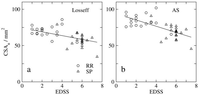
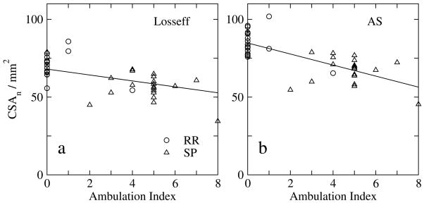
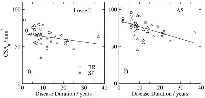
In MS patients, after adjusting for age and sex, there were strong correlations between the EDSS score and the normalized cord area measured using both the Losseff method at C2 (r = −0.51; p < 0.001) and the AS method between C2–C5 (r = −0.59; p < 0.001) (Fig. 4). Similarly, there were strong correlations between the ambulation index and the normalized cord area measured using both the Losseff method (r = −0.58; p < 0.001) and the AS method between C2–C5 (r = −0.648; p < 0.001) (Fig. 5). Correlations between the disease duration and the normalized cord area measured using the Losseff method (r = −0.17; p = 0.32) and the AS method between C2–C5 (r = −0.30; p = 0.074) were not statistically significant (Fig. 6).
Figure 4.
Relationship between normalized mean cord cross-sectional area (CSAn) and EDSS score, for a) the Losseff method at C2 and b) the new AS method between C2 and C5. Relapsing-remitting (RR; n=18) and secondary-progressive (SP; n=20) MS patients are shown separately, but for the pooled data r = −0.51 (p < 0.001) for the Losseff method and r = −0.59 (p < 0.001) for the AS method after adjusting for age and sex.
Figure 5.
Relationship between normalized mean cord cross-sectional area (CSAn) and ambulation index, for a) the Losseff method at C2 and b) the new AS method between C2 and C5. Relapsing-remitting (RR; n=18) and secondary-progressive (SP; n=20) MS patients are shown separately, but for the pooled data r = −0.58 (p < 0.001) for the Losseff method and r = −0.65 (p < 0.001) for the AS method, after adjusting for age and sex.
Figure 6.
Relationship between normalized mean cord cross-sectional area (CSAn) and disease duration, for a) the Losseff method at C2 and b) the new AS method between C2 and C5. Relapsing-remitting (RR; n=18) and secondary-progressive (SP; n=20) MS patients are shown separately, but for the pooled data, statistically-significant relationships are seen for neither the Losseff method (r = −0.17; p =0.32) nor the AS method (r = −0.30; p = 0.074), after adjusting for age and sex.
Using the AS method, the controls subjects had a mean (standard deviation) normalized cord area between C2 and C5 of 85.16 (6.57) mm2, while the RR patients had an area of 84.05 (7.61) mm2 and the SP patients an area of 68.23 (10.12) mm2. There was no difference between the normalized cord areas of the controls and the relapsing-remitting patients. However, the SP group had significantly lower normalized cord area than both the controls (p < 0.001) and the RR patients (p < 0.001).
Unwrapped Image
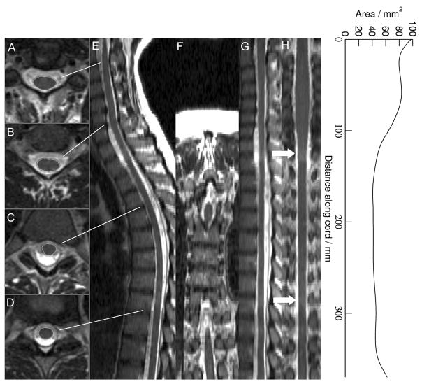
Fig. 7 shows sagittal and coronal reconstructions of the axial T2-weighted images acquired using three slice blocks. The left column shows the cord outlines found using the AS method at the stations indicated, while the right hand columns show the unwrapped cord image in sagittal and coronal views at the cord center line. Note that there are intensity artifacts in the areas where the slice blocks abut each other, since the patient table was moved between slice blocks to bring the center of each slice block in turn to magnet iso-center. Despite the high-intensity flow-related artifacts in the CSF, the cord has been successfully segmented from the foramen magnum to L1.
Figure 7.
Segmentation and unwrapping of the cord in an axial 2-D T2-weighted image. E and F show the original images reconstructed to sagittal and coronal views, while A (C2/C3 level), B (C6/C7), C (T4/T5) and D (T9/T10) show the cord segmentation at various representative levels down the cord as indicated. G and H show the unwrapped cord image in sagittal and coronal views over approximately the same section of cord, while the graph on the right indicates the cord cross-sectional area, measured in a plane perpendicular to the cord center-line. Note the high intensity artifacts seen at the T4/T5 and T9/T10 levels (arrows), which are caused by fact that the table moved between slice blocks. See Methods for more details of the scan acquisition parameters.
DISCUSSION
We have demonstrated a rapid method for segmenting the spinal cord from MR images, and have shown its high inter- and intra-observer reproducibility on 3-D T1-weighted images of the human cervical cord. The method needs only modest user interaction, requiring the user to identity landmarks at the extremes of the region of cord to be assessed, and to mark the approximate center-line of the cord on several slices between the landmarks. The algorithm is based on a constrained active surface model of the cord surface with a compact parameterization as a center-line and radius generators. This allows rapid segmentation, taking on the order of one minute, and the cord cross-sectional area can be assessed along the length of the extracted surface.
The intra- and inter-observer reproducibilities were compared to those of a well-established method for assessing cord atrophy at the C2 level, published by Losseff et al. (Losseff et al., 1996). The new algorithm compared favorably with Losseff’s method, at least in part because the cord area was assessed over a larger extent of the cord (approximately 80 mm, compared to the 15 mm of the Losseff method). Nevertheless, when the cord areas were measured over the same anatomical range as is used in the Losseff method, both the intra-and inter-observer variabilities were still considerably better for the AS method (see Table 2), indicating an intrinsic benefit of using a surface model with smoothness constraints.
The inter-observer CoV was approximately 750% greater and the intra-observer CoV approximately 500% greater for the Losseff method compared to the AS method over the extended cord region. The CoVs we measured for the Losseff method were, however, considerably greater than those found by Losseff where, for example the intra-observer CoV was only 0.73% compared to 2.15% in our work. This may be because of the relative inexperience of our observers with both techniques, or because our data is of somewhat poorer quality than that acquired by Losseff. In the original Losseff paper, a correction for non-uniformity of the receiver coil sensitivity was also performed, and this was not done with our data, possibly adversely affecting the reproducibility. However, we would expect the reproducibilities of both analysis methods to improve with experience and data quality. The reproducibility of our method is very similar to that of another semi-automatic method that can measure the cord area in the C2/C3 region, where the intra-observer CoV was reported to be 0.42% (Lin et al., 2003).
We evaluated the intra-observer reproducibilities by having two observers evaluate a subset of the scans twice, with an interval of at least two weeks between evaluations. In order to assess the true reproducibility of the whole method, including data acquisition and analysis, we would have needed to scan the subjects twice with a time interval between scans that would not have allowed any significant cord atrophy to occur. The work of Losseff showed that the intra-observer scan-rescan CoV (0.79%) was very similar to that when observers evaluate the same image twice (intra-observer same-scan CoV) (0.73%) and since both the Losseff method and the AS method have at their heart a detection of the intensity that separates cord from CSF, we would not expect a large increase in the scan-rescan CoV compared to our intra-observer CoV. This will need to be confirmed with a further study involving scanning the same subjects twice, to see whether for example, changes in neck flexion significantly impact on the scan-rescan reproducibility. We would not expect this to be case, because the cord cross-sectional area is always computed perpendicular to the cord axis.
Because the AS method is capable of assessing the cord cross-sectional area over an extended range of the cord, this improves the overall measurement reproducibility, and should lead to a greater sensitivity to real physiological change. However, it is possible that the cord at C2 is particularly sensitive to MS-related pathological damage and atrophy, which could lead the assessment of a greater length of cord being no more sensitive to disease-related change than a simple measure at C2. In MS, there is evidence of a higher atrophy rate for the white matter of the cord compared to the gray matter (Gilmore et al., 2005), and that the lower cord does not suffer from overall atrophy to the same extent as the upper cord (Evangelou et al., 2005). The possibility of differential atrophy rates along the cord should now be assessed using the AS method both in MS and in other diseases where spinal cord atrophy is a feature.
The AS method offers a considerable reduction in user interaction time, with identification of the landmarks and marking of the cord center-line taking typically five minutes per subject. This compared to approximately 20 minutes per subject to perform the Losseff analysis. If the images are acquired in a coronal or sagittal plane, then the scans can be automatically reformatted into a pure axial orientation to allow the center-line to be marked. The AS method does not require manual reformatting of the data so that the image plane is perpendicular to the cord axis; indeed such a reformatting cannot be done if the cord area is measured over a range of more a few tens of millimeters, because of the curvature of the cord axis. This removes one manual step in the analysis chain, thereby helping to improve reproducibility.
At the C2 level, the AS method overestimated the cord cross-sectional area by approximately 14% compared to manual outlining of the cord, while the Losseff method underestimated the area by approximately 4.5%. In the original paper by Losseff, the authors scanned a spinal cord phantom consisting of acrylic rods in a water bath, and found that the imaging and analysis procedure overestimated the cross-sectional area of the rods by between 4.3% and 10.8%, depending on the diameter of the rods, with a greater percentage error for the smallest rod. Of course, we cannot know whether the manual outline of the cord truly represents the cord surface. However, there was a very strong correlation between the cord cross-sectional area measured manually and assessed by both the Losseff and AS methods, thus indicating that both these techniques generate valid surrogates of the manually-assessed cord area. In both cross-sectional and follow-up studies, a consistent bias between the measured and actual cord areas would have no impact the comparison between patient groups or on the estimates of atrophy rates.
We found a significant reduction in the cord area for the SP patients compared to the control subjects, but no reduction in area for the patients with RR MS. This was also seen in the group of RR patients in Losseff’s work, which had similar patient demographics to our own subjects. More recently, Rashid et al. and Mann et al. (Rashid et al., 2006b; Mann et al., 2007) used normalized cord area measures (as was done in our study) in larger groups of RR patients and controls, and again found no significant differences in cross-sectional area. For the whole patient population, the correlations between cord area and clinical status were similar for both the AS and Losseff methods, and are similar to those reported by Losseff. Using an active surface model method for cord segmentation that is similar to our own, Hickman et al. (Hickman et al., 2003) also showed a similar degree of correlation between cord volume measured over the whole cervical region and EDSS, but without normalization to head size. With our data, there is considerable scatter in the plots of cord area vs. EDSS and AI (Fig. 4 and Fig. 5), even though the cross-sectional area was normalized against patient head size. Thus, even though we have removed some of the biological variability related to patient size, there remains, not surprisingly, a considerable component of disability that is not explained by cord pathology and atrophy.
The AS method can be applied to axially-acquired 2-D multi-slice T2-weighted cord images with coarser resolution in the head-foot direction compared to 3-D acquired images, though we have yet to perform an analysis of the relative reproducibility in comparison to 3-D T1-weighted imaging. Since the method can handle cases where the CSF is either hypo- or hyperintense relative to the cord, this suggests the AS method could equally be applied to 3-D T2-weighted images, or to 2-D or 3-D balanced gradient sequences (Ramli et al., 2001). However, it is possible that signal hyperintensities caused by focal or diffuse abnormalities in the cord would impact on the quality and reliability of the segmentation; this would need to be carefully assessed. Although our reproducibility study concentrated on the cervical cord region, between C2 and C5, we have shown in principle that the cord can be segmented over an extended region, as far down as L1. However, the reproducibility would need to be carefully assessed, since it may be that as the cord narrows towards the conus, and the nerve roots come off from the cord with increasing frequency, it may not be possible to assess the cord area in a meaningful way in the lumbar region.
Since the cord center-line is parameterised by the model, it is a straightforward matter to resample the original spine image with the curved center-line parameter as the new z-coordinate in the re-sampled image, in way similar to that done by Stroman et al. (Stroman et al., 2005). Image planes are generated that are always perpendicular to the center-line along its length, resulting in a 3-D image in which the cord appears as a straight structure with the center-line remaining at a fixed position between slices. This allows simple visualization of the cord structure and pathology, and this cord ‘unwrapping’ procedure could find wider application in standard radiological practice.
The AS method imposes continuity constraints on both the cord center-line and on the surface smoothness in the longitudinal and circumferential directions around the cord. Because of these constraints, the AS model is able to ‘bridge’ gaps in the image data caused by, for example, poor image contrast in certain regions, or image artifacts. Poor contrast commonly occurs in the region of C5, where the CSF space is very narrow, while CSF flow can cause regions of very bright CSF at various points along the cord, as can be seen in Fig. 7.
In conclusion, we have developed a method for assessing the cross-sectional area of the spinal cord using an active surface model that is constrained to give a smooth variation of cord radius both longitudinally and circumferentially around the cord. The compact parameterization of the cord surface allows rapid cord segmentation with minimal user interaction, which involves marking the cord center-line on a few slices along the cord. The cord cross-sectional area can be measured over an extended section of the cord, which improves the reproducibility and should give greater sensitivity to change compared to existing established methods. Furthermore, it permits the investigation regional differences in cord atrophy along the cord.
Supplementary Material
Acknowledgments
Dr. Bakshi acknowledges support of this project by research grants from the National Multiple Sclerosis Society (RG3705A1 and RG3798A2) and National Institutes of Health (1R01NS055083-01).
Footnotes
Publisher's Disclaimer: This is a PDF file of an unedited manuscript that has been accepted for publication. As a service to our customers we are providing this early version of the manuscript. The manuscript will undergo copyediting, typesetting, and review of the resulting proof before it is published in its final citable form. Please note that during the production process errors may be discovered which could affect the content, and all legal disclaimers that apply to the journal pertain.
References
- do Carmo M. Differential Geometry of Curves and Surfaces. Prentice-Hall; New Jersey, USA: 1976. [Google Scholar]
- Bakshi R, Thompson AJ, Rocca MA, Pelletier D, Dousset V, Barkhof F, Inglese M, Guttmann CR, Horsfield MA, Filippi M. MRI in multiple sclerosis: current status and future prospects. Lancet Neurol. 2008;7:615–625. doi: 10.1016/S1474-4422(08)70137-6. [DOI] [PMC free article] [PubMed] [Google Scholar]
- Coulon O, Hickman SJ, Parker GJM, Barker GJ, Miller DH, Arridge SR. Quantification of spinal cord atrophy from magnetic resonance images via a B-spline surface model. Magn Reson Med. 2002;47:1176–1185. doi: 10.1002/mrm.10162. [DOI] [PubMed] [Google Scholar]
- Duda R, Hart P. Pattern Classification and Scene Analysis. John Wiley and Sons; New York, USA: 1973. pp. 271–272. [Google Scholar]
- Evangelou N, DeLuca GC, Owens T, Esiri MM. Pathological study of spinal cord atrophy in multiple sclerosis suggests limited role of local lesions. Brain. 2005;128:29–34. doi: 10.1093/brain/awh323. [DOI] [PubMed] [Google Scholar]
- Filippi M, Campi A, Colombo B, Pereira C, Martinelli V, Baratti C, Comi G. A spinal cord MRI study of benign and secondary progressive multiple sclerosis. J Neurol. 1996;243:502–505. doi: 10.1007/BF00886870. [DOI] [PubMed] [Google Scholar]
- Filippi M, Rocca MA, Arnold DL, Bakshi R, Barkhof F, De Stefano N, Fazekas F, Frohman E, Wolinsky JS. EFNS guidelines on the use of neuroimaging in the management of multiple sclerosis. Eur J Neurol. 2006;13:313–325. doi: 10.1111/j.1468-1331.2006.01543.x. [DOI] [PubMed] [Google Scholar]
- Gilmore CP, DeLuca GC, Bö L, Owens T, Lowe J, Esiri MM, Evangelou N. Spinal cord atrophy in multiple sclerosis caused by white matter volume loss. Arch Neurol. 2005;62:1859–1862. doi: 10.1001/archneur.62.12.1859. [DOI] [PubMed] [Google Scholar]
- Goodkin DE, Ross JS, Medendorp SV, Konesci JJ, Rudick RA. Magnetic resonance imaging lesion enlargement in multiple sclerosis. Disease related activity, chance occurrence, or measurement artifact? Arch Neurol. 1999;49:261–263. doi: 10.1001/archneur.1992.00530270075021. [DOI] [PubMed] [Google Scholar]
- Hauser SL, Dawson DM, Lehrich JR, Beal MF, Kevy SV, Propper RD, Mills JA, Weiner HL. Intensive immunosuppression in progressive multiple sclerosis. A randomized, three-arm study of high-dose intravenous cyclophosphamide, plasma exchange, and ACTH. N Engl J Med. 1983;308:173–80. doi: 10.1056/NEJM198301273080401. [DOI] [PubMed] [Google Scholar]
- Henkelman RM. Measurement of signal intensities in the presence of noise in MR images. Med Phys. 1984;12:232–233. doi: 10.1118/1.595711. [DOI] [PubMed] [Google Scholar]
- Hickman SJ, Coulon O, Parker GJM, Barker GJ, Stevenson VL, Chard DT, Arridge SR, Thompson AJ, Miller DH. Application of a B-spline active surface technique to the measurement of cervical cord volume in multiple sclerosis from three-dimensional MR images. J Magn Reson Imaging. 2003;18:368–371. doi: 10.1002/jmri.10354. [DOI] [PubMed] [Google Scholar]
- Kalkers NF, Barkhof F, Bergers E, van Schijndel R, Polman CH. The effect of the neuroprotective agent riluzole on MRI parameters in primary progressive multiple sclerosis: a pilot study. Mult Scler. 2002;8:532–533. doi: 10.1191/1352458502ms849xx. [DOI] [PubMed] [Google Scholar]
- Kass M, Witkin A, Terzopoulos D. Snakes: active contour models. Int J Comput Vis. 1987;1:321–331. [Google Scholar]
- Kidd D, Thorpe JW, Thompson AJ, Kendall BE, Moseley IF, MacManus DG, McDonald WI, Miller DH. Spinal cord MRI using multi-array coils and fast-spin echo. II Finding in multiple sclerosis. Neurology. 1993;43:2632–2637. doi: 10.1212/wnl.43.12.2632. [DOI] [PubMed] [Google Scholar]
- Kurtzke JF. Rating neurological impairment in multiple sclerosis: an expanded disability status scale (EDSS) Neurology. 1983;33:1444–1452. doi: 10.1212/wnl.33.11.1444. [DOI] [PubMed] [Google Scholar]
- Lin X, Tench CR, Turner B, Blumhardt LD, Constantinescu CS. Spinal cord atrophy and disability in multiple sclerosis over four years: application of a reproducible automated technique in monitoring disease progression in a cohort of the interferon beta-1a (Rebif) treatment trial. J Neurol Neurosurg Psychiatry. 2003;74:1090–1094. doi: 10.1136/jnnp.74.8.1090. [DOI] [PMC free article] [PubMed] [Google Scholar]
- Lin X, Tench CR, Evangelou N, Jaspan T, Constantinescu CS. Measurement of spinal cord atrophy in multiple sclerosis. J Neuroimaging. 2004;14(3 Suppl):20S–26S. doi: 10.1177/1051228404266265. [DOI] [PubMed] [Google Scholar]
- Losseff NA, Webb SL, O’Riordan JI, Page R, Wang L, Barker GJ, Tofts PS, McDonald WI, Miller DH, Thompson AJ. Spinal cord atrophy and disability in multiple sclerosis. A new reproducible and sensitive MRI method with potential to monitor disease progression. Brain. 1996;119:701–708. doi: 10.1093/brain/119.3.701. [DOI] [PubMed] [Google Scholar]
- Lublin FD, Reingold SC. Defining the clinical course of multiple sclerosis: results of an international survey. National Multiple Sclerosis Society (USA) Advisory Committee on Clinical Trials of New Agents in Multiple Sclerosis. Neurology. 1996;46:907–11. doi: 10.1212/wnl.46.4.907. [DOI] [PubMed] [Google Scholar]
- Mann RS, Constantinescu CS, Tench CR. Upper cervical spinal cord cross-sectional area in relapsing remitting multiple sclerosis: application of a new technique for measuring cross-sectional area on magnetic resonance images. J Magn Reson Imag. 2007;26:61–65. doi: 10.1002/jmri.20959. [DOI] [PubMed] [Google Scholar]
- McIntosh C, Hamarneh G. Spinal crawlers: deformable organisms for spinal cord segmentation and analysis. Comput Comput Assist Interv Int Conf Med Image Comput Comput Assist Interv. 2006;9:808–815. doi: 10.1007/11866565_99. [DOI] [PubMed] [Google Scholar]
- Ramli N, Cooper A, Jaspan T. High resolution CISS imaging of the spine. Br J Radiol. 2001;74:862–873. doi: 10.1259/bjr.74.885.740862. [DOI] [PubMed] [Google Scholar]
- Rashid W, Davies GR, Chard DT, Griffin CM, Altmann DR, Gordon R, Thompson AJ, Miller DH. Increasing cord atrophy in early relapsing-remitting multiple sclerosis: a 3 year study. J Neurol Neurosurg Psychiatry. 2006;77:51–55. doi: 10.1136/jnnp.2005.068338. [DOI] [PMC free article] [PubMed] [Google Scholar]
- Rashid W, Davies GR, Chard DT, Griffin CM, Altmann DR, Gordon R, Kapoor R, Thompson AJ, Miller DH. Upper cervical cord area in early relapsing-remitting multiple sclerosis: cross-sectional study of factors influencing cord size. J Magn Reson Imag. 2006b;23:473–476. doi: 10.1002/jmri.20545. [DOI] [PubMed] [Google Scholar]
- Stroman PW, Kornelsen BA, Lawrence J. An improved method for spinal cord functional MRI with large volume coverage of the spinal cord. J Magn Reson Imag. 2005;21:520–526. doi: 10.1002/jmri.20315. [DOI] [PubMed] [Google Scholar]
- Tench CR, Morgan PS, Constantinescu CS. Measurement of cervical spinal cord cross-sectional area by MRI using edge detection and partial volume correction. J Magn Reson Imag. 2005;21:197–203. doi: 10.1002/jmri.20253. [DOI] [PubMed] [Google Scholar]
- Vaithianathar L, Tench CR, Morgan PS, et al. Magnetic resonance imaging of the cervical spinal corde in multiple sclerosis. A quantitative T1 relaxation time mapping approach. J Neurol. 2003;250:307–315. doi: 10.1007/s00415-003-1001-8. [DOI] [PubMed] [Google Scholar]
Associated Data
This section collects any data citations, data availability statements, or supplementary materials included in this article.


