Abstract
Background
Chorioretinal folds typically involve the choroid, Bruch membrane, retinal pigment epithelium (RPE), and sometimes overlying neurosensory retina. von Winning hypothesized that the alternate banding pattern of choroidal folds shown by fluorescein angiography is explained by RPE density. To our knowledge, autofluorescence imaging of chorioretinal folds has not been previously described.
Methods
Case report.
Patient
A 47-year-old healthy hyperopic man had best-corrected visual acuity of 20/30 in the right eye and 20/25 in the left eye. Posterior segment examination revealed bilateral chorioretinal folds with subtle streaks of RPE hyperpigmentation and hypopigmentation emanating from both optic nerve heads.
Results
Early-phase fluorescein angiography revealed the characteristic pattern of alternating light and dark bands. Autofluorescence imaging disclosed a similar pattern as well as peripapillary mottling. The alternating patterns of light and dark bands observed using autofluorescence imaging and fluorescein angiography were found to be precisely in register but inverted.
Conclusions
Autofluorescence imaging noninvasively demonstrates the pathognomonic pattern of alternating light and dark bands shown by fluorescein angiography diagnostic of choroidal folds but in an inverse fashion. This observation provides independent support of von Winning’s hypothesis regarding the etiopathogenesis of the banding pattern.
Keywords: chorioretinal folds, autofluorescence, fluorescein angiography, hyperopia, retinal pigment epithelium
Chorioretinal folds may be idiopathic or can be caused by a variety of conditions, including hypotony, hyperopia, elevated intracranial pressure, choroidal neovascularization, neoplasia, orbital conditions, and inflammation.1-4 Chorioretinal folds typically involve the choroid, Bruch membrane, retinal pigment epithelium (RPE), and sometimes overlying neurosensory retina. We describe autofluorescence imaging findings for a patient with long-standing chorioretinal folds.
Case Report
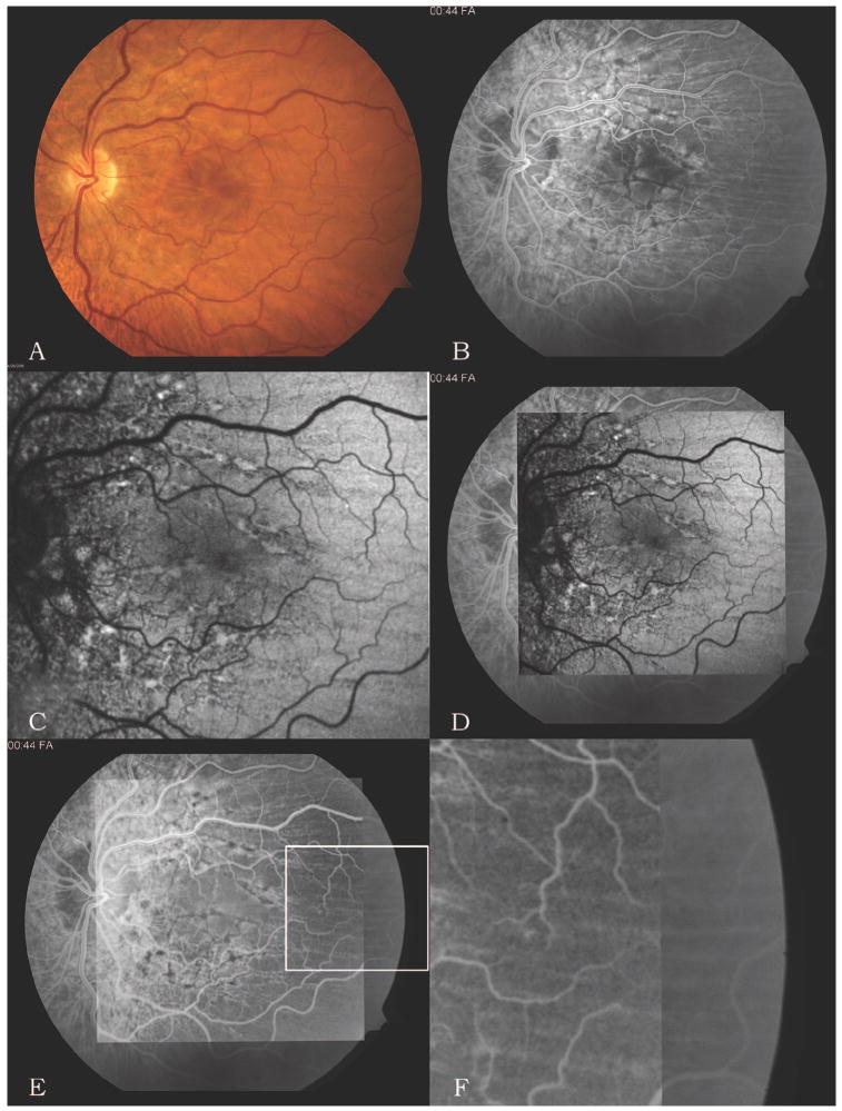
A 47-year-old man in excellent health was referred to the retina service for evaluation of posterior pole findings. Best-corrected visual acuity was 20/30 in the right eye and 20/25 in the left eye, with spherical equivalents of +6.75 and +5.25, respectively. Results of anterior segment and neuroophthalmic examinations were unremarkable. Posterior segment examination revealed bilateral chorioretinal folds in the posterior pole oriented predominantly in a horizontal orientation. Subtle streaks of RPE hyperpigmentation and hypopigmentation were noted emanating from both optic nerves (Fig. 1A). Early-phase fluorescein angiography revealed a horizontal pattern of alternating light and dark bands, characteristic of chorioretinal folds (Fig. 1B). Autofluorescence imaging revealed a similar pattern of alternating light and dark bands as well as peripapillary mottling (Fig. 1C). On close inspection, the alternating patterns of light and dark bands observed using autofluorescence imaging and fluorescein angiography were found to be precisely in register but inverted (Fig. 1, D–F).
Fig. 1.
A, Color photograph of the left fundus demonstrates predominantly horizontal chorioretinal folds, most prominent in the macula, with subtle peripapillary changes of the retinal pigment epithelium. Findings for the right eye were similar. B, Early-phase fluorescein angiogram of the left eye demonstrates a characteristic pattern of alternating light and dark bands, diagnostic of chorioretinal folds. Linear beadlike areas of hypofluorescence are present around the optic nerve and macula. Findings for the right eye were similar. C, Autofluorescence image of the left eye also demonstrates an alternating pattern of light and dark bands as well as linear beadlike areas of hyperautofluorescence in a distribution similar to that on the angiogram. Findings for the right eye were similar. D, Resulting image after the autofluorescence image (C) is superimposed over the early-phase fluorescein angiogram (B) and aligned in register. E, Resulting image after inverting the autofluorescence image. F, Magnified view of the area in the box in E showing the overlap between the inverted autofluorescence image (on left side) and the fluorescein angiogram (on right side), demonstrating precise alignment but inversion of the alternating banding pattern.
Discussion
This case of long-standing chorioretinal folds in a patient with hyperopia demonstrates that autofluorescence imaging may noninvasively demonstrate alterations similar to the alternating light and dark bands shown by fluorescein angiography. We superimposed the autofluorescence image and the early-phase fluorescein angiogram in register using a method previously described by Smith et al5 and found that the folding patterns revealed by autofluorescence imaging and fluorescein angiography were in precise alignment but inverted.
von Winning6 theorized that the pattern of light and dark bands shown by fluorescein angiography may be explained by differences in RPE density. This theory is consistent with the finding that the banding patterns were inverted between fluorescein angiographic and autofluorescence images: rarefied RPE cells at the peak of a chorioretinal fold are dark on autofluorescence images from sparse lipofuscin but bright on angiograms due to a relative window defect. Conversely, compacted RPE cells at the trough of a chorioretinal fold are bright on autofluorescence images from increased lipofuscin density but dark on angiograms due to blocked choroidal fluorescence. In addition, damage to compressed RPE cells at the trough of a fold, as demonstrated histopathologically,7 may contribute to increased autofluorescence in eyes with long-standing folds.
Newell8 described both linear beadlike and diffuse RPE pigmentary changes in long-standing chorioretinal folds. These areas are subtle on color photographs but correspond to the striking pattern of diffuse mottled peripapillary hyperautofluorescence and hypoautofluorescence and linear hyperautofluorescence emanating from the optic nerve head.
To our knowledge, autofluorescence imaging of long-standing chorioretinal folds has not been previously described in the literature. This case independently supports the von Winning hypothesis regarding the etiopathogenesis of the alternating banding pattern of chorioretinal folds shown by fluorescein angiography and, in addition, provides a noninvasive approach for visualizing such changes.
Acknowledgments
Supported in part by the Heed Foundation Fellowship.
References
- 1.Newell FW. Fundus changes in persistent and recurrent choroidal folds. Br J Ophthalmol. 1984;68:32–35. doi: 10.1136/bjo.68.1.32. [DOI] [PMC free article] [PubMed] [Google Scholar]
- 2.Gass JD. Radial chorioretinal folds. A sign of choroidal neovascularization. Arch Ophthalmol. 1981;99:1016–1018. doi: 10.1001/archopht.1981.03930011016006. [DOI] [PubMed] [Google Scholar]
- 3.Friberg TR, Grove AS., Jr Choroidal folds and refractive errors associated with orbital tumors. An analysis. Arch Ophthalmol. 1983;101:598–603. doi: 10.1001/archopht.1983.01040010598014. [DOI] [PubMed] [Google Scholar]
- 4.Singh G. Choroidal folds in posterior scleritis. Arch Ophthalmol. 1989;107:168–169. doi: 10.1001/archopht.1989.01070010174009. [DOI] [PubMed] [Google Scholar]
- 5.Smith RT, Nagasaki T, Sparrow JR, et al. Photographic patterns in macular images: representation by a mathematical model. J Biomed Opt. 2004;9:162–172. doi: 10.1117/1.1630604. [DOI] [PubMed] [Google Scholar]
- 6.von Winning CH. Proceedings: fluorography of choroidal folds. Ophthalmologica. 1973;167:436–439. doi: 10.1159/000306990. [DOI] [PubMed] [Google Scholar]
- 7.Shields JA, Shields CL, Rashid RC. Clinicopathologic correlation of choroidal folds: secondary to massive cranioorbital hemangiopericytoma. Ophthal Plast Reconstr Surg. 1992;8:62–68. doi: 10.1097/00002341-199203000-00011. [DOI] [PubMed] [Google Scholar]
- 8.Newell FW. Fundus changes in persistent and recurrent choroidal folds. Br J Ophthalmol. 1984;68:32–35. doi: 10.1136/bjo.68.1.32. [DOI] [PMC free article] [PubMed] [Google Scholar]


