

Abstract
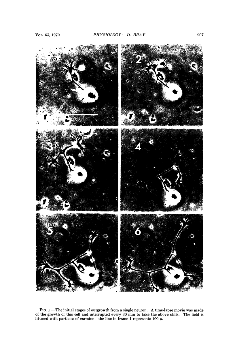
The processes of isolated rat sympathetic neurons growing in culture were marked with glass or carmine particles and observed with timelapse microphotography. Particles on the processes moved with the cell in relation to the dish and underwent continual small jerky movements. They did not, however, show any over-all distal motion and for long periods, during which the growth cone progressed more than 100 μ, the particles remained at about the same distance from the cell body. The most obvious explanation for this result is that new fiber surface, and perhaps the plasma membrane, is deposited in the region of the growing tip.
Full text
PDF


Images in this article


Selected References
These references are in PubMed. This may not be the complete list of references from this article.
- HUGHES A. The growth of embryonic neurites; a study of cultures of chick neural tissues. J Anat. 1953 Apr;87(2):150–162. [PMC free article] [PubMed] [Google Scholar]
- Ingram V. M. A side view of moving fibroblasts. Nature. 1969 May 17;222(5194):641–644. doi: 10.1038/222641a0. [DOI] [PubMed] [Google Scholar]
- LEVI-MONTALCINI R., ANGELETTI P. U. Essential role of the nerve growth factor in the survival and maintenance of dissociated sensory and sympathetic embryonic nerve cells in vitro. Dev Biol. 1963 Mar;6:653–659. doi: 10.1016/0012-1606(63)90149-0. [DOI] [PubMed] [Google Scholar]
- Varon S., Nomura J., Shooter E. M. Subunit structure of a high-molecular-weight form of the nerve growth factor from mouse submaxillary gland. Proc Natl Acad Sci U S A. 1967 Jun;57(6):1782–1789. doi: 10.1073/pnas.57.6.1782. [DOI] [PMC free article] [PubMed] [Google Scholar]