Abstract
RGC-5 cells are transformed cells that express several surface markers characteristic of neuronal precursor cells, but resemble glial cells morphologically and divide in culture. When treated with the apoptosis-inducing agent staurosporine, RGC-5 cells assume a neuronal morphology, extend neurites, stop dividing, and express ion channels without acute signs of apoptosis. This differentiation with staurosporine is similar to what has been described for certain other neuronal cell lines, and occurs by a mechanism not yet understood. Inhibition of several kinases known to be inhibited by staurosporine fails to differentiate RGC-5 cells, and examination of the kinome associated with staurosporine-dependent differentiation has been unhelpful so far. To better understand the mechanism of staurosporine-mediated differentiation of neuronal precursor cells, we studied the effects of the following structurally similar molecules on differentiation of neuronal and non-neuronal cell lines, comparing them to staurosporine: 9,12-Epoxy-1H-diindolo[1,2,3-fg:3',2',1'-kl]pyrrolo[3,4-i][1,6]benzodiazocine-10-carboxylic acid, 2,3,9,10,11,12-hexahydro-10-hydroxy-9-methyl-1-oxo-, methyl ester, (9S,10R,12R)- (K252a), (5R,6S,8S)-6-hydroxy-5-methyl-13-oxo-6,7,8,13,14,15-hexahydro-5H-16-oxa-4b,8a,14-triaza-5,8-methanodibenzo[b,h]cycloocta[jkl]cyclopenta[e]-as-indacene-6-carboxylic acid (K252b), staurosporine aglycone (K252c), 7-hydroxystaurosporine (UCN-01), and 4'-N-benzoylstaurosporine (PKC-412). Morphological differentiation, indicated by neurite extension and somal rounding, was quantitatively assessed with NeuronJ. We found that the critical structural component for differentiation in RGC-5 cells is a basic amine adjacent to an accessible methoxy group at the 3’ carbon. Given that UCN-01 and similar compounds are potent anti-cancer drugs, examination of molecules that share similar structural features may yield insights into the design of other drugs for differentiation.
The RGC-5 cell line expresses some neuronal markers characteristic of retinal ganglion cells (RGCs), but morphologically resembles glial cells and divides in culture (Krishnamoorthy et al., 2001). Unexpectedly, the phenotypic similarity between RGC-5 cells and primary RGCs can be increased by treating cells with the broad-spectrum kinase inhibitor staurosporine (Frassetto et al., 2006), which normally is used to induce apoptosis. Differentiation with staurosporine causes RGC-5 cells to stop dividing, express some ion channels, and assume a neuronal morphology. The somas become round and elevated, and neurites are extended (Frassetto et al., 2006). These neurites immunostain for microtubule-associated protein 2, tau, and growth-associated protein 43, and axon-like and dendrite-like processes can be distinguished (Lieven et al., 2007). Given that differentiated RGC-5 cells more closely resemble primary RGCs in morphology and physiology, they serve as a more relevant model cell population for primary RGCs, than undifferentiated RGC-5 cells. The use of differentiated RGC-5 cells also has advantages over the use of primary RGCs, namely the comparative ease of maintaining a population of RGC-5 cells in culture, and the homogeneity of the population of cells obtained.
Differentiation with staurosporine is surprising, because this kinase inhibitor is typically used as a potent inducer of apoptosis. RGC-5 cells can also be differentiated by inhibiting histone deacetylase with trichostatin A (Schwechter et al., 2007). However, this mechanism of differentiation differs from that seen with staurosporine in that the extension of neurites induced by HDAC inhibition requires RNA transcription and the cells produced are neurotrophic factor-dependent (Schwechter et al., 2007).
The presumed target(s) of kinase inhibition mediating staurosporine-induced differentiation are unknown. Treatment of RGC-5 cells with a variety of specific kinase inhibitors alone or in combination does not induce similar phenotypic changes (Frassetto et al., 2006). Treatment of RGC-5 cells with H-1152, a Rho-kinase inhibitor, or H-89, a nonspecific protein kinase A inhibitor, results in some process formation, but not the somal rounding seen with staurosporine-induced differentiation (Frassetto et al., 2006). Although phosphorylation state changes in RGC-5 cells treated with staurosporine can be studied, it is not clear which phosphorylation targets signal differentiation and which are secondary or unrelated to differentiation (Frassetto et al., 2006). Yet staurosporine has been shown to differentiate other neuronal precursor cell lines: PC-12 (Hashimoto and Hagino, 1989), NB-1 (Morioka et al., 1985), SK-N-SH (Lombet et al., 2001), and SHSY5Y (Shea and Beermann, 1991).
Given the difficulty in understanding the specificity and molecular mechanism of staurosporine differentiation of neuronal precursors into neurons via a functional (kinase-based) approach, we focused on the molecular structure of staurosporine. We examined the morphological effects on RGC-5 cells of the structurally related molecules K252a, K252b, K252c, UCN-01, and PKC-412, and quantitatively and qualitatively correlated differentiation with the presence of specific chemical groups on those molecules. To examine how specific the differentiation process was to RGC-5 cells, we also examined differentiation of two other neuron precursor cell lines (PC-12 and PC6-3) and the non-neuronal 3T3 line. PC-12 and PC6-3 are pheochromocytoma cell lines which when differentiated with NGF have features of sympathetic neurons. PC6-3 cells are a subline of PC-12 cells, and are significantly more dependent on nerve growth factor for survival (Pittman et al., 1993). 3T3 is a fibroblast cell line derived from mouse embryonic tissue (Todaro and Green, 1963). We found that the presence of specific structural elements found in some of the kinase inhibtors tested were associated with cellular differentiation to a neuron-like phenotype.
Experimental procedures
Materials
Staurosporine (isolated from Streptomyces staurosporeus) was obtained from Alexis Biochemical (San Diego, CA). Fetal bovine serum was obtained from Gemini Bio-products (West Sacramento, CA). Other cell culture reagents were obtained from Mediatech (Herndon, VA) unless otherwise noted. Dimethyl sulfoxide (DMSO), K252a, K252b, K252c (staurosporine aglycone), cycloheximide, and actinomycin D were obtained from Sigma-Aldrich (St. Louis, MO). UCN-01 (7-hydroxystaurosporine) was obtained from Calbiochem (San Diego, CA). PKC-412 (midostaurin, 4'-N-benzoylstaurosporine) was obtained from LC Laboratories (Woburn, MA). Paraformaldehyde (16% solution) was obtained from Electron Microscopy Sciences (Hatfield, PA). All other materials were obtained from Fisher Scientific (Pittsburgh, PA).
Cell culture
RGC-5 cells were cultured in Dulbecco's modified Eagle's medium (DMEM) with 1 g/L glucose and L-glutamine, supplemented with 10% fetal bovine serum (FBS), 100 U/mL penicillin, and 100 μg/mL streptomycin. 3T3 cells were grown in DMEM with 4.5 g/L glucose and L-glutamine, supplemented with 10% calf serum. PC-12 cells were cultured in DMEM with 1 g/L glucose and L-glutamine, supplemented with 10% horse serum, 5% FBS, 100 U/mL penicillin, and 100 μg/mL streptomycin. PC6-3 cells were cultured in RPMI-1640 with L-glutamine, supplemented with 10 % horse serum, 5% FBS, 100 U/mL penicillin, and 100 μg/mL streptomycin. Cells to be treated with kinase inhibitors were plated onto 12-mm round cover glass slips in 24-well plates, at a density of approximately 8,000 cells in 450 μL growth media per well. All cells were incubated at 37°C in humidified 5% CO2.
Treatment with kinase inhibitors
All kinase inhibitors were stored as 1 mM stock solutions in dimethyl sulfoxide (DMSO). Stock solutions were then diluted in growth media and added to cultures to final concentrations of 0, 100 nM, 316 nM, or 1μM. In some conditions transcription and translation were inhibited with actinomycin D (4 μM) or cycloheximide (100 μM).
After 24 hours in culture, pharmacological agents or additional media were added to a total volume of 500 μL per well. After 24 hours of treatment, cells were fixed in 4% paraformaldehyde for 10 minutes and ice-cold methanol for 5 minutes. Coverslips were then mounted on slides and sealed. All conditions were tested in duplicate.
Cell morphology
Digital photomicrographs of the mounted cells were taken at 400x total magnification on a Zeiss Axiophot microscope and stored as TIFF images. Photomicrographs were analyzed and the number of primary neurites per cell counted by an observer masked to the experimental group. Primary neurites were classified as projections that originated at the cell soma and were at least as long as the soma was long or wide. Neurite counts were averaged for each condition. Student's t-tests were used to compare neurite counts.
The NeuronJ plug-in for NIH ImageJ software was used to assess neurite length and branching. Neurites were traced, and labeled as primary, secondary, tertiary, or quaternary. Percentages of each type of neurite were tabulated for each condition. Length measurements from individual neurites were converted to microns, and the value for total neurite field length for each cell was calculated.
Cell viability
Cells were grown in a 96-well tissue culture plate at a density of approximately 2,000 cells/well, and treated in triplicate. Viability was assessed 24 hours after treatment. Growth media was removed, and replaced with calcein-AM (10 μM) and propidium iodide (1.5 μM) in phosphate-buffered saline. Cells were then incubated for a further 30 minutes at room temperature in the dark. Digital photomicrographs of the wells were taken at a total magnification of 200x on a Zeiss Axiovert inverted microscope under epifluorescence. ImageJ software was used to analyze the number of cells stained with calcein (living) and propidium iodide (dead).
Statistical analysis
Comparisons between 2 groups was by Student t test. Comparisons across 2 or more groups with a control group was by ANOVA followed by Bonferroni correction for multiple comparisons. All procedures were run using the Data Analysis add-in in Microsoft Excel 2004.
Results
Morphologic Changes in RGC-5 Cells
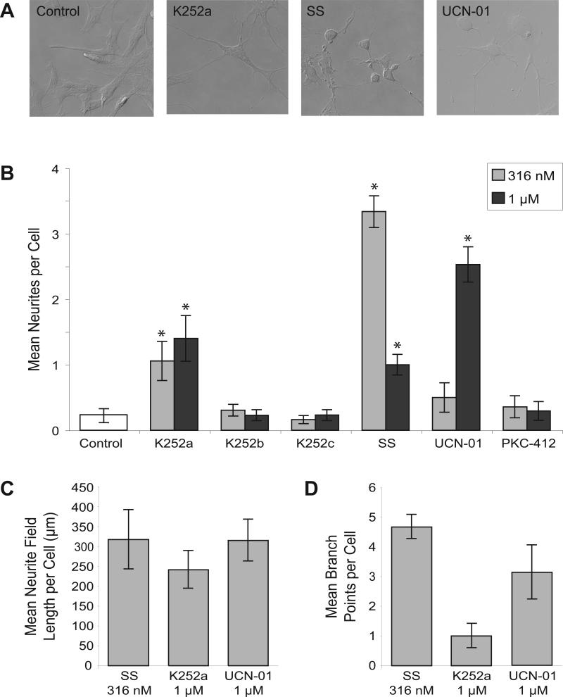
RGC-5 cells treated with staurosporine for 24 hours had a morphology similar to that of primary RGCs, including somal rounding and extension of neurites, as previously reported (Frassetto et al., 2006). When treated with 316 nM staurosporine, RGC-5 cells had 3.3 ± 0.2 primary neurites per cell, compared to 0.19 ± 0.1 primary neurites per cell seen in cells treated with vehicle control (p < 0.001). Twenty-four hours of treatment with 1 μM UCN-01 also induced morphological differentiation, which was not seen at lower concentrations. Treatment with UCN-01 caused both the extension of neurites (2.5 ± 0.3 at 1 μM vs. 0.19 ± 0.1 with vehicle; p = 0.012) and somal rounding. Staurosporine induced extension of a greater number of neurites than UCN-01 when 316 nM treatments of the compounds were observed (3.3 ± 0.2 vs. 0.5 ± 0.2 respectively, p < 0.001). However, when 1 μM concentrations of staurosporine and UCN-01 were compared, the cells treated with UCN-01 extended a greater number of neurites (1.0 ± 0.2 average neurites per cell with 1 μM staurosporine, 2.5 ± 0.3 with UCN-01, p < 0.001) (Figure 1B).
Figure 1. Staurosporine (SS), K252a, and UCN-01 induce morphological differentiation in RGC-5 cells.
(A) Cells treated with SS or UCN-01 exhibit a morphology similar to primary RGCs, including neurites and rounded somas. Treatment with K252a induces extension of broader, flatter neurites than those seen in SS-differentiated cells, and no somal rounding. (B) SS differentiates RGC-5 cells most effectively at a concentration of 316 nM (resulting in 3.3 ± 0.2 primary neurites per cell), while UCN-01 is most effective at a 1 μM concentration (2.5 ± 0.3 primary neurites per cell). (C) Cells treated with K252a extended neurites with a mean field length slightly but statistically insignificantly shorter than mean field lengths per cell of staurosporine or UCN-01 treatment. (D) Neurites induced by K252a were noticeably but insignificantly less complex than those induced by staurosporine or UCN-01 (1.0 ± 0.4 branch points per cell after K252a treatment vs. 4.7 ± 1.3 and 3.1 ± 0.9 branch points per cell after staurosporine or UCN-01 treatment respectively, p = 0.11).
Treatment of RGC-5 cells with K252a resulted in a subset of the morphological changes seen with staurosporine or UCN-01. Cells treated with K252a extended neurites, but the somas did not become round and elevated. Neurites appeared broader than those seen with staurosporine (Figure 1A). Cells treated with K252a extended fewer neurites than cells treated with staurosporine (1.4 ± 0.3 with 1 μM K252a vs. 3.3 ± 0.2 with 316 nM staurosporine; p < 0.001).
Further analysis of cell photomicrograhs with NeuronJ software showed that neurites induced by staurosporine and UCN-01 were morphologically similar, while neurites induced by K252a were slightly shorter and significantly less branched (Figure 1C, 1D). The neurites compared were those induced by treatment of cells with each kinase inhibitor at the concentration at which it most effectively induced morphological changes (316 nM staurosporine, 1 μM K252a, and 1 μM UCN-01). RGC-5 cells treated with K252a extended neurites with a mean field length per cell of 242 ± 47 μm, a length noticeably but statistically insignificantly shorter than the mean field lengths per cell of staurosporine or UCN-01 treatment (317 ± 75 μm and 315 ± 53 μm respectively). Similarly, neurites induced by K252a treatment showed noticeably but statistically insignificantly less branching than neurites induced by staurosporine or UCN-01 (1.0 ± 0.4 branch points per cell after K252a treatment vs. 4.7 ± 1.3 and 3.1 ± 0.9 branch points per cell after staurosporine or UCN-01 treatment respectively, p = 0.11). These differences are consistent with staurosporine and UCN-01 inducing morphological differentiation, while K252a induces only some of the morphological changes observed with differentiation.
Treatment of RGC-5 cells with K252b, K252c, or PKC-412 did not cause morphological changes of differentiation.
Effects on Other Neuronal and Non-Neuronal Cell Lines
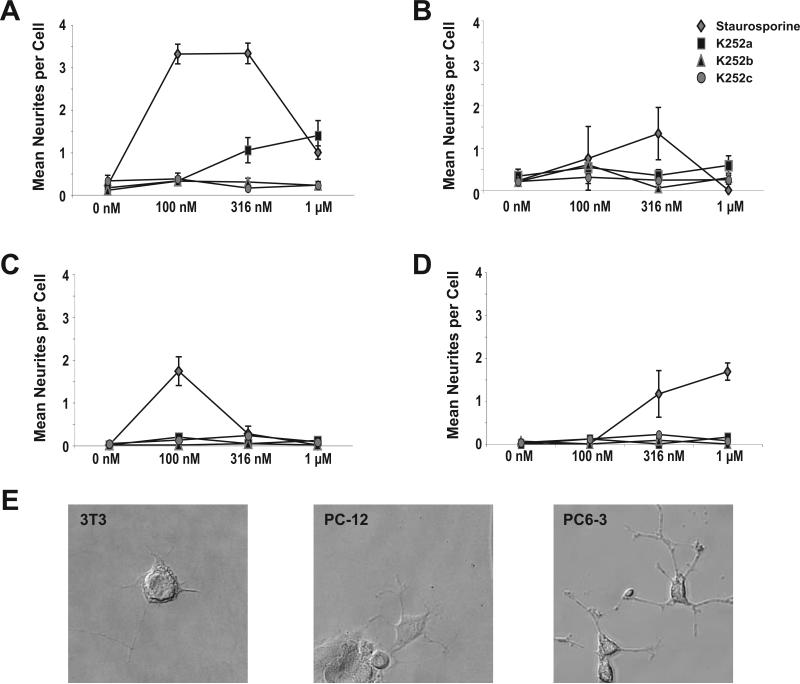
PC-12 and PC6-3 cells are pheochromocytoma-derived cells that can be differentiated into a neuronal phenotype with nerve growth factor (Greene and Tischler, 1976; Pittman et al., 1993). Staurosporine also induced morphological differentiation of these cell lines (Figure 2). The greatest mean number of primary neurites seen in this study in PC-12 cells was observed at a staurosporine concentration of 100 nM (1.7 ± 0.3 per cell vs. 0 in untreated cells; p = 0.005). In PC6-3 cells, the greatest mean number of primary neurites was observed at 1 μM (1.7 ± 0.2 per cell vs. 0 in untreated cells; p < 0.0001). Neurites extended by PC-12 and PC6-3 cells were similar in structure and shape to those extended by differentiated RGC-5 cells.
Figure 2. Effects of staurosporine and related compounds on neuronal and non-neuronal differentiation.
Treatment of RGC-5 (A), 3T3 (B), PC-12 (C), and PC6-3 (D) cells with staurosporine caused significant levels of differentiation. K252b and K252c had no effect on neurite extension. Treatment of RGC-5 cells with K252a caused significant primary neurites to extend (1.0 ± 0.3 at 316 nM vs. 0.17 ± 0.07 at 0 nM; p < 0.001), but RGC-5 cells treated with K252a extended fewer neurites than cells treated with staurosporine (1.0 ± 0.3vs. 3.3 ± 0.2 respectively at 316 nM; p < 0.001). Similarly, process formation was seen with staurosporine, but not K252a, K252b, or K252c in 3T3, PC-12, and PC6-3 cells. Photomicrographs (E) illustrate process extension of 3T3 cells treated with 316 nM SS, PC-12 cells treated with 316 nM SS, and PC6-3 cells treated with 1 μM SS.
Surprisingly, 3T3 fibroblasts also extended processes when treated with staurosporine for 24 hours. The greatest mean number of primary processes was seen at 316 nM (1.3 ± 0.6 per cell vs. 0.2 ± 0.1 in untreated cells; p = 0.001). The processes seen in 3T3 cells treated with staurosporine were flatter and more delicate than those observed with RGC-5, PC-12, or PC6-3 cells. Treatment of PC-12, PC6-3, and 3T3 cells with K252a, K252b, or K252c at any concentration did not result in significant extension of neurites or change in the shape of the cell soma.
Transcriptional and Translational Dependence
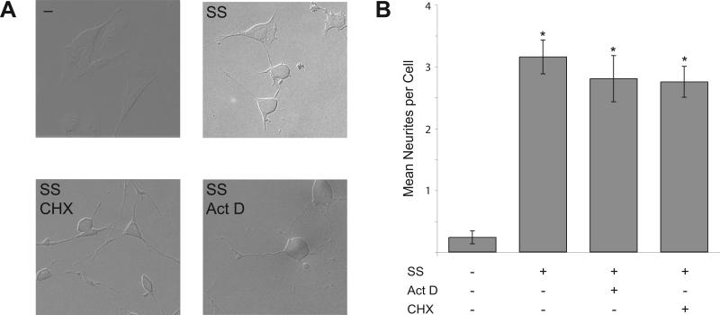
The dependence of RGC-5 differentiation on mRNA and protein synthesis was examined. Cells were treated with staurosporine, K252a, K252b, or K252c in combination with actinomycin D (4 μM) to inhibit transcription or cycloheximide (100 μM) to inhibit translation. Neither treatment prevented the extension of neurites by RGC-5 cells treated with staurosporine (Figure 3A), and treatment with either cycloheximide or actinomycin D did not result in a significant reduction in the number of neurites extended (3.2 ± 0.3 neurites per cell with 316 nM staurosporine alone vs. 2.8 ± 0.2 with staurosporine and cycloheximide and 2.8 ± 0.4 with staurosporine and actinomycin D; p = 0.5 and p = 0.3, respectively) (Figure 3B) indicating that process formation induced by staurosporine is initially neither transcription- nor translation-dependent. However, significant cell loss was observed in conditions combining high levels of staurosporine (316 nM or 1μM) with cycloheximide treatment. Cells treated with K252a, K252b, or K252c and cycloheximide or actinomycin D did not extend processes, and no abnormal cell loss was observed with these combinations (data not shown). These results were similar to previous experiments demonstrating that staurosporine-induced differentiation was not blocked by the RNA-polymerase II inhibitor α-amanitin (Schwechter et al., 2007).
Figure 3. Effects of transcription and translation inhibition on differentiation by staurosporine.
(A) RGC-5 cells treated with 100 nM staurosporine (SS) extend neurites. Treating cells with 100 nM SS and either actinomycin D to inhibit transcription (Act D, 4 μM) or cycloheximide to inhibit translation (CHX, 100 μM) did not prevent this extension of neurites. (B) When treated with 316 nM SS, cells had an average of 3.2 ± 0.3 primary neurites per cell. Treatment of cells with SS and either 4 μM Act D or 100 μM CHX did not result in a significantly different number of neurites per cell (2.8 ± 0.4 neurites per cell, p = 0.3, and 2.8 ± 0.5 neurites per cell, p = 0.5, respectively.)
Effect of Kinase Inhibition on RGC-5 Cell Viability
Although staurosporine induces apoptosis in a broad range of cell types (Bertrand et al., 1994), it causes differentiation in RGC-5 cells (Frassetto et al., 2006). To study this further, RGC-5 cells were incubated with varying concentrations of staurosporine, UCN-01, K252a, K252b, K252c, or PKC-412 for 24 hours and stained with calcein-AM and propidium iodide to assess viability. RGC-5 cells treated with staurosporine for 24 hours at concentrations from 100 nM to 1 μM had 98.8 ± 0.9% to 84.4 ± 4.6% viability, compared with 99.6 ± 0.3% in untreated cells. A similar small decrease in viability was seen with UCN-01 at concentrations of 316 nM (86.6 ± 3.6% viability) and 1 μM (83.6 ± 3.7% viability). Insignificant levels of death were seen when cells were treated with K252a, K252b, K252c, or PKC-412, compared to untreated cells (Figure 4).
Figure 4. Staurosporine and related compounds have minimal effects of cell viability.
The number of viable cells compared to untreated cells did not change when RGC-5 cells were treated with K252a, K252b, K252c, or PKC-412 at 100 nM, 316 nM, or 1 μM concentrations. The percentage of viable cells decreased slightly when cells were treated with 1 μM staurosporine (SS) (84.4 ± 4.6%, p = 0.007), 316 nM UCN-01 (86.6 ± 3.6%, p = 0.002), or 1 μM UCN-01 (83.6 ± 3.7%, p = 0.0006).
Discussion
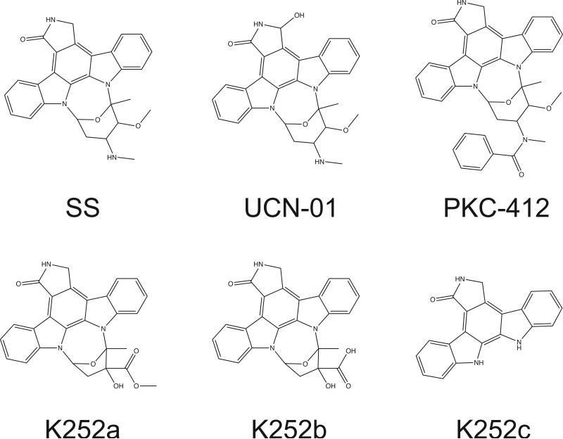
Although the molecular structures of staurosporine, K252a, K252b, K252c, UCN-01, and PKC-412 are similar (Figure 5), differences among these six kinase inhibitors are significant enough that only staurosporine and UCN-01 induce differentiation, evidenced by both process formation and somal rounding in RGC-5 cells. Although treatment with K252a leads to process formation, it does not cause the cells to assume the round, raised soma shape characteristic of primary RGCs and differentiated RGC-5 cells. The fact that that UCN-01 causes the same morphological changes as staurosporine is not surprising, since this compound (7-hydroxystaurosporine) is the most structurally similar to staurosporine of the kinase inhibitors tested here.
Figure 5. Molecular structures of staurosporine (SS), UCN-01, PKC-412, K252a, K252b, and K252c are similar, but the compounds differ at or near the 3' carbon.
Staurosporine and UCN-01 contain a methoxy group at this position, while K252a contains a structurally similar methyl ester. While PKC-412 does contain a methoxy group at the 3' carbon, the nearby benzoyl group containing a phenyl ring would sterically hinder the methoxy group.
Both staurosporine and UCN-01 contain a methoxy group at the 3’ carbon. A similar methoxy group is not seen in K252a, K252b, or K252c. K252a, which is able to induce some process formation but not somal rounding in RGC-5 cells, does however have a structurally similar methyl ester at the same carbon where the methoxy group is found in staurosporine and UCN-01. It therefore is likely that an accessible 3’-methoxy group is necessary for differentiation signaling, since it is found in the two compounds tested here which differentiate RGC-5 cells, and a similar group is seen in a compound that induces process formation. Additionally, staurosporine and UCN-01 both contain an amine adjacent to the 3’-methoxy group, a feature seen in no other kinase inhibitors studied here. The nitrogen would be positively charged at physiological pH, and this may relate to why only staurosporine and UCN-01 were capable of inducing morphological differentiation in RGC-5 cells. PKC-412, which is inactive in neuronal differentiation, also contains a 3’-methoxy group but does not have the adjacent nitrogen. There is also a large benzoyl group containing a phenyl ring attached to an adjacent carbon, which might sterically hinder the methoxy group.
The disparate effects of these compounds on RGC-5 cells could be due either to different levels and specificities of protein kinase inhibition or effects on other cellular processes independent of kinase inhibition. Staurosporine is a broad-spectrum protein kinase inhibitor, and strongly inhibits, among others, protein kinase C (PKC), protein kinase A (PKA), and protein kinase G (PKG) (Tamaoki et al., 1986). K252a (Kase et al., 1987) and UCN-01 (Ruegg and Burgess, 1989) also inhibit PKC, PKA, and PKG, and are similarly non-specific. K252b (Ruegg and Burgess, 1989) and K252c (Horton et al., 1994) have some selectivity for PKC over PKA or PKG. Finally, PKC-412 is a strong PKC inhibitor, but does have some activity against other protein kinases (Fabbro et al., 2000).
No specific inhibitor for any one target of staurosporine has so far been shown capable of inducing neuronal morphology (Frassetto et al., 2006). Beyond the kinases mentioned above, staurosporine has also been shown to inhibit cyclin-dependent kinase 1, cyclin-dependent kinase 2, cyclin-dependent kinase 5, Janus kinase 2, epidermal growth factor receptor kinase, platelet-derived growth factor receptor kinase, myosin light-chain kinase, calmodulin-dependent protein kinase, Rho kinase, MAP/ERK kinase, and Akt (Bain et al., 2003). Treatment with more specific inhibitors of these kinases fails to induce morphological differentiation of RGC-5 cells similar to staurosporine (Frassetto et al., 2006). Inhibition of Rho kinase with H-1152 or inhibition of PKA with H-89 causes some process extension, but less than that seen with staurosporine. Neither of these kinase inhibitors induced somal rounding (Frassetto et al., 2006). It is therefore likely that either inhibition of multiple kinases is necessary for differentiation of RGC-5 cells, differentiation depends on the inhibition of an as of yet unidentified protein kinase, or staurosporine has an effect on some other cellular process.
Although staurosporine has been shown to induce apoptosis in several cell lines, including human promyelocytic leukemia (HL-60), Burkitt lymphoma (CA46), follicular lymphoma (SUDHL6), colon adenocarcinoma (HT29), small cell lung carcinoma (SCL209), and lung fibroblast cells (DC3F) (Bertrand et al., 1994) and UCN-01 has been shown to induce apoptosis in colon carcinoma cells (Bhonde et al., 2005), in RGC-5 cells these kinase inhibitors induce differentiation without obvious induction of apoptosis (Frassetto et al., 2006). Drugs that induce apoptosis, e.g. rotenone, ionomycin, thapsigargin, and etoposide, do not induce differentiation (unpublished data). Pre-treatment with the apoptosis inhibitors minocycline or ZVAD-fmk does not prevent differentiation (Frassetto et al., 2006). In the present study, 24 hour treatment with 316 nM staurosporine, the concentration that causes the extension of the greatest number of neurites, did not lead to significant cell death compared to cells treated with vehicle control. Together, these findings all indicate that the effect of staurosporine on differentiation is not related to induction of apoptosis.
It is not surprising that staurosporine and UCN-01 induce neuritogenesis in PC-12 and PC6-3 cells, since PC-12 cells are pheochromocytoma-derived and have previously been shown to stop dividing and extend neurites when treated with 100 nM staurosporine (Hashimoto and Hagino, 1989), similar to what is seen with nerve growth factor (Greene and Tischler, 1976). Differentiation of PC-12 cells by staurosporine has been shown to be independent of PKC activity (Rasouly et al., 1996), and may instead be linked to the activation of a novel c-Jun N-terminal kinase isoform (Yao et al., 1997). The effect of K252a on PC-12 cells has also been previously studied. Unlike staurosporine, K252a does not induce differentiation in PC-12 cells. In fact, treatment with K252a will prevent extension of processes induced by NGF (Koizumi et al., 1988), but will not prevent staurosporine-induced differentiation (Tischler et al., 1990). Staurosporine has been shown to induce differentiation in other neuronal cell lines in addition to PC-12 cells, including NB-1 (Morioka et al., 1985), SK-N-SH (Lombet et al., 2001), and SH-SY5Y human neuroblastoma cells (Shea and Beermann, 1991). In SH-SY5Y cells, treatment with staurosporine resulted in the extension of neurites, increased levels of mRNA for the axonal protein GAP-43 (Jalava et al., 1992), and the formation of nerve terminal-like varicosities on neurites that exhibited voltage-sensitive intracellular Ca+2 levels and expressed localized neuropeptide Y (Kukkonen et al., 1997). This differentiation process was not translation-dependent; cycloheximide treatment did not prevent process extension. Additionally, treatment of SH-SY5Ycells with HA-1004, which inhibits PKA and PKG, did not induce differentiation (Shea and Beermann, 1991) Process extension in non-neuronal 3T3 fibroblasts was unexpected, but is consistent with the fact that 3T3 cells have been shown to extend thin processes following Rho-associated protein kinase (ROCK) or Cbl inhibition (Scaife et al., 2003). ROCK inhibition in 3T3 cells has been shown to induce rounded cell bodies and true extension of actively elongating processes, not simply retraction of fibers (Hirose et al., 1998).
In summary, treatment of RGC-5 cells with low concentrations of the kinase inhibitors staurosporine and UCN-01 induces differentiation, manifested by neurite extension and somal rounding. Treatment with K252a causes neurite extension only. K252b, K252c, and PKC-412 treatment did not lead to morphological change. A common feature that correlates with degree of differentiation is a basic amine adjacent to an accessible methoxy group at the 3’ carbon. This may be a structural motif necessary for this class of compounds to act as a differentiating agent. Understanding the molecular mechanism of kinase inhibition-dependent RGC-5 differentiation could lead to the identification of a new class of differentiation-inducing compounds.
Acknowledgments
Grant Support: NIH R01EY012492, R21EY017970, and P30EY016665, Retina Research Foundation, and an unrestricted departmental grant from Research to Prevent Blindness, Inc. LAL is a Canada Research Chair.
Footnotes
Publisher's Disclaimer: This is a PDF file of an unedited manuscript that has been accepted for publication. As a service to our customers we are providing this early version of the manuscript. The manuscript will undergo copyediting, typesetting, and review of the resulting proof before it is published in its final citable form. Please note that during the production process errors may be discovered which could affect the content, and all legal disclaimers that apply to the journal pertain.
Proprietary Interest: A patent on RGC-5 cell differentiation has been assigned to the Wisconsin Alumni Research Foundation.
References
- Bain J, McLauchlan H, Elliott M, Cohen P. The specificities of protein kinase inhibitors: an update. Biochem J. 2003;371:199–204. doi: 10.1042/BJ20021535. [DOI] [PMC free article] [PubMed] [Google Scholar]
- Bertrand R, Solary E, O'Connor P, Kohn KW, Pommier Y. Induction of a common pathway of apoptosis by staurosporine. Exp Cell Res. 1994;211:314–321. doi: 10.1006/excr.1994.1093. [DOI] [PubMed] [Google Scholar]
- Bhonde MR, Hanski ML, Magrini R, Moorthy D, Muller A, Sausville EA, Kohno K, Wiegand P, Daniel PT, Zeitz M, Hanski C. The broad-range cyclin-dependent kinase inhibitor UCN-01 induces apoptosis in colon carcinoma cells through transcriptional suppression of the Bcl-x(L) protein. Oncogene. 2005;24:148–156. doi: 10.1038/sj.onc.1207842. [DOI] [PubMed] [Google Scholar]
- Fabbro D, Ruetz S, Bodis S, Pruschy M, Csermak K, Man A, Campochiaro P, Wood J, O'Reilly T, Meyer T. PKC412--a protein kinase inhibitor with a broad therapeutic potential. Anticancer Drug Des. 2000;15:17–28. [PubMed] [Google Scholar]
- Frassetto LJ, Schlieve CR, Lieven CJ, Utter AA, Jones MV, Agarwal N, Levin LA. Kinase-dependent differentiation of a retinal ganglion cell precursor. Invest Ophthalmol Vis Sci. 2006;47:427–438. doi: 10.1167/iovs.05-0340. [DOI] [PubMed] [Google Scholar]
- Greene LA, Tischler AS. Establishment of a noradrenergic clonal line of rat adrenal pheochromocytoma cells which respond to nerve growth factor. Proc Natl Acad Sci U S A. 1976;73:2424–2428. doi: 10.1073/pnas.73.7.2424. [DOI] [PMC free article] [PubMed] [Google Scholar]
- Hashimoto S, Hagino A. Staurosporine-induced neurite outgrowth in PC12h cells. Exp Cell Res. 1989;184:351–359. doi: 10.1016/0014-4827(89)90334-0. [DOI] [PubMed] [Google Scholar]
- Hirose M, Ishizaki T, Watanabe N, Uehata M, Kranenburg O, Moolenaar WH, Matsumura F, Maekawa M, Bito H, Narumiya S. Molecular dissection of the Rho-associated protein kinase (p160ROCK)-regulated neurite remodeling in neuroblastoma N1E-115 cells. J Cell Biol. 1998;141:1625–1636. doi: 10.1083/jcb.141.7.1625. [DOI] [PMC free article] [PubMed] [Google Scholar]
- Horton PA, Longley RE, McConnell OJ, Ballas LM. Staurosporine aglycone (K252-c) and arcyriaflavin A from the marine ascidian, Eudistoma sp. Experientia. 1994;50:843–845. doi: 10.1007/BF01956468. [DOI] [PubMed] [Google Scholar]
- Jalava A, Heikkila J, Lintunen M, Akerman K, Pahlman S. Staurosporine induces a neuronal phenotype in SH-SY5Y human neuroblastoma cells that resembles that induced by the phorbol ester 12-O-tetradecanoyl phorbol-13 acetate (TPA). FEBS Lett. 1992;300:114–118. doi: 10.1016/0014-5793(92)80176-h. [DOI] [PubMed] [Google Scholar]
- Kase H, Iwahashi K, Nakanishi S, Matsuda Y, Yamada K, Takahashi M, Murakata C, Sato A, Kaneko M. K-252 compounds, novel and potent inhibitors of protein kinase C and cyclic nucleotide-dependent protein kinases. Biochem Biophys Res Commun. 1987;142:436–440. doi: 10.1016/0006-291x(87)90293-2. [DOI] [PubMed] [Google Scholar]
- Koizumi S, Contreras ML, Matsuda Y, Hama T, Lazarovici P, Guroff G. K-252a: a specific inhibitor of the action of nerve growth factor on PC 12 cells. J Neurosci. 1988;8:715–721. doi: 10.1523/JNEUROSCI.08-02-00715.1988. [DOI] [PMC free article] [PubMed] [Google Scholar]
- Krishnamoorthy RR, Agarwal P, Prasanna G, Vopat K, Lambert W, Sheedlo HJ, Pang IH, Shade D, Wordinger RJ, Yorio T, Clark AF, Agarwal N. Characterization of a transformed rat retinal ganglion cell line. Brain Res Mol Brain Res. 2001;86:1–12. doi: 10.1016/s0169-328x(00)00224-2. [DOI] [PubMed] [Google Scholar]
- Kukkonen JP, Shariatmadari R, Courtney MJ, Akerman KE. Localization of voltage-sensitive Ca2+ fluxes and neuropeptide Y immunoreactivity to varicosities in SH-SY5Y human neuroblastoma cells differentiated by treatment with the protein kinase inhibitor staurosporine. Eur J Neurosci. 1997;9:140–150. doi: 10.1111/j.1460-9568.1997.tb01362.x. [DOI] [PubMed] [Google Scholar]
- Lieven CJ, Millet LE, Hoegger MJ, Levin LA. Induction of axon and dendrite formation during early RGC-5 cell differentiation. Exp Eye Res. 2007;85:678–683. doi: 10.1016/j.exer.2007.08.001. [DOI] [PMC free article] [PubMed] [Google Scholar]
- Lombet A, Zujovic V, Kandouz M, Billardon C, Carvajal-Gonzalez S, Gompel A, Rostene W. Resistance to induced apoptosis in the human neuroblastoma cell line SK-N-SH in relation to neuronal differentiation. Role of Bcl-2 protein family. Eur J Biochem. 2001;268:1352–1362. doi: 10.1046/j.1432-1327.2001.02002.x. [DOI] [PubMed] [Google Scholar]
- Morioka H, Ishihara M, Shibai H, Suzuki T. Staurosporine-induced differentiation in a human neuroblastoma cell line, NB-1. Agric. Biol. Chem. 1985;49:1959–1963. [Google Scholar]
- Pittman RN, Wang S, DiBenedetto AJ, Mills JC. A system for characterizing cellular and molecular events in programmed neuronal cell death. J Neurosci. 1993;13:3669–3680. doi: 10.1523/JNEUROSCI.13-09-03669.1993. [DOI] [PMC free article] [PubMed] [Google Scholar]
- Rasouly D, Shavit D, Zuniga R, Elejalde RB, Unsworth BR, Yayon A, Lazarovici P, Lelkes PI. Staurosporine induces neurite outgrowth in neuronal hybrids (PC12EN) lacking NGF receptors. J Cell Biochem. 1996;62:356–371. doi: 10.1002/(sici)1097-4644(199609)62:3<356::aid-jcb6>3.0.co;2-q. [DOI] [PubMed] [Google Scholar]
- Ruegg UT, Burgess GM. Staurosporine, K-252 and UCN-01: potent but nonspecific inhibitors of protein kinases. Trends Pharmacol Sci. 1989;10:218–220. doi: 10.1016/0165-6147(89)90263-0. [DOI] [PubMed] [Google Scholar]
- Scaife RM, Job D, Langdon WY. Rapid microtubule-dependent induction of neurite-like extensions in NIH 3T3 fibroblasts by inhibition of ROCK and Cbl. Mol Biol Cell. 2003;14:4605–4617. doi: 10.1091/mbc.E02-11-0739. [DOI] [PMC free article] [PubMed] [Google Scholar]
- Schwechter BR, Millet LE, Levin LA. Histone deacetylase inhibition-mediated differentiation of RGC-5 cells and interaction with survival. Invest Ophthalmol Vis Sci. 2007;48:2845–2857. doi: 10.1167/iovs.06-1364. [DOI] [PMC free article] [PubMed] [Google Scholar]
- Shea TB, Beermann ML. Staurosporine-induced morphological differentiation of human neuroblastoma cells. Cell Biol Int Rep. 1991;15:161–168. doi: 10.1016/0309-1651(91)90107-t. [DOI] [PubMed] [Google Scholar]
- Tamaoki T, Nomoto H, Takahashi I, Kato Y, Morimoto M, Tomita F. Staurosporine, a potent inhibitor of phospholipid/Ca++dependent protein kinase. Biochem Biophys Res Commun. 1986;135:397–402. doi: 10.1016/0006-291x(86)90008-2. [DOI] [PubMed] [Google Scholar]
- Tischler AS, Ruzicka LA, Perlman RL. Mimicry and inhibition of nerve growth factor effects: interactions of staurosporine, forskolin, and K252a in PC12 cells and normal rat chromaffin cells in vitro. J Neurochem. 1990;55:1159–1165. doi: 10.1111/j.1471-4159.1990.tb03120.x. [DOI] [PubMed] [Google Scholar]
- Todaro GJ, Green H. Quantitative studies of the growth of mouse embryo cells in culture and their development into established lines. J Cell Biol. 1963;17:299–313. doi: 10.1083/jcb.17.2.299. [DOI] [PMC free article] [PubMed] [Google Scholar]
- Yao R, Yoshihara M, Osada H. Specific activation of a c-Jun NH2-terminal kinase isoform and induction of neurite outgrowth in PC-12 cells by staurosporine. J Biol Chem. 1997;272:18261–18266. doi: 10.1074/jbc.272.29.18261. [DOI] [PubMed] [Google Scholar]


