

Abstract
The central portion of chronic wounds is often hypoxic and relatively hypothermic, representing a deficient energy supply of the tissue, which impedes wound healing or even makes it impossible. Water-filtered infrared-A (wIRA) is a special form of heat radiation with a high tissue penetration and a low thermal load to the skin surface. wIRA produces a therapeutically usable field of heat and increases temperature, oxygen partial pressure and perfusion of the tissue. These three factors are decisive for a sufficient tissue supply with energy and oxygen and consequently as well for wound healing, especially in chronic wounds, and infection defense. wIRA acts both by thermal and thermic as well as by non-thermal and non-thermic effects. wIRA can advance wound healing or improve an impaired wound healing process and can especially enable wound healing in non-healing chronic wounds. wIRA can considerably alleviate the pain and diminish wound exudation and inflammation and can show positive immunomodulatory effects.
In a prospective, randomized, controlled study of 40 patients with chronic venous stasis ulcers of the lower legs irradiation with wIRA and visible light (VIS) accelerated the wound healing process (on average 18 vs. 42 days until complete wound closure, residual ulcer area after 42 days 0.4 cm² vs. 2.8 cm²) and led to a reduction of the required dose of pain medication in comparison to the control group of patients treated with the same standard care (wound cleansing, wound dressing with antibacterial gauze, and compression garment therapy) without the concomitant irradiation.
Another prospective study of 10 patients with non-healing chronic venous stasis ulcers of the lower legs included extensive thermographic investigation. Therapy with wIRA(+VIS) resulted in a complete or almost complete wound healing in 7 patients and a marked reduction of the ulcer size in another 2 of the 10 patients, a clear reduction of pain and required dose of pain medication, and a normalization of the thermographic image.
In a current prospective, randomized, controlled, blinded study patients with non-healing chronic venous stasis ulcers of the lower legs are treated with compression garment therapy, wound cleansing, wound dressings and 30 minutes irradiation five times per week over 9 weeks. A preliminary analysis of the first 23 patients of this study has shown in the group with wIRA(+VIS) compared to a control group with VIS an advanced wound healing, an improved granulation and in the later phase of treatment a decrease of the bacterial burden.
Some case reports have demonstrated that wIRA can also be used for mixed arterial-venous ulcers or arterial ulcers, if irradiation intensity is chosen appropriately low and if irradiation is monitored carefully. wIRA can be used concerning decubital ulcers both in a preventive and in a therapeutic indication. wIRA can improve the resorption of topically applied substances also on wounds.
An irradiation with VIS and wIRA presumably acts with endogenous protoporphyrin IX (or protoporphyrin IX of bacteria) virtually similar as a mild photodynamic therapy (endogenous PDT-like effect). This could lead to improved cell regeneration and wound healing and to antibacterial effects.
In conclusion, these results indicate that wIRA generally should be considered for the treatment of chronic wounds.
Keywords: water-filtered infrared-A (wIRA); wound healing; chronic wounds; chronic venous stasis ulcers of the lower legs; prospective, randomized, controlled, blinded study; reduction of pain; problem wounds; wound infections; infection defense; wound exudation; inflammation; energy supply; oxygen supply; tissue oxygen partial pressure; tissue temperature; tissue blood flow; thermal and non-thermal effects; thermic and non-thermic effects; visual analog scales (VAS); infrared thermography; thermographic image analysis; quality of life
Abstract
Das Zentrum von chronischen Wunden ist oft hypoxisch und relativ hypotherm. Dies entspricht einer defizitären Energiebereitstellung im Gewebe, die die Wundheilung behindert oder unmöglich macht. Wassergefiltertes Infrarot A (wIRA) ist eine spezielle Form der Wärmestrahlung mit hohem Eindringvermögen in das Gewebe bei geringer thermischer Oberflächenbelastung. wIRA erzeugt ein therapeutisch nutzbares Wärmefeld und steigert Temperatur, Sauerstoffpartialdruck sowie die Durchblutung im Gewebe. Diese drei Faktoren sind entscheidend für eine ausreichende Versorgung des Gewebes mit Energie und Sauerstoff und deshalb auch für die Wundheilung, speziell bei chronischen Wunden, und die Infektionsabwehr. wIRA wirkt sowohl über thermische und temperaturabhängige als auch über nicht-thermische und temperaturunabhängige Effekte. wIRA kann die Wundheilung beschleunigen oder einen stagnierenden Wundheilungsprozess verbessern und insbesondere bei nicht-heilenden chronischen Wunden eine Wundheilung ermöglichen. wIRA vermag Schmerzen deutlich zu mindern und die Wundsekretion sowie Entzündung zu reduzieren sowie positive immunmodulierende Effekte zu zeigen.
In einer prospektiven, randomisierten, kontrollierten Studie mit 40 Patienten mit chronischen venösen Unterschenkelulzera führte eine Bestrahlung mit wIRA und sichtbarem Licht (VIS) zu einer schnelleren Wundheilung (im Durchschnitt 18 vs. 42 Tage bis zum kompletten Wundschluss, Restulkusfläche nach 42 Tagen 0,4 cm² vs. 2,8 cm²) und einem geringeren Schmerzmittelverbrauch gegenüber einer in gleicher Form (Wundsäuberung, antibakterielle Wundauflagen und Kompressionstherapie) therapierten, aber nicht bestrahlten Kontrollgruppe.
Eine weitere prospektive Studie mit 10 Patienten mit aufwändiger thermographischer Verlaufskontrolle ergab unter Therapie mit wIRA(+VIS) eine vollständige oder fast vollständige Abheilung therapierefraktärer chronischer Unterschenkelulzera bei 7 sowie eine deutliche Ulkusverkleinerung bei 2 weiteren der 10 Patienten, eine ausgeprägte Minderung der Schmerzen und des Schmerzmittelverbrauchs und eine Normalisierung des thermographischen Bildes.
In einer laufenden prospektiven, randomisierten, kontrollierten, verblindeten Studie werden Patienten mit nicht-heilenden chronischen venösen Unterschenkelulzera mit Kompressionstherapie, Wundsäuberung und nicht-adhäsiven Wundauflagen sowie 30 Minuten Bestrahlung fünfmal pro Woche über 9 Wochen behandelt. Eine vorläufige Auswertung der ersten 23 Patienten zeigte, dass die Gruppe mit wIRA(+VIS) verglichen mit einer Kontrollgruppe mit VIS eine schnellere Wundheilung, eine bessere Granulation und in der späteren Phase der Behandlung eine Abnahme der bakteriellen Last der Wunden aufwies.
Einige Fallberichte haben gezeigt, dass wIRA selbst bei gemischt arteriell-venösen Ulzera oder arteriellen Ulzera eingesetzt werden kann, wenn die Bestrahlungsstärke angemessen niedrig gewählt und die Bestrahlung sorgfältig überwacht wird. wIRA kann bei Dekubitalulzera sowohl präventiv als auch therapeutisch eingesetzt werden. wIRA kann die Resorption topisch applizierter Substanzen auch auf Wunden verbessern.
Eine Bestrahlung mit VIS und wIRA wirkt vermutlich in Verbindung mit endogenem Protoporphyrin IX (oder Protoporphyrin IX von Bakterien) quasi ähnlich wie eine milde photodynamische Therapie (endogener PDT-ähnlicher Effekt). Dies kann die Zellregeneration und Wundheilung fördern und antibakteriell wirken.
Zusammengefasst zeigen die Ergebnisse, dass wIRA generell für die Behandlung chronischer Wunden erwogen werden sollte.
General aspects of water-filtered infrared-A (wIRA) for the improvement of wound healing in dermatology and surgery
Principles and working mechanisms (thermal and thermic effects, non-thermal and non-thermic effects) of wIRA related to wound healing and fundamental recommendations for the clinical use of wIRA as well as safety aspects are described in detail in [1], [2].
General aspects of wIRA for the improvement of wound healing in dermatology and surgery are presented in [3]. This includes the following typical main clinical effects of wIRA (both in acute and in chronic wounds) [3]:
wIRA increases
tissue temperature
tissue oxygen partial pressure
tissue perfusion.
wIRA decreases
pain (and consequently the required dose of pain medication)
inflammation
hypersecretion.
wIRA has
positive immunomodulatory/antiinfective effects.
wIRA improves and advances
wound healing (even the normal wound healing process).
wIRA shortens
time until complete wound healing and hospital stay.
The central portion of chronic wounds is often markedly hypoxic and relatively hypothermic [1], [2], [4], [5], [6], [7], [8], [9], [10], [11], [12], [13], [14], [15], [16], representing a deficient energy supply of the tissue, which impedes wound healing, as wound healing and infection defense are processes with an extremely high energy demand [1], [2], [4], [5]. Tissue temperature [4], [5], [17], [18], tissue oxygen partial pressure [4], [5], [6], [7], [8], [9], [10], [11], [12], [13], [14], [15], [16], and tissue perfusion are decisive factors for a sufficient tissue supply with energy and oxygen and consequently as well for wound healing, especially in chronic wounds [1], [2]. Water-filtered infrared-A (wIRA) as a special form of heat radiation with a high tissue penetration and with a low thermal load to the skin surface produces a therapeutically usable field of heat in the tissue and increases tissue temperature [4], [5], [19], [20], [21], [22], [23], [24], tissue oxygen partial pressure [4], [25], and tissue perfusion [22], [23], [24], [25], see review in [1], [2]. wIRA can advance wound healing or improve an impaired wound healing both in acute and in chronic wounds including infected wounds [1], [2].
wIRA in clinical use at appropriate irradiances has been described as helpful and safe [4], [5], [9], [19], [20], [21], [22], [23], [24], [25], [26], [27], [28], [29], [30], [31], [32], [33], [34], [35], [36], [37], [38], [39], [40], [41], [42], [43], [44], review in [1], [2], [3], and with possible protective cellular effects [45], [46], [47], [48], [49], [50].
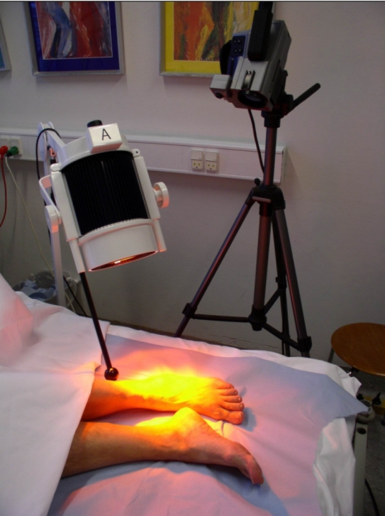
Acute wounds and especially chronic wounds, non-healing wounds or infected problem wounds should be irradiated with wIRA at least three times per week and ideally once or twice per day for 20-30 minutes each [1], [2], [3]. Longer irradiation times are possible and often helpful. wIRA does not replace other sensible/necessary therapeutic procedures (such as compression garment therapy of chronic venous stasis ulcers of the lower legs [51], [52], [53]) but complements them [1], [2], [3]. Correspondingly the therapy with wIRA has to be embedded in an overall therapeutic concept [1], [2], [3]. wIRA can be used independently from therapy preferences concerning wound management (e.g. moist wound management) [1], [2], [3]. Typically during wIRA irradiation the wound has to be uncovered, as most bandages or wound dressings (with the exception of e.g. some tested transparent foils) are not adequately permeable for wIRA [1], [3].
Up to now 6 prospective clinical studies with wIRA concerning wound healing have been performed, 3 with acute wounds (presented in [3]) and 3 with chronic wounds (presented here), one of these is still in progress. Experiences with wIRA in wound healing especially of chronic wounds exist since approximately 1989 [54]. One of the authors has experiences in this field since 1991 [9]. Already from different results of these early publications the importance of moderate, limited irradiances could be deduced. Typical irradiances are 80-160 mW/cm² wIRA and visible light (VIS), corresponding to approximately 60-120 mW/cm² wIRA [1]. In the year 2000 the idea to treat as well acute wounds like surgical wounds with wIRA was introduced (first study with acute wounds [4], reviewed in [3]).
wIRA for chronic venous stasis ulcers of the lower legs (Study in Basel)
40 patients with chronic venous stasis ulcers of the lower legs were treated in a prospective, randomized, controlled study in Basel, Switzerland, with standard care including wound cleansing, wound dressing with antibacterial gauze, and compression garment therapy. Additional application of three times a week 30 minutes irradiation with wIRA(+VIS) over a maximum of 6 weeks resulted in a significantly and relevantly accelerated wound healing process (on average 18 vs. 42 days until complete wound closure, residual ulcer area after 42 days 0.4 cm² vs. 2.8 cm²) as well as in a statistically significant (p<0.001) and relevant reduction of the required dose of pain medication in relation to the control group of patients treated with the same standard care without the concomitant irradiation [2], [28]. Figure 1 (Fig. 1) depicts a successful course of treatment.
Figure 1. Example of a healing process of a chronic venous stasis ulcer of the lower leg under therapy with wIRA .

(three times a week 30 minutes irradiation with water-filtered infrared-A (wIRA) and visible light (VIS)) (Study Basel, Switzerland) ([28], from [2]): Initial findings, result after 2 weeks, after 4 weeks and after 6 weeks (healed)
wIRA for chronic venous stasis ulcers of the lower legs (Study of the University Tromsø/Norway and the Hospital in Hillerød/Denmark)
Another prospective, primarily designed randomized, controlled study (University Tromsø/Norway and the Hospital in Hillerød/Denmark, performed in Hillerød/Denmark) of 10 patients with non-healing chronic venous stasis ulcers of the lower legs (one of the inclusion criteria: ulcer size up to 5 cm in diameter) including extensive thermographic investigations (see Figure 2 (Fig. 2)) resulted under therapy with wIRA(+VIS) (maximum total irradiance 185 mW/cm², approximately 140 mW/cm² (75%) wIRA and 45 mW/cm² (25%) VIS) in an accelerated wound healing process with complete or almost complete wound healing (96-100% reduction of wound area) in 7 of 10 patients and a reduction of the ulcer size in another 2 of 10 patients [2], [5].
Figure 2. Infrared thermography before, during and after irradiation with wIRA .
(Study of the University Tromsø/Norway and the Hospital in Hillerød/Denmark) (from [5])
An example of a successful course of therapy with wIRA(+VIS) irradiation is demonstrated in Figure 3 (Fig. 3) with normal view, thermographic image and temperature profile across the ulcer, in each case before therapy and after completion of therapy [2], [5].
Figure 3. Example of a healing process of a chronic venous stasis ulcer of the lower leg under therapy with wIRA .

(28 times 30 minutes irradiation with water-filtered infrared-A (wIRA) and visible light (VIS) within 52 days = approximately 7 weeks) with normal view, thermographic image, and temperature profile across the ulcer, in each case to the left before therapy and to the right after completion of the course of therapy. The arrow and the long arm of the piece of wire in the thermography image point to the place where the wound has been. Diameter of the red circles: 16 mm. (Study of the University Tromsø/Norway and the Hospital in Hillerød/Denmark) (adapted from [5], [2])
Among the 6 patients without concomitant problems (peripheral occlusive arterial disease, smoking or lacking compression garment therapy) a complete or almost complete wound healing (96-100% reduction of wound area) was achieved without any exception. In contrast only in 1 of 5 ulcers of the 4 patients with peripheral occlusive arterial disease, smoking or lacking compression garment therapy a complete or almost complete wound closure was accomplished [2], [5]. However, even in these 4 patients with 5 ulcers clear reductions of wound area were reached in 4 of the 5 ulcers. Only one patient, lacking compression garment therapy, had a slight increase of wound size, showing the importance of a compression garment therapy in the treatment of venous stasis ulcers. In one patient a randomized controlled side comparison was possible (therapy of one leg with an ulcer with wIRA(+VIS), therapy of the other leg with another ulcer with a control group radiator, emitting only VIS without wIRA) and revealed marked differences in favor of wIRA [2], [5].
In addition the study showed under therapy with wIRA(+VIS) a clear reduction of pain and required pain medication (e.g. from 15 to 0 pain tablets per day) and a normalization of the thermographic image. Prior to the start of therapy typically the ulcer border was hyperthermic, accompanied with a relative hypothermic ulcer base, partly associated with several degrees of temperature gradiance. At the end of the course of therapy the temperature differences were mostly balanced [2], [5]. All assessments using visual analog scales (VAS: pain sensation of the patient in the wound area, overall rating of the effect of the irradiation by the patient and by the clinical investigator, overall assessment of the feeling of the patient of the wound area, overall evaluation of the wound healing process by the clinical investigator, overall assessment of the cosmetic appearance by the patient and by the clinical investigator) improved remarkably during the period of irradiation therapy and commensurated with the improvement of the quality of life [2], [5].
wIRA for chronic venous stasis ulcers of the lower legs (Study of the University Freiburg, Department of Dermatology)
Currently a prospective, randomized, controlled, blinded study with a planned cohort of approximately 50 patients with non-healing chronic venous stasis ulcers (one of the inclusion criteria: ulcer size 1-200 cm²) of the lower legs is performed at the University Medical Center Freiburg, Germany, Department of Dermatology. A descriptive interim evaluation of the first 23 patients with compression garment therapy, wound cleansing and non-adhesive wound dressings (standardized regarding wound conditions) and 30 minutes irradiation five times per week over 9 weeks (and additional 4 weeks without irradiation) demonstrated an advanced wound healing and better granulation in the group with wIRA(+VIS) (maximum total irradiance 185 mW/cm², approximately 140 mW/cm² (75%) wIRA and 45 mW/cm² (25%) VIS) compared to a control group with VIS.
Concerning wound bacteria the group with wIRA(+VIS) showed – compared to the control group with only VIS – after a temporary increase from the beginning of therapy until week 5 a decrease of the bacterial load in the wounds from week 7 over week 9 until the end of the study at week 13. Bacteriological differentiation revealed in the time period with decreasing bacterial level of the wounds both a decrease of physiologic flora of the skin, called contamination flora (coagulase negative staphylococci, micrococcus spp., corynebacterium spp., streptococcus spp.) and a decrease of pathogenic flora (S. aureus, P. aeruginosa, hemolytic streptococci, K. pneumoniae, K. oxytoca, E. coli). The early phase with an increase in the size of the bacterial inoculum (until week 3) is mainly due to nonpathogenic contaminants. In summary, after a phase with increased bacterial growth for the first 3 weeks (mainly with nonpathogenic bacteria), the group with wIRA(+VIS) showed a decrease of bacterial growth. The better healing of the wounds might explain the impeded growth of bacteria, which vice versa results in an accelerated healing of these lesions even in the presence of significant and severe comorbidities and challenges.
wIRA for chronic venous stasis ulcers of the lower legs (Examples)
88 year old woman with an infected (lightly malodorous) crustaceous ulcer (of the right distal medial lower leg), which had existed for 13 months and had increased despite of conservative dermatological therapy including local antisepsis, systemic antibiotic, and non-adhesive wound dressing up to 10 cm in diameter. Chronic venous insufficiency with marked stasis related edemas of the lower legs and extensive stasis dermatitis, diabetes mellitus type II (orally treated), slight overweight, and decreased amount of daily motion. Under irradiation with wIRA(+VIS) 30 minutes once daily, compression garment therapy, local antisepsis, non-adhesive wound dressing and possibility to end the systemic antibiotic therapy a complete wound closure was reached within approximately five months. The course of the treatment is depicted in Figure 4 (Fig. 4) [2].
Figure 4. Example of a healing process of a chronic venous stasis ulcer of the lower leg under therapy with wIRA .

(once daily 30 minutes irradiation with water-filtered infrared-A (wIRA) and visible light (VIS)):
initial findings, result after 3½ months, result after 4½ months (healed) (from [2])
77 year old woman with a pruriginous, erosive stasis dermatitis for 8 years. Contact allergy to scent-mixture. Intermittent topical therapies with hydrocortisone and tacrolimus. In November 2005 development of an ulcer of the lower leg. Local treatments with povidon iodine, ammoniumbituminosulfonat and compression garment therapy of the lower leg resulted in a reduction of ulcer size, but no healing of the ulcer was achieved. In September 2006 irradiations with wIRA(+VIS) for 30 minutes each were started, see initial situation of Figure 5 (Fig. 5). The first 7 irradiations were performed within the first 3 weeks and additional 6 irradiations within the next 5 weeks. After 13 irradiations the ulcer and the stasis dermatitis in the treated area were healed, see course of the treatment in Figure 5 (Fig. 5). Up to now (approximately 1 year follow-up) the patient is free of complaints.
Figure 5. Example of a healing process of a chronic venous stasis ulcer of the lower leg under therapy with wIRA .

(13 irradiations of 30 minutes with water-filtered infrared-A (wIRA) and visible light (VIS) within 8 weeks, the first 7 irradiations within the first 3 weeks, additional 6 irradiations within the next 5 weeks):
Initial findings (day of first irradiation with wIRA(+VIS)), result after 3 days, after 5 weeks, after 6 weeks, after 8 weeks (day of last irradiation with wIRA(+VIS)) and after 14 weeks.
After 13 irradiations the ulcer and the stasis dermatitis in the treated area were healed.
wIRA for mixed arterial-venous ulcers of the lower legs
Approximately 80% of ulcers of the lower legs are venous stasis ulcers (or ulcers at least dominated by venous problems) and systematic studies about the use of wIRA concerning ulcers of the lower legs have been performed up to now in venous stasis ulcers (or ulcers at least dominated by venous problems). From a theoretical point of view undesired effects like an arterial steal effect might be considered when thinking about the use of wIRA for mixed arterial-venous ulcers or even for arterial ulcers. The following examples show that wIRA can be used even in these indications, if irradiance is chosen appropriately low and if irradiation is monitored carefully.
Example 1: the size of an already for years existing mixed arterial-venous ulcer of the lower leg of a smoker in a nursing home decreased within 16 days with 14 wIRA(+VIS) treatments of 15 minutes from 4.2 x 2.5 cm to 1.5 x 0.5 cm, the overall evaluation of the wound healing process (visual analog scale VAS 0-100) by the nurse improved within these 16 days from 6 to 75, the pain in the wound area (VAS 0-100) decreased from 48 to 33, the assessment of the feeling of the patient of the irradiation (VAS -50/+50, 0 as indifferent point) improved from +4 to +16, and the overall rating of the effect of the irradiation (VAS -50/+50, 0 as indifferent point) by the nurse improved from 0 to +26 [2].
Example 2: chronic venous insufficiency (third degree to Widmer) combined with peripheral occlusive arterial disease (second degree to Fontaine) and poorly controlled diabetes mellitus (type II). Already within approximately 10 days with wIRA(+VIS) therapy a clear trend to heal with nearly complete healing of three smaller ulcers of the lower leg was achieved [2], [25].
Example 3: chronic venous insufficiency combined with peripheral occlusive arterial disease and diabetes mellitus with microangiopathy; approximately 7 cm large ulcer of the lower leg in the area of the tibia (with small second ulcer): under wIRA(+VIS) therapy within 15 days clear reduction of scabs, begin of granulation, nearly complete reduction of inflammatory readness of the surrounding skin [2]. A marked increase of oxygen saturation of hemoglobin between before and after a single wIRA treatment, especially in the depth of tissue, could be measured with an external probe [2], [25].
wIRA for arterial ulcers of the lower legs
Under special precautions (monitoring of oxygen saturation of the hemoglobin, of relative hemoglobin quantity in the tissue, of blood flow velocity and blood flow with an external measuring probe acral to the arterial ulcer of the lower leg during irradiation and on the ulcer base following the irradiation) an arterial ulcer of the lower leg was irradiated with a low irradiance with wIRA(+VIS): despite of the peripheral occlusive arterial disease there was a good subjective tolerance, no steal effect occurred and the measurement on the ulcer base after the end of the irradiation with wIRA(+VIS) showed a marked increase of the superficial and deep blood flow [2], [25].
wIRA for decubitus
wIRA can be used both in a preventive and a therapeutic indication concerning decubital ulcers e.g. in nursing homes. A decubitus existing for 6 weeks without tendency to recover on the buttocks clearly improved (less furred, visible granulation) under therapy with wIRA(+VIS) irradiation 30 minutes twice daily over 5 weeks and the diameter decreased from 6 to approximately 3.5 cm [2].
wIRA for the resorption improvement of topically applied substances also for wounds
wIRA causes – as shown in several investigations with different methods [33], [34], [35] – an amplified effect of topically applied substances, e.g. increased effect of a topically applied local anaesthetic (tetracain) with a more rapid, more intensive and longer effect [33] or of topical corticosteroids [34]. Thus, wIRA can be used as an alternative to an occlusive dressing.
As an additional effect in wounds, wIRA can be used to increase the penetration of a wound cream, as used for a part of the treatment period in the female patient depicted in Figure 4 (Fig. 4).
Photodynamic therapy (PDT) and wIRA
Photodynamic therapy (PDT) uses photosensitizing agents. These agents are taken up by the target tissue, preferentially by hyperproliferative tissue or by tissue with neoangiogenesis [55]. The photosensitizer is a chemical compound that can be excited by light of a specific wavelength. The PDT involves typically the photosensitizer, (light-) radiation, oxygen, and cell substances (e.g. lipids). This leads by different mechanisms including oxygen radicals, hydroxyl radicals, superoxide anions, and singlet oxygen to the desired cytotoxic or cell metabolism modulating effect [26], [56], [57], [58].
An irradiation of skin or wounds with VIS+wIRA without a topically administered photosensitizer presumably acts with endogenous protoporphyrin IX (and/or protoporphyrin IX of bacteria) virtually similar as a mild PDT (endogenous PDT-like effect) [2], [59], being able to modulate the immune system and/or to induce necrosis/apoptosis of damaged cells and of bacteria [59]. This can improve cell regeneration and thus wound healing [2], [60]. From clinical experience under irradiation with VIS+wIRA superficial wound infections are often overcome without antibiotic or antiseptic usage within days. For excitation of protoporphyrin IX it is possible to use within the visible light only one absorption band at approximately 629 nm (see orange curve in Figure 6 (Fig. 6) [26]) or all 5 absorption bands [55] (see blue curve in Figure 6 (Fig. 6)) or any other single absorption band or combination of absorption bands. The choice of the absorption bands has influence on the gradient of the effective integral irradiance of absorption bands from the surface into the depth [26]. wIRA might amplify the PDT effect. The above mentioned point of view of an endogenous PDT-like effect [2], [59] is supported by current publications which describe the photoinactivation of bacteria by visible light and endogenously formed and accumulated porphyrins without an administered substance (most effective blue violet light, 405 nm, corresponding to an absorption band of protoporphyrin IX [61], [62], or 420 nm, phototoxic effect on the porphyrin metabolism of Propionibacterium acnes [63]).
Figure 6. Spectra of a wIRA radiator and of protoporphyrin IX (PPIX) .

wIRA radiator: Calculated for Hydrosun® 501 with 4 mm water cuvette and orange filter OG590 at 250 mW/cm² (= 2.5 x 10³ W/m²) total irradiance (approximately 190 mW/cm² water-filtered infrared-A (wIRA) and 60 mW/cm² visible light (VIS)), from Measurement of University of Applied Sciences Munich, dated 30th June 1999 (from [26])
PPIX: Qualitative presentation (e.g. only relative scaling of ordinate scale) for comparison of absorption diameter of protoporphyrin IX (PPIX) monomeres in DMSO. (from [26], adapted from [55])
Endogenous PDT-like effects might be a part of the explanation of the clinically positive effects of VIS+wIRA on wounds with regard to both healing and infection. It seems plausible that a topically applied photosensitizer could increase the PDT effect. The prodrugs delta aminolevulinic acid (5-ALA) and the methyl derivative methyl-amino-oxo-pentanoat (MAOP) are frequently used for PDT in dermatology. They are metabolized endogenously into the photosensitizing substance protoporphyrin IX. For example, a massive reduction of the bacterial colonisation of burns in mice infected with Staphylococcus aureus by means of PDT with a photosensitizing porphyrin and visible (red) light has been published [64].
Perspectives for wIRA for the improvement of healing of chronic wounds
Positive effects of wIRA on chronic wounds have been demonstrated in three studies and already in routine clinical use: especially improved wound healing and enabling of the healing process in non-healing wounds combined with antiinfective effects and decreased pain, inflammation, and hypersecretion. Concerning patients with chronic wounds wIRA is a highly valuable therapeutic option and should be generally taken into account. Prevention of decubital ulcers by wIRA in long-term care facilities or home care is an additional valuable indication. The above described combination with photodynamic therapy might additionally improve the treatment of infected wounds.
References
- 1.Hoffmann G. Principles and working mechanisms of water-filtered infrared-A (wIRA) in relation to wound healing [review] GMS Krankenhaushyg Interdiszip. 2007;2(2):Doc54. Available from: http://www.egms.de/en/journals/dgkh/2007-2/dgkh000087.shtml. [PMC free article] [PubMed] [Google Scholar]
- 2.Hoffmann G. Wassergefiltertes Infrarot A (wIRA) zur Verbesserung der Wundheilung [Übersichtsarbeit] [Water-filtered infrared A (wIRA) for the improvement of wound healing [review]]. GMS Krankenhaushyg Interdiszip. 2006;1(1):Doc20. (Ger). Available from: http://www.egms.de/en/journals/dgkh/2006-1/dgkh000020.shtml. [Google Scholar]
- 3.Hartel M, Illing P, Mercer JB, Lademann J, Daeschlein G, Hoffmann G. Therapy of acute wounds with water-filtered infrared-A (wIRA) [review] GMS Krankenhaushyg Interdiszip. 2007;2(2):Doc53. Available from: http://www.egms.de/en/journals/dgkh/2007-2/dgkh000086.shtml. [PMC free article] [PubMed] [Google Scholar]
- 4.Hartel M, Hoffmann G, Wente MN, Martignoni ME, Büchler MW, Friess H. Randomized clinical trial of the influence of local water-filtered infrared A irradiation on wound healing after abdominal surgery. Br J Surg. 2006;93(8):952–960. doi: 10.1002/bjs.5429. Available from: http://dx.doi.org/10.1002/bjs.5429. [DOI] [PubMed] [Google Scholar]
- 5.Mercer JB, Nielsen SP, Hoffmann G. Improvement of wound healing by water-filtered infrared-A (wIRA) in patients with chronic venous leg ulcers including evaluation using infrared thermography. GMS Ger Med Sci. 2008;6 Publication in preparation. [PMC free article] [PubMed] [Google Scholar]
- 6.Kivisaari J, Vihersaari T, Renvall S, Niinikoski J. Energy metabolism of experimental wounds at various oxygen environments. Ann Surg. 1975;181:823–828. doi: 10.1097/00000658-197506000-00011. [DOI] [PMC free article] [PubMed] [Google Scholar]
- 7.Kühne HH, Ullmann U, Kühne FW. New aspects on the pathophysiology of wound infection and wound healing - the problem of lowered oxygen pressure in the tissue. Infection. 1985;13:52–56. doi: 10.1007/BF01660413. [DOI] [PubMed] [Google Scholar]
- 8.Niinikoski J, Gottrup F, Hunt TK. The role of oxygen in wound repair. In: Janssen H, Rooman R, Robertson JIS, editors. Wound healing. Petersfield: Wrightson Biomedical Publishing; 1991. [Google Scholar]
- 9.Hoffmann G. Improvement of wound healing in chronic ulcers by hyperbaric oxygenation and by waterfiltered ultrared A induced localized hyperthermia. Adv Exp Med Biol. 1994;345:181–188. doi: 10.1007/978-1-4615-2468-7_24. [DOI] [PubMed] [Google Scholar]
- 10.Buslau M, Hoffmann G. Hyperbaric oxygenation in the treatment of skin diseases [review] In: Fuchs J, Packer L, editors. Oxidative stress in dermatology. New York: Marcel Dekker; 1993. pp. 457–485. [Google Scholar]
- 11.Buslau M, Hoffmann G. Die hyperbare Oxygenation (HBO) - eine adjuvante Therapie akuter und chronischer Wundheilungsstörungen [Review] [Hyperbaric oxygenation - an adjuvant therapy of acute and chronic wound healing impairments [review]]. Dermatol Monatsschr. 1993;179:39–54. (Ger). [Google Scholar]
- 12.Hoffmann G, Buslau M. Treatment of skin diseases by hyperbaric oxygenation. In: Cramer FS, editor. Proceedings of the Eleventh International Congress on Hyperbaric Medicine. Flaggstaff, USA: Best Publishing Company; 1995. pp. 20–21.pp. 153–159. [Google Scholar]
- 13.Wright J. Hyperbaric oxygen therapy for wound healing. World Wide Wounds. 2001. Available from: http://www.worldwidewounds.com/2001/april/Wright/|HyperbaricOxygen.html.
- 14.Knighton DR, Silver IA, Hunt TK. Regulation of wound-healing angiogenesis - effect of oxygen gradients and inspired oxygen concentration. Surgery. 1981;90:262–270. [PubMed] [Google Scholar]
- 15.Barnikol WKR, Teslenko A, Pötzschke H. Eine neue topische Behandlung chronischer Wunden mit Haemoglobin und Sauerstoff: Verfahren und erste Ergebnisse. [A new topic treatment of chronic wounds with haemoglobin and oxygen: procedere and first results]. Z Wundheilung - J Wound Healing. 2005;10(3):98–108. (Ger). [Google Scholar]
- 16.Jünger M, Hahn M, Klyscz T, Steins A. Role of microangiopathy in the development of venous leg ulcers. Vol. 23. Basel: Karger; 1999. pp. 180–193. (Progr. Appl. Microc.). [Google Scholar]
- 17.Melling AC, Ali B, Scott EM, Leaper DJ. Effects of preoperative warming on the incidence of wound infection after clean surgery: a randomised controlled trial. Lancet. 2001;358:876–880. doi: 10.1016/S0140-6736(01)06071-8. [DOI] [PubMed] [Google Scholar]
- 18.Plattner O, Akca O, Herbst F, Arkilic CF, Függer R, Barlan M, Kurz A, Hopf H, Werba A, Sessler DI. The influence of 2 surgical bandage systems on wound tissue oxygen tension. Arch Surg. 2000;135:818–822. doi: 10.1001/archsurg.135.7.818. [DOI] [PubMed] [Google Scholar]
- 19.Vaupel P, Stofft E. Wassergefilterte Infrarot-A-Strahlung im Vergleich zu konventioneller Infrarotstrahlung oder Fango-Paraffin-Packungen: Temperaturprofile bei lokaler Wärmetherapie. In: Vaupel P, Krüger W, editors. Wärmetherapie mit wassergefilterter Infrarot-A-Strahlung. 2. Aufl. [Thermal therapy with water-filtered infrared-A radiation]. Stuttgart: Hippokrates; 1995. pp. 135–147. (Ger). [Google Scholar]
- 20.Vaupel P, Rzeznik J, Stofft E. Wassergefilterte Infrarot-A-Strahlung versus konventionelle Infrarotstrahlung: Temperaturprofile bei lokoregionaler Wärmetherapie. [Water-filtered infrared-A radiation versus conventional infrared-A radiation: temperature profiles in local thermal therapy]. Phys Med Rehabilitationsmed Kurortmed. 1995;5:77–81. (Ger). [Google Scholar]
- 21.Stofft E, Vaupel P. Wassergefilterte Infrarot-A-Strahlung versus Fango-Paraffin-Packung: Temperaturprofile bei lokoregionaler Wärmetherapie. [Water-filtered infrared-A radiation versus fango paraffine packages: temperature profiles in local thermal therapy]. Phys Med Rehabilitationsmed Kurortmed. 1996;6:7–11. (Ger). [Google Scholar]
- 22.Mercer JB, de Weerd L. The effect of water-filtered infrared-A (wIRA) irradiation on skin temperature and skin blood flow as evaluated by infrared thermography and scanning laser Doppler imaging. Thermology Int. 2005;15(3):89–94. [Google Scholar]
- 23.Pascoe DD, Mercer JB, de Weerd L. Biomedical Engineering Handbook. 3rd edition. Boca Raton (Florida/USA): Taylor and Francis Group, CRC press; 2006. Physiology of thermal signals; pp. 21-1–21-20. [Google Scholar]
- 24.Hellige G, Becker G, Hahn G. Temperaturverteilung und Eindringtiefe wassergefilterter Infrarot-A-Strahlung. In: Vaupel P, Krüger W, editors. Wärmetherapie mit wassergefilterter Infrarot-A-Strahlung. 2. Aufl. [Thermal therapy with water-filtered infrared-A radiation]. Stuttgart: Hippokrates; 1995. pp. 63–79. (Ger). [Google Scholar]
- 25.Schumann H, Schempp CM. wIRA in der Wundtherapie - erste Erfahrungen in der Anwendung bei chronischen Wunden in der Universitäts-Hautklinik Freiburg. Lecture presented at the symposium entitled "Water-filtered infrared-A (wIRA) in dermatology" of the Dr. med. h.c. Erwin Braun Foundation in Liestal/Basel, Switzerland, November 20, 2004. 2004. (Ger).
- 26.Fuchs SM, Fluhr JW, Bankova L, Tittelbach J, Hoffmann G, Elsner P. Photodynamic therapy (PDT) and waterfiltered infrared A (wIRA) in patients with recalcitrant common hand and foot warts. Ger Med Sci. 2004;2:Doc08. Available from: http://www.egms.de/en/gms/2004-2/000018.shtml. [PMC free article] [PubMed] [Google Scholar]
- 27.Möckel F, Hoffmann G, Obermüller R, Drobnik W, Schmitz G. Influence of water-filtered infrared-A (wIRA) on reduction of local fat and body weight by physical exercise. GMS Ger Med Sci. 2006;4:Doc05. Available from: http://www.egms.de/en/gms/2006-4/000034.shtml. [PMC free article] [PubMed] [Google Scholar]
- 28.Biland L, Barras J. Die wassergefilterte Infrarot-A-Hyperthermie zur Behandlung venöser Ulcera. [Water-filtered infrared-A induced hyperthermia used as therapy of venous ulcers]. Hefte Wundbehand. 2001;5:41. (Ger). [Google Scholar]
- 29.Hoffmann G. Water-filtered infrared A (wIRA) for the improvement of wound healing in acute and chronic wounds. Wassergefiltertes Infrarot A (wIRA) zur Verbesserung der Wundheilung bei akuten und chronischen Wunden. Z Wundheilung - J Wound Healing. 2005;(special issue 2):130. [Google Scholar]
- 30.Hoffmann G. Wassergefiltertes Infrarot A (wIRA) zur Verbesserung der Wundheilung bei akuten und chronischen Wunden. [Water-filtered infrared-A (wIRA) for the improvement of wound healing of acute and chronic wounds]. MedReport. 2005;29(34):4. (Ger). Available from: http://www.medreports.de/medpdf05/mreport34_05.pdf. [Google Scholar]
- 31.von Felbert V, Streit M, Weis J, Braathen LR. Anwendungsbeobachtungen mit wassergefilterter Infrarot-A-Strahlung in der Dermatologie. [Application observations with water-filtered infrared-A radiation in dermatology]. Dermatol Helvetica. 2004;16(7):32–33. (Ger). [Google Scholar]
- 32.Illing P, Gresing T. Improvement of wound healing in severely burned children by water-filtered infrared-A (wIRA) GMS Ger Med Sci. 2008;6 [Google Scholar]
- 33.Haupenthal H. Mainz: Johannes Gutenberg-Universität; 1997. In vitro- und in vivo-Untersuchungen zur temperaturgesteuerten Arzneistoff-Liberation und Permeation [Thesis] (Ger). [Google Scholar]
- 34.Bankova L, Heinemann C, Fluhr JW, Hoffmann G, Elsner P. Improvement of penetration of a topical corticoid by waterfiltered infrared A (wIRA). 1st Joint Meeting, 14th International Congress for Bioengineering and the Skin & 8th Congress of the International Society for Skin Imaging; 2003 May 21-24; Hamburg. 2003. p. P96. [Google Scholar]
- 35.Otberg N, Grone D, Meyer L, Schanzer S, Hoffmann G, Ackermann H, Sterry W, Lademann J. Water-filtered infrared-A (wIRA) can act as a penetration enhancer for topically applied substances. GMS Ger Med Sci. 2008;6 [PMC free article] [PubMed] [Google Scholar]
- 36.Meffert H, Buchholtz I, Brenke A. Milde Infrarot-A-Hyperthermie zur Behandlung der systemischen Sklerodermie. [Mild infrared-A hyperthermia for treatment of systemic scleroderma]. Dermatol Monatsschr. 1990;176(11):683–686. (Ger). [PubMed] [Google Scholar]
- 37.Foss P. Einsatz eines patentierten, wassergefilterten Infrarot-A-Strahlers (Hydrosun) zur photodynamischen Therapie aktinischer Dyskeratosen der Gesichts- und Kopfhaut. [Application of a patented water-filtered infrared-A radiator (Hydrosun) for photodynamic therapy of actinic keratosis of the skin of the face and the scalp]. Z naturheilkundl Onkologie krit Komplementärmed. 2003;6(11):26–28. (Ger). [Google Scholar]
- 38.Hübner K. Die Photo-dynamische Therapie (PDT) der aktinischen Keratosen, Basalzellkarzinome und Plantarwarzen. [The photodynamic therapy (PDT) of the actinic keratoses, basal cell carcinomas and plantar warts]. derm - Praktische Dermatologie. 2005;11(4):301–304. (Ger). [Google Scholar]
- 39.Dickreiter B. Phototherapie - Therapeutische Möglichkeiten von Infrarotstrahlung und sichtbarem Licht. [Phototherapy - therapeutic possibilities of infrared radiation and visible light]. Gesundes Leben. 2002;79(6):52–57. (Ger). [Google Scholar]
- 40.Meffert H, Müller GM, Scherf HP. Milde Infrarot-A-Hyperthermie zur Behandlung von Erkrankungen des rheumatischen Formenkreises. Anhaltende Verminderung der Aktivität polymorphkerniger Granulozyten. [Mild infrared-A-hyperthermia for the treatment of diseases of the rheumatic disorders circle. Persistent decrease of the activity of granulocytes with polymorph nuclei]. Intern Sauna-Arch. 1993;10:125–129. (Ger). [Google Scholar]
- 41.Falkenbach A, Dorigoni H, Werny F, Gütl S. Wassergefilterte Infrarot-A-Bestrahlung bei Morbus Bechterew und degenerativen Wirbelsäulenveränderungen: Effekte auf Beweglichkeit und Druckschmerzhaftigkeit. [Water-filtered infrared-A irradiation in Morbus Bechterew and degenerative vertebral column diseases: effects on flexibility and feeling of pressure]. Österr Z Physikal Med Rehab. 1996;6(3):96–102. (Ger). [Google Scholar]
- 42.Hoffmann G. Improvement of regeneration by local hyperthermia induced by waterfiltered infrared A (wIRA) Int J Sports Med. 2002;23(Suppl 2):S145. [Google Scholar]
- 43.Singer D, Schröder M, Harms K. Vorteile der wassergefilterten gegenüber herkömmlicher Infrarot-Strahlung in der Neonatologie. [Advantages of water filtered over conventional infrared irradiation in neonatology]. Z Geburtshilfe Neonatol. 2000;204(3):85–92. doi: 10.1055/s-2000-10202. (Ger). [DOI] [PubMed] [Google Scholar]
- 44.Rowe E, Vinogradova I, Meffert H. Neue Methoden gegen Warzen: wIRA - effektiv und wirtschaftlich interessant. [New methods against warts: wIRA - effective and commercially interesting]. Dtsch Dermatologe. 2004;52(7):487–489. (Ger). [Google Scholar]
- 45.Applegate LA, Scaletta C, Panizzon R, Frenk E, Hohlfeld P, Schwarzkopf S. Induction of the putative protective protein ferritin by infrared radiation: implications in skin repair. Int J Mol Med. 2000;5(3):247–251. doi: 10.3892/ijmm.5.3.247. [DOI] [PubMed] [Google Scholar]
- 46.Burri N, Gebbers N, Applegate LA. Chronic infrared-A radiation repair: Implications in cellular senescence and extracellular matrix. In: Pandalai SG, editor. Recent Research Developments in Photochemistry & Photobiology. Vol. 7. Trivandrum: Transworld Research Network; 2004. pp. 219–231. [Google Scholar]
- 47.Gebbers N, Hirt-Burri N, Scaletta C, Hoffmann G, Applegate LA. Water-filtered infrared-A radiation (wIRA) is not implicated in cellular degeneration of human skin. GMS Ger Med Sci. 2007;5:Doc08. Available from: http://www.egms.de/en/gms/2007-5/000044.shtml. [PMC free article] [PubMed] [Google Scholar]
- 48.Menezes S, Coulomb B, Lebreton C, Dubertret L. Non-coherent near infrared radiation protects normal human dermal fibroblasts from solar ultraviolet toxicity. J Invest Dermatol. 1998;111(4):629–633. doi: 10.1046/j.1523-1747.1998.00338.x. [DOI] [PubMed] [Google Scholar]
- 49.Frank S, Menezes S, Lebreton-De Coster C, Oster M, Dubertret L, Coulomb B. Infrared radiation induces the p53 signaling pathway: role in infrared prevention of ultraviolet B toxicity. Exp Dermatol. 2006;15(2):130–137. doi: 10.1111/j.1600-0625.2005.00397.x. [DOI] [PubMed] [Google Scholar]
- 50.Danno K, Horio T, Imamura S. Infrared radiation suppresses ultraviolet B-induced sunburn-cell formation. Arch Dermatol Res. 1992;284(2):92–94. doi: 10.1007/BF00373376. [DOI] [PubMed] [Google Scholar]
- 51.Thomas S. Compression bandaging in the treatment of venous leg ulcers. World Wide Wounds. 1997. [First publication 1997, last modification 2001]. Available from: http://www.worldwidewounds.com/1997/september/Thomas-Bandaging/bandage-paper.html.
- 52.Johnson S. Compression hosiery in the prevention and treatment of venous leg ulcers. World Wide Wounds. 2002. Available from: http://www.worldwidewounds.com/2002/september/|Johnson/Compression-Hosiery-Leg-Ulcers.html. [DOI] [PubMed]
- 53.Thomas S, Fram P, Phillips P. The importance of compression on dressing performance. World Wide Wounds. 2007. Available from: http://www.worldwidewounds.com/2007/November/Thomas-Fram-Phillips/Thomas-Fram-Phillips-Compression-WRAP.html.
- 54.Staudt R, Ippen H. Erfahrungen mit einem neuartigen Infra-Rot-Strahler - eine Entwicklung des Erwin Braun Institutes, Basel. Geriatrie und Rehabilitation. 1989;2:71–73. [Google Scholar]
- 55.Ackermann G. Photophysikalische Grundlagen zur Fluoreszenzdiagnostik von Tumoren der Haut [Thesis] [Photophysical fundamentals of fluorescence diagnosis of skin tumors [thesis]]. Regensburg: University Regensburg; 2001. (Ger). Available from: http://www.bibliothek.uni-regensburg.de/opus/volltexte/2001/27/ [Google Scholar]
- 56.Kalka K, Merk H, Mukhtar H. Photodynamic therapy in dermatology. J Am Acad Dermatol. 2000;42(3):389–413. doi: 10.1016/s0190-9622(00)90209-3. [DOI] [PubMed] [Google Scholar]
- 57.Wolf P. Photodynamische Therapie: Grundlagen und klinische Anwendung in der Dermatologie. [Photodynamic therapy: fundamentals and clinical application in dermatology]. Dtsch Ärztebl. 1999;96:1493–1498. (Ger). [Google Scholar]
- 58.Fritsch C, Ruzicka T. Fluorescence diagnosis and photodynamic therapy of skin diseases. Wien: Springer; 2003. [Google Scholar]
- 59.Hoffmann G, Meffert H. Apparent contradiction between negative effects of UV radiation and positive effects of sun exposure. GMS Ger Med Sci. 2005;3:Doc01. Available from: http://www.egms.de/en/gms/2005-3/000019.shtml. [PMC free article] [PubMed] [Google Scholar]
- 60.Hoffmann G. Wassergefiltertes Infrarot A (wIRA) In: Kramer A, Assadian O, editors. Wallhäußers Praxis der Sterilisation, Desinfektion, Antiseptik und Konservierung. Qualitätssicherung der Hygiene in medizinischen und industriellen Bereichen. Stuttgart: Thieme; 2008. (Ger). [Google Scholar]
- 61.Hamblin MR, Viveiros J, Yang C, Ahmadi A, Ganz RA, Tolkoff MJ. Helicobacter pylori accumulates photoactive porphyrins and is killed by visible light. Antimicrob Agents Chemother. 2005;49(7):2822–2827. doi: 10.1128/AAC.49.7.2822-2827.2005. [DOI] [PMC free article] [PubMed] [Google Scholar]
- 62.Ganz RA, Viveiros J, Ahmad A, Ahmadi A, Khalil A, Tolkoff MJ, Nishioka NS, Hamblin MR. Helicobacter pylori in patients can be killed by visible light. Lasers Surg Med. 2005;36(4):260–265. doi: 10.1002/lsm.20161. [DOI] [PMC free article] [PubMed] [Google Scholar]
- 63.Shnitkind E, Yaping E, Geen S, Shalita AR, Lee WL. Anti-inflammatory properties of narrow-band blue light. J Drugs Dermatol. 2006;5(7):605–610. [PubMed] [Google Scholar]
- 64.Lambrechts SA, Demidova TN, Aalders MC, Hasan T, Hamblin MR. Photodynamic therapy for Staphylococcus aureus infected burn wounds in mice. Photochem Photobiol Sci. 2005;4(7):503–509. doi: 10.1039/b502125a. [DOI] [PMC free article] [PubMed] [Google Scholar]